. 2023 Sep 26;13(19):3017.
doi: 10.3390/ani13193017.

A Comparison of Oocyte Yield between Ultrasound-Guided and Laparoscopic Oocyte Retrieval in Rhesus Macaques

Affiliations

A Comparison of Oocyte Yield between Ultrasound-Guided and Laparoscopic Oocyte Retrieval in Rhesus Macaques

Nadine Piekarski et al. Animals (Basel). .

Abstract

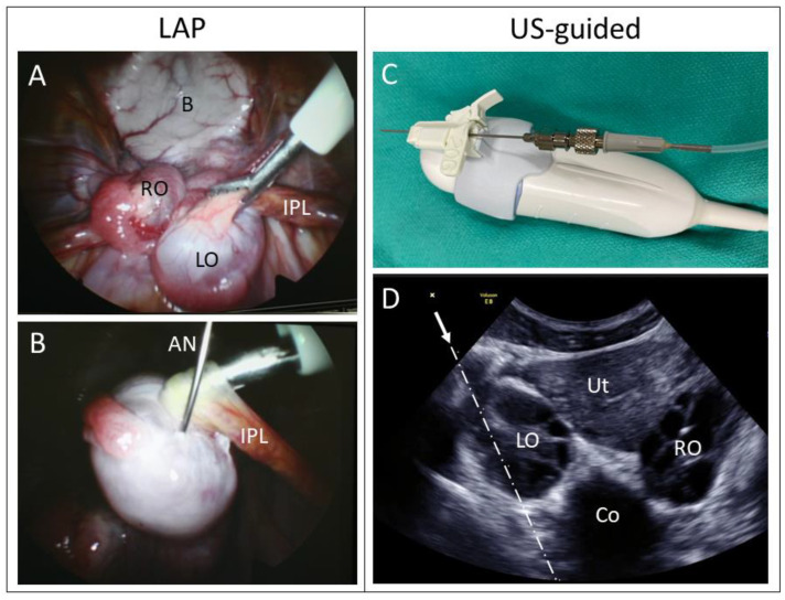
Obtaining quality oocytes is a prerequisite for ART-based studies. Here we describe a method for transabdominal ultrasound-guided (US) oocyte retrieval in rhesus macaques (Macaca mullata) and compare it to the standard surgical approach using laparoscopy (LAP). We analyzed oocyte yield from six continuous reproductive seasons (2017-2023) that included n = 177 US-guided and n = 136 laparoscopic oocyte retrievals. While the ultrasound-guided technique retrieved significantly fewer oocytes on average (LAP: 40 ± 2 vs. US: 27 ± 1), there was no difference in the number of mature metaphase II oocytes (MII) between the two techniques (LAP: 17 ± 1 vs. US: 15 ± 1). We show that oocytes retrieved by the ultrasound-guided approach fertilize at the same rates as those obtained via the laparoscopic procedure (LAP Fert Rate: 84% ± 2% vs. US Fert Rate: 83% ± 2%). In conclusion, minimally invasive ultrasound-guided oocyte retrieval improves animal welfare while delivering equivalent numbers of mature oocytes, which are ideal for ART. Furthermore, we show that oocyte competency, as represented by fertilization rate, is not affected by retrieval technique. Therefore, the Oregon National Primate Research Center (ONPRC) has adopted the ultrasound-guided approach as the standard technique for oocyte retrieval.

Keywords: animal welfare; assisted reproductive technology; laparoscopy; non-human primate; oocyte retrieval; ultrasound-guided.


Conflict of interest statement

The authors declare no conflict of interest. The funders had no role in the design of this study, in the collection, analysis, or interpretation of data, in the writing of the manuscript, or in the decision to publish the results.

Figures

Figure 1
Figure 1
(AC) Different stages of oocytes from Rhesus macaques were found at the time of retrieval in order of their stages of maturation. (A) An immature oocyte with a clearly visible germinal vesicle (arrow). (B) An immature oocyte that has resumed meiosis to metaphase 1 (MI). The germinal vesicle is broken down, and there are no other morphological markers. (C) The mature oocyte called MII is identified by an extruded polar body (arrow) and is susceptible to fertilization. (D) A fertilized oocyte (2pN) with two pronuclei indicated by an arrow.
Figure 2
Figure 2
(A,B) Laparoscopic views into the Rhesus macaque pelvis:. (A) Forceps grasp the infundibulopelvic ligament (IPL) to stabilize the stimulated left ovary (LO). The right ovary (RO) and bladder (B) can be seen in the background. (B) The aspiration needle (AN) within a follicle of a stimulated ovary. (C,D) Pictures from an ultrasound-guided procedure. (C) The 3D/4D ultrasound transducer with an attached needle guide adaptor. The 20 ga. aspiration needle is inserted through the needle guide. (D) Ultrasound image of the uterus (Ut), right ovary (RO), left ovary (LO), and colon (Co). Note the honeycomb appearance of the multiple sonolucent follicles within the stimulated rhesus ovary. The arrow indicates the trajectory (dashed line) of the aspiration needle.
Figure 3
Figure 3
Comparison of oocyte yield from laparoscopic (LAP, n = 136) and ultrasound-guided (US, n = 177) retrievals. The mean number of total oocytes retrieved is significantly less using the US-guided approach. However, this result is only manifested by the numbers of immature oocytes (MI and GV) retrieved, while the mean number of mature MII oocytes is similar between both retrieval techniques. (* indicates a significant p value < 0.05).

References

    1. Nakamura T., Fujiwara K., Saitou M., Tsukiyama T. Non-Human Primates as a Model for Human Development. Stem Cell Rep. 2021;16:1093–1103. doi: 10.1016/j.stemcr.2021.03.021. - DOI - PMC - PubMed
    1. Chan A.W.S. Progress and Prospects for Genetic Modification of Nonhuman Primate Models in Biomedical Research. ILAR J. 2013;54:211–223. doi: 10.1093/ilar/ilt035. - DOI - PMC - PubMed
    1. Ryu J., Statz J.P., Chan W., Burch F.C., Brigande J.V., Kempton B., Porsov E.V., Renner L., McGill T., Burwitz B.J., et al. CRISPR/Cas9 Editing of the MYO7A Gene in Rhesus Macaque Embryos to Generate a Primate Model of Usher Syndrome Type 1B. Sci. Rep. 2022;12:1–13. doi: 10.1038/s41598-022-13689-x. - DOI - PMC - PubMed
    1. Wikland M., Enk L., Hammarberg K., Nilsson L. Use of a Vaginal Transducer for Oocyte Retrieval in an IVF/ET Program. J. Clin. Ultrasound. 1987;15:245–251. doi: 10.1002/jcu.1870150405. - DOI - PubMed
    1. Wiseman D.A., Short W.B., Pattinson H.A., Taylor P.J., Fleetham J.A., Mortimer S.T. Oocyte Retrieval 4 Methods in Human. Radiology. 1989;173:99–102. doi: 10.1148/radiology.173.1.2528782. - DOI - PubMed