. 2023 Sep 27;13(19):3030.
doi: 10.3390/ani13193030.

Intravenous Injection of Sodium Hyaluronate Diminishes Basal Inflammatory Gene Expression in Equine Skeletal Muscle

Affiliations

Intravenous Injection of Sodium Hyaluronate Diminishes Basal Inflammatory Gene Expression in Equine Skeletal Muscle

Savannah R Gregg et al. Animals (Basel). .

Abstract

Following strenuous exercise, skeletal muscle experiences an acute inflammatory state that initiates the repair process. Systemic hyaluronic acid (HA) is injected to horses routinely as a joint anti-inflammatory. To gain insight into the effects of HA on skeletal muscle, adult Thoroughbred geldings (n = 6) were injected with a commercial HA product weekly for 3 weeks prior to performing a submaximal exercise test. Gluteal muscle (GM) biopsies were obtained before and 1 h after exercise for gene expression analysis and HA localization. The results from RNA sequencing demonstrate differences in gene expression between non-injected controls (CON; n = 6) and HA horses. Prior to exercise, HA horses contained fewer (p < 0.05) transcripts associated with leukocyte activity and cytokine production than CON. The performance of exercise resulted in the upregulation (p < 0.05) of several cytokine genes and their signaling intermediates, indicating that HA does not suppress the normal inflammatory response to exercise. The transcript abundance for marker genes of neutrophils (NCF2) and macrophages (CD163) was greater (p < 0.05) post-exercise and was unaffected by HA injection. The anti-inflammatory effects of HA on muscle are indirect as no differences (p > 0.05) in the relative amount of the macromolecule was observed between the CON and HA fiber extracellular matrix (ECM). However, exercise tended (p = 0.10) to cause an increase in ECM size suggestive of muscle damage and remodeling. The finding was supported by the increased (p < 0.05) expression of CTGF, TGFβ1, MMP9, TIMP4 and Col4A1. Collectively, the results validate HA as an anti-inflammatory aid that does not disrupt the normal post-exercise muscle repair process.

Keywords: equine; exercise; hyaluronic acid; inflammation; skeletal muscle.


Conflict of interest statement

The authors declare no conflict of interest.

Figures

Figure 1
Figure 1
DEGs were identified between CON and HA treatment groups. The majority of genes were common between groups. Here, 155 genes were unique to HA and 446 genes were unique to CON.
Figure 2
Figure 2
Gene ontology (GO) terms associated with inflammatory DEGs affected by HA treatment. Enrichment analysis was performed with clusterProfiler and the GO terms with the greatest numbers of DEGs are shown. The gene count histogram is scaled to size, with the largest blue dots containing at least 60 DEGs and the smallest fewer than 20 DEGs per GO term.
Figure 3
Figure 3
Inflammatory DEGs downregulated by HA are not refractile to exercise-induced stress. A heat map was constructed comparing pre- (PR_1-5) and post-exercise (PT_1-5) reads per kilobase million for selected DEGs (right column) in HA-treated horses. Row Z-scores range from −2-fold lower (red) to +2-fold greater (green). Dendrogram denotes average linkage between genes with distance measured by Spearman rank correlation.
Figure 4
Figure 4
Neutrophil and macrophage invasion was apparent in the muscle tissue at 1 h post-exercise. An increased expression of cell markers NCF2 ((A); p = 0.05) and CD163 ((B); p < 0.01) was observed. No change in dendritic cell marker FLT3 (p > 0.05) was observed (C).
Figure 5
Figure 5
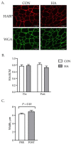
Exercise affects the structure of the basal lamina. HA was localized to the basal lamina (WGA) of muscle fibers with HABP (A). The ratio of HABP:WGA was calculated and used as an estimate of basal lamina composition (HA:ECM) (B). The width of the basal lamina was measured throughout the GM samples of HA and CON horses before and after exercise (C). HA injection did not alter the relative quantity of HA within the ECM of muscle fibers before or after exercise. Exercise tended to increase (p = 0.10) the size of the ECM.
Figure 6
Figure 6
Genes associated with ECM remodeling are upregulated by exercise. Select genes for growth factors (CTGF, TGFβ1, decorin), ECM proteases (MMP9, MMP14, TIMP4) and collagens (Col3A1, Col4A1) involved in remodeling were examined before (PRE) and 1 h after (POST) exercise. ** denotes significance at p < 0.01; *** denotes significance at p < 0.001.

References

    1. Gupta R.C., Lall R., Srivastava A., Sinha A. Hyaluronic Acid: Molecular Mechanisms and Therapeutic Trajectory. Front. Vet. Sci. 2019;6:192. doi: 10.3389/fvets.2019.00192. - DOI - PMC - PubMed
    1. Salathia S., Gigliobianco M.R., Casadidio C., Martino P.D., Censi R. Hyaluronic Acid-Based Nanosystems for CD44 Mediated Anti-Inflammatory and Antinociceptive Activity. Int. J. Mol. Sci. 2023;24:7286. doi: 10.3390/ijms24087286. - DOI - PMC - PubMed
    1. Yasuda T. Hyaluronan Inhibits Cytokine Production by Lipopolysaccharide-Stimulated U937 Macrophages through down-Regulation of NF-ΚB via ICAM-1. Inflamm. Res. 2007;56:246–253. doi: 10.1007/s00011-007-6168-5. - DOI - PubMed
    1. Voynow J.A., Zheng S., Kummarapurugu A.B. Glycosaminoglycans as Multifunctional Anti-Elastase and Anti-Inflammatory Drugs in Cystic Fibrosis Lung Disease. Front. Pharmacol. 2020;11:1011. doi: 10.3389/fphar.2020.01011. - DOI - PMC - PubMed
    1. Babasola O., Rees-Milton K.J., Bebe S., Wang J., Anastassiades T.P. Chemically Modified N-Acylated Hyaluronan Fragments Modulate Proinflammatory Cytokine Production by Stimulated Human Macrophages. J. Biol. Chem. 2014;289:24779–24791. doi: 10.1074/jbc.M113.515783. - DOI - PMC - PubMed