. 2023 Sep 22;15(19):4101.
doi: 10.3390/nu15194101.

Maternal Iron Deficiency and Environmental Lead (Pb) Exposure Alter the Predictive Value of Blood Pb Levels on Brain Pb Burden in the Offspring in a Dietary Mouse Model: An Important Consideration for Cumulative Risk in Development

Affiliations

Maternal Iron Deficiency and Environmental Lead (Pb) Exposure Alter the Predictive Value of Blood Pb Levels on Brain Pb Burden in the Offspring in a Dietary Mouse Model: An Important Consideration for Cumulative Risk in Development

Janine Cubello et al. Nutrients. .

Abstract

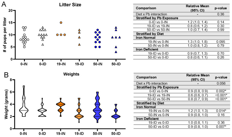
Maternal iron deficiency (ID) and environmental lead (Pb) exposure are co-occurring insults that both affect the neurodevelopment of offspring. Few studies have investigated how ID affects brain-region-specific Pb accumulations using human-relevant Pb concentrations. Furthermore, how these Pb exposures impact blood and brain Fe levels remains unclear. Importantly, we also wanted to determine whether the use of blood Pb levels as a surrogate for the brain Pb burden is affected by underlying iron status. We exposed virgin Swiss Webster female mice to one of six conditions differing by iron diet and Pb water concentration (0 ppm, 19 ppm, or 50 ppm lead acetate) and used Inductively Coupled Plasma Mass Spectrometry to measure the maternal and offspring circulating, stored, and brain Pb levels. We found that maternal ID rendered the offspring iron-deficient anemic and led to a region-specific depletion of brain Fe that was exacerbated by Pb in a dose-specific manner. The postnatal iron deficiency anemia also exacerbated cortical and hippocampal Pb accumulation. Interestingly, BPb levels only correlated with the brain Pb burden in ID pups but not in IN offspring. We conclude that ID significantly increases the brain Pb burden and that BPb levels alone are insufficient as a clinical surrogate to make extrapolations on the brain Pb burden.

Keywords: Pb burden; Swiss Webster; anemia; metals; micronutrients; neurodevelopment; pregnancy; risk assessment.


Conflict of interest statement

The authors declare no conflict of interest. The funders had no role in the design of the study; in the collection, analyses, or interpretation of data; in the writing of the manuscript; or in the decision to publish the results.

Figures

Figure 1
Figure 1
Schematic of animal exposure paradigm and micro-isolated tissues (Created with BioRender.com; accessed on 1 August 2023).
Figure 2
Figure 2
Effects of ID diet and Pb on maternal blood iron indices. Original data values are graphed for maternal hematocrits (A), hemoglobin concentrations (B), and venous blood Fe levels (C). Statistical analyses were performed using log-link gamma GLMs, separated by either iron deficiency or Pb exposure. The test for interaction was based on a single overarching log-link gamma GLM including both diet and Pb exposure plus their interaction; a significant interaction indicates evidence that the relative means for diet (ID vs. IN) differ by Pb exposure, and the relative means for Pb exposure (19 vs. 0, 50 vs. 0) differ by diet. Statistically significant differences are indicated with “*” in tables adjacent to graphs. Abbreviations: HCT = hematocrit; RBC = red blood cell; HGB = hemoglobin concentration; g/dL = grams per deciliter; µg/dL = micrograms per deciliter.
Figure 3
Figure 3
Effect of ID diet and Pb exposure on maternal blood and femoral Pb. Original data values are graphed for Pb levels in the femurs (A) and blood (B) of exposed dams. Statistical analyses were performed using log-link gamma GLMs, separated by either iron deficiency or Pb exposure and adjusted for sex and litter size. The test for interaction was based on a single overarching log-link gamma GLM including both diet and Pb exposure plus their interaction, as well as sex and litter size; a significant interaction indicates evidence that the relative means for diet (ID vs. IN) differ by Pb exposure, and the relative means for Pb exposure (50 vs. 19) differ by diet. Statistically significant differences are indicated with “*” in tables adjacent to graphs. Abbreviations: µg/g = micrograms per gram; µg/dL = micrograms per deciliter.
Figure 4
Figure 4
Effects of ID diet and Pb on litter size and offspring weights. Original data values are graphed for litter sizes (A) and weights of offspring (B). Statistical analyses were performed for both using log-link gamma GLMs, separated by either iron deficiency or Pb exposure, but weight analyses were additionally adjusted for sex and litter size. The test for interaction was based on a single overarching log-link gamma GLM including both diet and Pb exposure plus their interaction, as well as sex and litter size in weight analyses; a significant interaction indicates evidence that the relative means for diet (ID vs. IN) differ by Pb exposure, and the relative means for Pb exposure (19 vs. 0, 50 vs. 0) differ by diet. Within each treatment group is a minimum of 1 animal per sex across 4–14 (for litter size analyses) or 3–7 (for weights analyses) independent litters. Statistically significant differences are indicated with “*” in tables adjacent to graphs.
Figure 5
Figure 5
Effects of ID diet and Pb on offspring hematocrit and hemoglobin values. Original data values are graphed for hematocrits (A) and hemoglobin levels (B) in trunk blood of offspring. Statistical analyses were performed using log-link gamma GLMs, separated by either iron deficiency or Pb exposure, adjusted for sex and litter size. The test for interaction was based on a single overarching log-link gamma GLM including both diet and Pb exposure plus their interaction, as well as sex and litter size; a significant interaction indicates evidence that the relative means for diet (ID vs. IN) differ by Pb exposure, and the relative means for Pb exposure (19 vs. 0, 50 vs. 0) differ by diet. Within each treatment group is a minimum of 1 animal per sex across 3–7 independent litters. Statistically significant differences are indicated with “*” in tables adjacent to graphs. Abbreviations: HCT = hematocrit; RBC = red blood cell; HGB = hemoglobin concentration; g/dL = grams per deciliter.
Figure 6
Figure 6
Effects of ID diet and Pb on offspring circulating and brain Fe levels. Original data values are graphed for circulating (A) and brain Fe levels in the cerebral cortex (B) and hippocampus (C) of offspring. Statistical analyses were performed using log-link gamma GLMs, separated by either iron deficiency or Pb exposure, adjusted for sex and litter size. The test for interaction was based on a single overarching log-link gamma GLM including both diet and Pb exposure plus their interaction, as well as sex and litter size; a significant interaction indicates evidence that the relative means for diet (ID vs. IN) differ by Pb exposure, and the relative means for Pb exposure (19 vs. 0, 50 vs. 0) differ by diet. Within each treatment group is a minimum of 1 animal per sex across 3–7 independent litters. Statistically significant differences are indicated with “*” in tables adjacent to graphs. Abbreviations: µg/dL = micrograms per deciliter; µg/g = micrograms per gram.
Figure 7
Figure 7
Effects of ID diet and Pb on offspring circulating and stored Pb levels. Original data values are graphed for circulating blood (A) and stored femoral (B) Pb levels in offspring. Statistical analyses were performed amongst the 19 ppm and 50 ppm Pb acetate-exposed groups using log-link gamma GLMs, separated by either iron deficiency or Pb exposure, adjusted for sex and litter size. The test for interaction was based on a single overarching log-link gamma GLM including both diet and Pb exposure plus their interaction, as well as sex and litter size; a significant interaction indicates evidence that the relative means for diet (ID vs. IN) differ by Pb exposure, and the relative means for Pb exposure (50 vs. 19) differ by diet. Within each treatment group is a minimum of 1 animal per sex across 3–7 independent litters. Statistically significant differences are indicated with “*” in tables adjacent to graphs. Abbreviations: µg/dL: micrograms per deciliter; µg/g = micrograms per gram.
Figure 8
Figure 8
Effects of ID diet and Pb on offspring brain Pb burden. Original data values are graphed for brain Pb levels in the cerebral cortex (A) and hippocampus (B). Statistical analyses were performed amongst the 19 ppm and 50 ppm Pb acetate-exposed groups using log-link gamma GLMs, separated by either iron deficiency or Pb exposure, adjusted for sex and litter size. The test for interaction was based on a single overarching log-link gamma GLM including both diet and Pb exposure plus their interaction, as well as sex and litter size; a significant interaction indicates evidence that the relative means for diet (ID vs. IN) differ by Pb exposure, and the relative means for Pb exposure (50 vs. 19) differ by diet. Within each treatment group is a minimum of 1 animal per sex across 3–7 independent litters. Statistically significant differences are indicated with “*” in tables adjacent to graphs. Abbreviation: µg/g = micrograms per gram.
Figure 9
Figure 9
Log–log scale scatterplots of Pb concentrations in cerebral cortex and hippocampus by BPb concentration, stratified by diet. Least squares lines are superimposed on the data for each diet group, and the corresponding Pearson’s product–moment correlation coefficients (r) and p-values for the log-transformed Pb concentrations are annotated in the graph. Statistically significant differences are indicated with “*”. Abbreviations: µg/g = micrograms per gram; µg/dL = micrograms per deciliter.

Similar articles

Cited by

References

    1. WHO . The Global Prevalence of Anaemia in 2011. WHO; Geneva, Switzerland: 2015.
    1. Iglesias-Vazquez L., Gimeno M., Coronel P., Caspersen I.H., Basora J., Arija V. Maternal factors associated with iron deficiency without anaemia in early pregnancy: ECLIPSES study. Ann. Hematol. 2023;102:741–748. doi: 10.1007/s00277-023-05123-7. - DOI - PMC - PubMed
    1. Auerbach M., Munro M.G. Iron Deficiency in Pregnancy and Postpartum: It Is Time for a Change. Obstet. Gynecol. 2023;141:1046–1048. doi: 10.1097/AOG.0000000000005209. - DOI - PubMed
    1. Hua M., Shi D., Xu W., Zhu L., Hao X., Zhu B., Shu Q., Lozoff B., Geng F., Shao J. Differentiation between fetal and postnatal iron deficiency in altering brain substrates of cognitive control in pre-adolescence. BMC Med. 2023;21:167. doi: 10.1186/s12916-023-02850-6. - DOI - PMC - PubMed
    1. Barks A.K., Liu S.X., Georgieff M.K., Hallstrom T.C., Tran P.V. Early-Life Iron Deficiency Anemia Programs the Hippocampal Epigenomic Landscape. Nutrients. 2021;13:3857. doi: 10.3390/nu13113857. - DOI - PMC - PubMed