. 2023 Sep 25;28(19):6796.
doi: 10.3390/molecules28196796.

Discovery of Novel Mono-Carbonyl Curcumin Derivatives as Potential Anti-Hepatoma Agents

Affiliations

Discovery of Novel Mono-Carbonyl Curcumin Derivatives as Potential Anti-Hepatoma Agents

Weiya Cao et al. Molecules. .

Abstract

Curcumin possesses a wide spectrum of liver cancer inhibition effects, yet it has chemical instability and poor metabolic properties as a drug candidate. To alleviate these problems, a series of new mono-carbonyl curcumin derivatives G1-G7 were designed, synthesized, and evaluated by in vitro and in vivo studies. Compound G2 was found to be the most potent derivative (IC50 = 15.39 μM) compared to curcumin (IC50 = 40.56 μM) by anti-proliferation assay. Subsequently, molecular docking, wound healing, transwell, JC-1 staining, and Western blotting experiments were performed, and it was found that compound G2 could suppress cell migration and induce cell apoptosis by inhibiting the phosphorylation of AKT and affecting the expression of apoptosis-related proteins. Moreover, the HepG2 cell xenograft model and H&E staining results confirmed that compound G2 was more effective than curcumin in inhibiting tumor growth. Hence, G2 is a promising leading compound with the potential to be developed as a chemotherapy agent for hepatocellular carcinoma.

Keywords: AKT inhibition; anti-hepatoma activity; curcumin derivatives; molecular docking; xenograft model.


Conflict of interest statement

The authors declare that they have no known competing financial interests or personal relationships that could have appeared to influence the work reported in this paper.

Figures

Figure 1
Figure 1
The SAR analysis and the structures of the most important compounds.
Figure 2
Figure 2
Synthesis of the curcumin derivatives G1–G7. Reagents and conditions: (a) acetone, aqueous NaOH, ethanol, 48 h; (b) SOCl2, 80 °C, reflux, 2 h; (c) CHCl3, triethylamine, 0 °C to room temperature, 12 h.
Figure 3
Figure 3
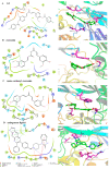
The interaction pattern between derivative G2 (A), curcumin (B), mono-carbonyl curcumin (C), endogenous ligand (D), and AKT protein shows the electrostatic surface in the 2D and 3D representations from molecular docking. G2 (green) and key residues (red) are represented as sticks and colored by atom type.
Figure 4
Figure 4
Anti-proliferation assay to HepG2 cells in vitro. (AC) Curcumin derivatives G1G7 at the concentrations of 5, 10, 15 μM for 48 h compared with curcumin and sorafenib. (D) G2 and curcumin at different concentrations (0.5, 1, 2, 4, 8, 16, 32, 64 μM) for 48 h. (E) Clone formation assay to HepG2 cells treatment with G2 (0, 1, 2, 4 μM) for 2 weeks. Data are represented by the mean ± SD of three independent experiments. *** p < 0.001, compared with the control group.
Figure 5
Figure 5
Wound healing assay (A) and transwell migration assay (B) to HepG2 cells treatment with DMEM medium containing the indicated concentrations (0, 2.5, 5, 10 μM) of G2 for 24 h and 48 h. Data are represented by the mean ± SD of three independent experiments. *** p < 0.001, ** p < 0.01, * p < 0.05, compared with the 0 μM group.
Figure 6
Figure 6
(A) Analysis of apoptotic HepG2 cells by JC-1 staining assay. The cell was treated with G2 (0, 2.5, 5, 10 μM) for 48 h, and then stained with JC-1 for 20 min at 37 °C. The representative images are shown of the corresponding fluorescent channel (40× g magnification). (B) Apoptosis-related proteins Bcl-2 and caspase 3 protein levels were determined by Western blotting with β-actin as the reference. Data are represented by the mean ± SD of three independent experiments. *** p < 0.001, * p < 0.05, compared with the 0 μM group.
Figure 7
Figure 7
Effect of derivative G2 (0, 2.5, 5, 10 μM) on Akt protein at the cell level via Western blot. Data are represented by the mean ± SD of three independent experiments. *** p < 0.001, compared with the 0 μM group.
Figure 8
Figure 8
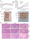
Derivative G2 inhibited HepG2 xenograft growth in vivo compared with control and curcumin. Body weight (A) and tumor volume (B) changes in mice were examined every 3 days for 21 days during treatment. (C) Visible tumor formation and photographs of representative tumors removed from mice after treatment. (D) The xenograft tumor tissues of nude mice were observed and photographed through H&E staining (100×, 200×, and 400×). Data are represented by the mean ± SD of three independent experiments.

Similar articles

Cited by

References

    1. Zhao S., Pi C., Ye Y., Zhao L., Wei Y. Recent advances of analogues of curcumin for treatment of cancer. Eur. J. Med. Chem. 2019;180:524–535. doi: 10.1016/j.ejmech.2019.07.034. - DOI - PubMed
    1. Agrawal D.K., Mishra P.K. Curcumin and its analogues: Potential anticancer agents. Med. Res. Rev. 2010;30:818–860. doi: 10.1002/med.20188. - DOI - PubMed
    1. Noorafshan A., Ashkani-Esfahani S. A review of therapeutic effects of curcumin. Curr. Pharm. Des. 2013;19:2032–2046. - PubMed
    1. Ireson C.R., Jones D.J.L., Orr S., Coughtrie M.W.H., Boocock D.J., Williams M.L., Farmer P.B., Steward W.P., Gescher A.J. Metabolism of the cancer chemopreventive agent curcumin in human and rat intestine. Cancer Epidemiol. Biomark. Prev. 2002;11:105–111. - PubMed
    1. Anand P., Kunnumakkara A.B., Newman R.A., Aggarwal B.B. Bioavailability of curcumin: Problems and promises. Mol. Pharm. 2007;4:807–818. doi: 10.1021/mp700113r. - DOI - PubMed