. 2023 Sep 30;28(19):6895.
doi: 10.3390/molecules28196895.

Antioxidant and Anti-Aging Phytoconstituents from Faucaria tuberculosa: In Vitro and In Silico Studies

Affiliations

Antioxidant and Anti-Aging Phytoconstituents from Faucaria tuberculosa: In Vitro and In Silico Studies

Hayam S Ahmed et al. Molecules. .

Abstract

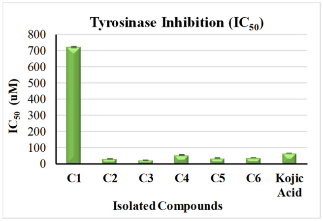
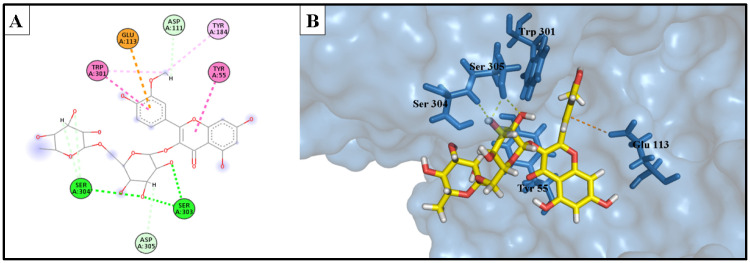
Research targeting natural cosmeceuticals is now increasing due to the safety and/or limited side effects of natural products that are highly valued in cosmetology. Within a research program exploring botanical sources for valuable skincare antioxidant components, the current study investigated the phytochemical content and the biological potential of Faucaria tuberculosa. Phytochemical investigation of F. tuberculosa extract resulted in purification and characterization of six phytoconstituents, including a new one. The structure of the new constituent was elucidated as (-) catechin-(2→1',4→2')-phloroglucinol (4). The structural identity of all isolated compounds were confirmed on the basis of extensive physical and spectral (1D, 2D-NMR and HRESIMS) investigations. The ethanolic extract exhibits a rich content of total phenolics (TPC) and total flavonoids (TFC), estimated as 32 ± 0.034 mg GAE/g and 43 ± 0.004 mg RE/g, respectively. In addition, the antioxidant (ABTS and FRAP), antihyaluronidase and antityrosinase activities of all purified phytoconstituents were evaluated. The results noted (-) catechin-(2→1',4→2') phloroglucinol (4) and phloroglucinol (1) for their remarkable antioxidant activity, while isorhamnetin 3-O-rutinoside (3) and 3,5-dihydroxyphenyl β-D-glucopyranoside (2) achieved the most potent inhibitory activity against tyrosinase (IC50 22.09 ± 0.7 µM and 29.96 ± 0.44 µM, respectively) and hyaluronidase enzymes (IC50 49.30 ± 1.57 µM and 62.58 ± 0.92, respectively) that remarkably exceeds the activity of the standard drugs kojic acid (IC50 = 65.21 ± 0.47 µM) and luteolin, (IC50 = 116.16 ± 1.69 µM), respectively. A molecular docking study of the two active compounds (3 and 2) highlighted their high potential to bind to the active sites of the two enzymes involved in the study.

Keywords: Faucaria tuberculosa; anioxidant; anti-aging; hyaluronidase enzyme; molecular docking simulation; tyrosinase enzyme.


Conflict of interest statement

The authors declare that they have no conflict of interest.

Figures

Figure 1
Figure 1
Structures of the phytoconstituents (1–6) from F. tuberculosa.
Figure 2
Figure 2
Key HMBC (blue colour) and NOESY (red colour) correlations.
Figure 3
Figure 3
Anti-tyrosinase activity of the phytoconstituents from F. tuberculosa: (C1) phloroglucinol, (C2) phlorin, (C3) isorhamnetin 3-O-rutinoside, (C4) (-) catechin-(2→1′,4→2′)-phloroglucinol, (C5) isorhamnetin 3-O-[α-rhamnopyranosyl-(1→4)-α-rhamnopyranosyl-(1→6)-β-glucopyranoside], (C6) β-sitosterol.
Figure 4
Figure 4
Anti-hyaluronidase activity of the phytoconstituents from F. tuberculosa: (C1) phloroglucinol, (C2) phlorin, (C3) isorhamnetin 3-O-rutinoside, (C4) (-) catechin-(2→1′,4→2′)-phloroglucinol, (C5) isorhamnetin 3-O-[α-rhamnopyranosyl-(1→4)-α-rhamnopyranosyl-(1→6)-β-glucopyranoside], (C6) β-sitosterol.
Figure 5
Figure 5
Phlorin, isorhamnetin 3-O-rutinoside and kojic acid bound to the active domain of tyrosinase enzyme (PDB: 2Y9X): (A) 3D binding presentation of tyrosinase enzyme; (B) magnified 3D binding mode showing the active site of a tyrosinase enzyme containing two copper ions and the three docked ligands (phlorinin in red, isorhamnetin 3-O-rutinoside in blue, and kojic acid in yellow). The figure was generated using BIOVIA Discovery Studio 2021.
Figure 6
Figure 6
(A) 2D interaction diagram of the top docking pose of phlorin into the active site of the tyrosinase enzyme; (B) 3D interaction diagram of the top docking pose of phlorin into the active site of the tyrosinase enzyme.
Figure 7
Figure 7
(A) Two-dimensional interaction diagram of the top docking pose of isorhamnetin 3-O-rutinoside into the active site of the tyrosinase enzyme; (B) Three-dimensional interaction diagram of the top docking pose of isorhamnetin 3-O-rutinoside into the active site of the tyrosinase enzyme.
Figure 8
Figure 8
(A) Two-dimensional interaction diagram of the top docking pose of kojic acid into the active site of the tyrosinase enzyme; (B) Three-dimensional interaction diagram of the top docking pose of kojic acid into the active site of the tyrosinase enzyme.
Figure 9
Figure 9
Phlorin, isorhamnetin 3-O-rutinoside and luteolin bound to the active domain of the hyaluronidase enzyme (PDB: 1FCV): (A) 3D binding presentation of the hyaluronidase enzyme; (B) magnified 3D binding mode showing active site of the hyaluronidase enzyme containing the three docked ligands (phlorin in red, isorhamnetin 3-O-rutinoside in blue, and luteolin in yellow). The figure was generated using BIOVIA Discovery Studio 2021.
Figure 10
Figure 10
(A) Two-dimensional interaction diagram of the top docking pose of phlorin into the active site of the hyaluronidase enzyme; (B) Three-dimensional interaction diagram of the top docking pose of phlorin into the active site of the hyaluronidase enzyme.
Figure 11
Figure 11
(A) Two-dimensional interaction diagram of the top docking pose of isorhamnetin 3-O-rutinoside into the active site of the hyaluronidase enzyme; (B) Three-dimensional interaction diagram of the top docking pose of F3 into the active site of the hyaluronidase enzyme.
Figure 12
Figure 12
(A) Two-dimensional interaction diagram of the top docking pose of luteolin into the active site of the hyaluronidase enzyme; (B) Three-dimensional interaction diagram of the top docking pose of luteolin into the active site of the hyaluronidase enzyme.
Figure 13
Figure 13
Tyrosinase enzyme: co-crystallized (light green) and re-docked (yellow) ligands’ superimposition.

Similar articles

References

    1. Abd El-Raouf H.S. Taxonomic significance of leaves in family Aizoaceae. Saudi J. Biol. Sci. 2021;28:512–522. doi: 10.1016/j.sjbs.2020.10.036. - DOI - PMC - PubMed
    1. Bakr R.O. A Comprehensive Review of the Aizoaceae Family: Phytochemical and Biological Studies. Nat. Prod. J. 2021;11:288–304. doi: 10.2174/2210315510666200206154000. - DOI
    1. Glavich T. Beginner’s Guide to Faucaria. Cactus Succul. J. 2016;88:290–292. doi: 10.2985/015.088.0605. - DOI
    1. Schwantes G., Higgins V. Flowering Stones and Mid-Day Flowers: A Book for Plant and Nature Lovers on the Mesembryanthemaceae. E. Benn; London, UK: 1957.
    1. Graf A.B. Pictorial Cyclopedia of Exotic Plants from Tropical and Near-Tropic Regions: Guide to Care of Plants Indoors, Horticultural Color Guide, Plant Geography. Roehr; Cincinnati, OH, USA: 1976.

LinkOut - more resources