. 2023 Dec;13(12):e3275.
doi: 10.1002/brb3.3275. Epub 2023 Oct 14.

Inhibitory effect and mechanism of Rosiglitazone on M1 type polarization of central microglia in intracerebral hemorrhage mice based on JNK/STAT3 signaling pathway

Affiliations

Inhibitory effect and mechanism of Rosiglitazone on M1 type polarization of central microglia in intracerebral hemorrhage mice based on JNK/STAT3 signaling pathway

Chenglei Chao et al. Brain Behav. 2023 Dec.

Abstract

Background: Intracerebral hemorrhage (ICH) seriously threatens the health of people. In addition, microglia M1 polarization was confirmed to be involved in the progression of ICH. Rosiglitazone was able to be used as an antidiabetic agent, which could activate PPAR-γ, and PPAR-γ was reported to inhibit inflammation in microglia. However, the detailed function of Rosiglitazone in ICH remains unclear.

Methods: In vivo and in vitro experiments were used to test the function of Rosiglitazone in ICH. In addition, RT-qPCR and western blot were performed to evaluate the mRNA and protein level of PPAR-γ, respectively. Immunofluorescence staining was performed to detect the levels of CD206 and CD86, and ELISA was used to measure the levels of pro-inflammatory cytokines.

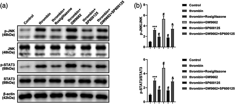
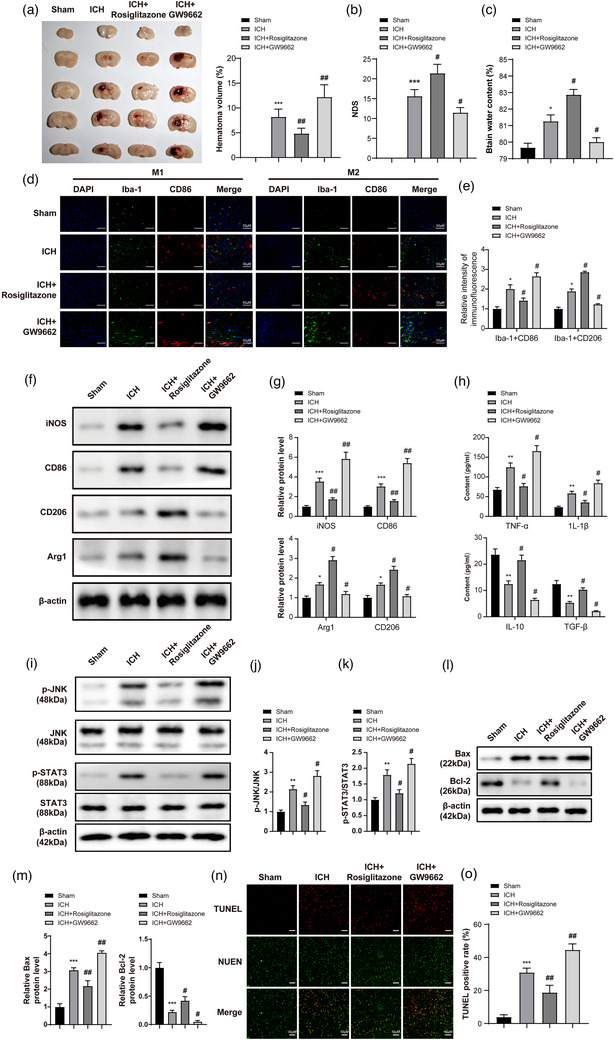
Results: PPAR-γ was downregulated in ICH mice, whereas p-JNK and p-STAT3 were upregulated. Thrombin notably downregulated the level of PPAR-γ in BV2 cells, whereas Rosiglitazone partially reversed this phenomenon. In addition, Rosiglitazone markedly reversed thrombin-induced microglia M1 polarization. Consistently, thrombin-induced inflammatory response in BV2 cells was abolished in the presence of Rosiglitazone. SP600125 (JNK/STAT3 inhibitor) greatly reversed thrombin-induced M1 polarization in microglia, and GW9662 abolished the effect of SP600125. Meanwhile, Rosiglitazone could inactivate JNK/STAT3 pathway through the upregulation of PPAR-γ. Furthermore, Rosiglitazone notably alleviated the symptom of ICH in vivo through inhibiting the apoptosis and mediating PPAR-γ/JNK/STAT3 axis.

Conclusion: Rosiglitazone could attenuate the inflammation in ICH through inhibiting microglia M1 polarization. Thus, our research would shed now lights on exploring new therapeutic strategies against ICH.

Keywords: JNK/STAT3; PPAR-γ; Rosiglitazone; intracerebral hemorrhage.


Conflict of interest statement

These authors declared no conflicts of interest in this study.

Figures

FIGURE 1
FIGURE 1
PPAR‐γ was downregulated in intracerebral hemorrhage (ICH), whereas JNK/STAT3 signaling was upregulated. (a) At the end of animal study, the brain tissues of mice were collected and pictured. The hematoma volume of tissues was detected. (b) The neurological deficit score (NDS) of mice was evaluated. (c) The brain water content of mice was calculated. (d) The protein levels of JNK, p‐JNK, STAT3, p‐STAT3, and PPAR‐γ in tissues of mice were assessed by western blot. (e) The relative expressions of p‐JNK and p‐STAT3 were quantified by normalizing to the total protein. The relative level of PPAR‐γ was quantified by normalizing to β‐actin. n = 5 per group. * p < .05, ** p < .01 compared to sham.
FIGURE 2
FIGURE 2
Rosiglitazone inhibited microglia M1 polarization through the upregulation of PPAR‐γ. BV2 cells were exposed to thrombin, thrombin + Rosiglitazone or thrombin + GW9662. (a and b) The levels of CD86 and CD206 in BV2 cells were assessed by immunofluorescence staining. The scale bar is 100 μm. (c) The protein levels of CD86, CD206, Arg1, and iNOS in BV2 cells were assessed by western blot. (d) The relative levels of CD86, CD206, Arg1, and iNOS were quantified by normalizing to β‐actin. (e) The levels of IL‐1β, TNF‐α, TGF‐β, and IL‐10 in BV2 cell supernatants were assessed by ELISA. * p < .05, ** p < .01, *** p < .001 compared to control. # p < .05, ## p < .01 compared to thrombin.
FIGURE 3
FIGURE 3
Downregulation of PPAR‐γ reversed JNK/STAT3 signaling‐induced microglia M1 polarization. BV2 cells were exposed to thrombin, thrombin + SP600125 or thrombin + SP600125 + GW9662. (a and b) The levels of CD86 and CD206 in BV2 cells were assessed by immunofluorescence staining. The scale bar is 100 μm. (c) The protein levels of CD86, CD206, Arg1, and iNOS in BV2 cells were assessed by western blot. (d) The relative levels of CD86, CD206, Arg1, and iNOS were quantified by normalizing to β‐actin. (e) The levels of IL‐1β, TNF‐α, TGF‐β, and IL‐10 in BV2 cell supernatants were assessed by ELISA. * p < .05, ** p < .01 compared to thrombin. # p < .05 compared to thrombin + SP600125.
FIGURE 4
FIGURE 4
Rosiglitazone inactivated JNK/STAT3 signaling through the upregulation of PPAR‐γ. BV2 cells were exposed to thrombin, thrombin + Rosiglitazone, thrombin + GW9662, thrombin + SP600125, or thrombin + SP600125 + GW9662. (a) The protein levels of JNK, p‐JNK, STAT3, and p‐STAT3 in BV2 cells were assessed by western blot. (b) The relative expressions of p‐JNK and p‐STAT3 were quantified by normalizing to the total protein. *** p < .001 compared to control. # p < .05 compared to thrombin. & p < .05 compared to thrombin + SP600125.
FIGURE 5
FIGURE 5
Rosiglitazone alleviated the progression of intracerebral hemorrhage (ICH) in vivo through mediation of PPAR‐γ/JNK/STAT3 axis. (a) At the end of animal study, the brain tissues of mice were collected and pictured. The hematoma volume of tissues was detected. (b) The neurological deficit score (NDS) of mice was evaluated. (c) The brain water content of mice was calculated. (d and e) The levels of CD86 and CD206 in mice were assessed by immunofluorescence staining. The scale bar is 100 μm. (f) The protein levels of CD86, CD206, Arg1, and iNOS in BV2 cells were assessed by western blot. (g) The relative levels of CD86, CD206, Arg1, and iNOS were quantified by normalizing to β‐actin. (h) The levels of IL‐1β, TNF‐α, TGF‐β, and IL‐10 in BV2 cell supernatants were assessed by ELISA. (i) The protein levels of JNK, p‐JNK, STAT3, p‐STAT3, and PPAR‐γ in mice were assessed by western blot. (j and k) The relative levels of p‐JNK and p‐STAT3 were quantified by normalizing to the total protein. (l and m) The protein levels of Bcl‐2 and Bax in mice were detected by western blotting. The relative levels of CD86, CD206, Arg1, and iNOS were quantified by normalizing to β‐actin. (n and o) The apoptosis in tissues of mice was tested by TUNEL staining. n = 5 per group. * p < .05, ** p < .01, *** p < .001 compared to sham. # p < .05, ## p < .01 compared to ICH.

References

    1. Cheng, W.‐L. , Zhang, Q. , Li, Bo. , Cao, J.‐L. , Jiao, L. , Chao, S.‐P. , Lu, Z. , & Zhao, F. (2021). PAK1 silencing attenuated proinflammatory macrophage activation and foam cell formation by increasing PPAR‐gamma expression. Oxidative Medicine and Cellular Longevity, 2021, 6957900. - PMC - PubMed
    1. Deng, S. , Chen, X. , Lei, Q. , & Lu, W. (2022). AQP2 promotes astrocyte activation by modulating the TLR4/NFkappaB‐p65 pathway following intracerebral hemorrhage. Frontiers in Immunology, 13, 847360. - PMC - PubMed
    1. Dicpinigaitis, A J. , Feldstein, E. , Shapiro, S D. , Kamal, H. , Bauerschmidt, A. , Rosenberg, J. , Amuluru, K. , Pisapia, J. , Dangayach, N S. , Liang, J W. , Bowers, C A. , Mayer, S A. , Gandhi, C D. , & Al‐Mufti, F. (2022). Cerebral vasospasm following arteriovenous malformation rupture: A population‐based cross‐sectional study. Neurosurgical Focus, [Electronic Resource], 53(1), E15. - PubMed
    1. Duan, S. , Wang, F. , Cao, J. , & Wang, C. (2020). Exosomes derived from microRNA‐146a‐5p‐enriched bone marrow mesenchymal stem cells alleviate intracerebral hemorrhage by inhibiting neuronal apoptosis and microglial M1 polarization. Drug Designs Development and Therapiae, 14, 3143–3158. - PMC - PubMed
    1. Gao, X. , Cao, Z. , Tan, H. , Li, P. , Su, W. , Wan, T. , & Guo, W. (2022). LncRNA, an emerging approach for neurological diseases treatment by regulating microglia polarization. Frontiers in Neuroscience, 16, 903472. - PMC - PubMed

Publication types

MeSH terms