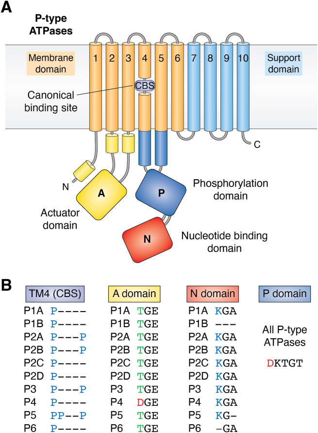
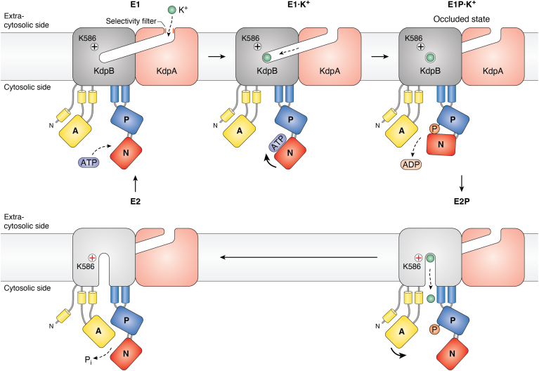
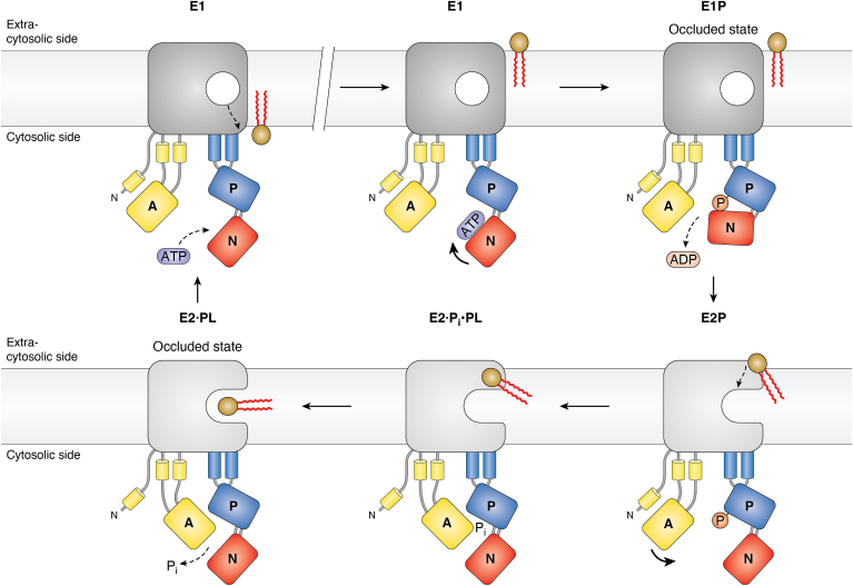
Review
. 2023 Nov;299(11):105352.
doi: 10.1016/j.jbc.2023.105352. Epub 2023 Oct 12.

P-type ATPases: Many more enigmas left to solve

Affiliations
Review

P-type ATPases: Many more enigmas left to solve

Michael Palmgren. J Biol Chem. 2023 Nov.

Abstract

P-type ATPases constitute a large ancient super-family of primary active pumps that have diverse substrate specificities ranging from H+ to phospholipids. The significance of these enzymes in biology cannot be overstated. They are structurally related, and their catalytic cycles alternate between high- and low-affinity conformations that are induced by phosphorylation and dephosphorylation of a conserved aspartate residue. In the year 1988, all P-type sequences available by then were analyzed and five major families, P1 to P5, were identified. Since then, a large body of knowledge has accumulated concerning the structure, function, and physiological roles of members of these families, but only one additional family, P6 ATPases, has been identified. However, much is still left to be learned. For each family a few remaining enigmas are presented, with the intention that they will stimulate interest in continued research in the field. The review is by no way comprehensive and merely presents personal views with a focus on evolution.

Keywords: P-type ATPases; evolution; mechanism; primary active transport.


Conflict of interest statement

Conflict of interest The author declares to have no conflicts of interest with the contents of this article.

Figures

Figure 1
Figure 1
Overview of domain organization in P-type ATPases. A, the catalytic machinery consists of three cytosolic domains (A, P, and N) and two membrane-located domains (M and S). All P-type ATPases are phosphorylated and dephosphorylated during the catalytic cycle at an invariant aspartate residue (red letter in protein sequence) in the phosphorylation (P) domain (shaded dark blue). The phosphorylation reaction is carried out by the nucleotide-binding (N) domain (shaded red), which binds ATP and is an inbuilt protein kinase. The dephosphorylation reaction is carried out by the actuator (A) domain (shaded yellow), which is an in-built protein phosphatase. Phosphorylation and dephosphorylation of the pump cause conformational changes in the membrane domain (M) (orange helices) where the ligand(s) to be transported are bound. This domain comprises six transmembrane helices among which transmembrane helix 4 (TM4) is broken by one or more proline residues, which give room for a cavity and provide a saddle for the ligand to rest on. This ligand binding site is commonly referred to as the CBS. The support (S) domain (light blue helices) delivers structural support for the M domain and varies with respect to the number of helices and location at either the N- or C-terminal end. An autoinhibitory regulatory (R) domain may also be present at either terminal (not shown). B, conserved sequence motifs in P-type ATPase domains. The phosphorylatable aspartate (D) residue is present in a signature motif of P-type ATPases: DKTGT. The CBS in TM4 of the M domain results from helix-breaking proline (P) residues, the number of which varies from one to three depending on the P-type ATPase subfamily. The phosphatase motif in the A domain includes a negatively charged glutamate (E) residue in all pumps but in P4 ATPases also an aspartate (D) residue. The ATP binding site in the N domain includes a conserved lysine (K) residue, which is absent in P1B and P6 ATPases. CBS, canonical binding site; DKTGT, Asp-Lys-Thr-Gly-Thr.
Figure 2
Figure 2
Catalytic cycle and mechanism of a P-type ATPase with the Na+/K+-ATPase used as an example. According to the Post-Albers scheme, which has been confirmed through numerous structures in different states, P-type ATPases alternate between two major conformations termed E1 and E2. The E1 conformation has one or more high-affinity membrane-located ion binding site(s) exposed to the cytosolic side of the membrane (in this case for Na+; the size of ions is enlarged for clarity). The site(s) have a low affinity for the counter-transported ligand (here K+). Phosphorylation of the pump by ATP causes a transition to the E2 conformation, which has the ion binding site(s) exposed to the extra-cytosolic side of the membrane. Now the site(s) have low affinity for the ligand to be exported (Na+) and high affinity for the counter-transported ligand (K+) and an exchange reaction takes place. Phosphorylation of the pump is triggered by binding of the (last in case there is more than one) ligand to be transported. Dephosphorylation is triggered by the (last) ligand to be counter-transported. In this way, coupling between ATP hydrolysis and transport is assured. During the reaction cycle, it is the cytosolic phosphorylation (P) domain that is phosphorylated. See text and Figure 1 for more details.
Figure 3
Figure 3
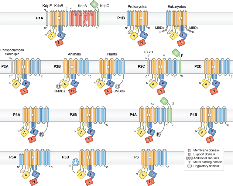
Evolution and function of P-type ATPases.A, phylogenetic tree based on core sequences of 159 P-type ATPases. The sequences used and their numbers used for constructing the phylogenetic tree are the same as in Axelsen and Palmgren (1998). Clades that represent different P-type ATPase families and sub-families are named P1A to P6. A question mark indicates that it is uncertain to which family a sequence(s) belong. The tree was constructed using the maximum likelihood method using the program RAxML and Bayesian inference analysis using the program MrBayes as described (Palmgren, 2023). Shown is the best RAxML tree after 1000 bootstrap rounds. Number at major nodes indicate bootstrap values of ≥90. A separate Bayesian inference analysis was carried out, which resulted in a similar tree. The Bayesian inference analysis was run for 3,000,000 generations with a resulting average standard deviation of split frequencies at 0.01. Black dots at nodes in the RAxML tree indicate maximum statistical support (p = 1) in the Bayesian inference analysis; open circles at nodes indicate that p ≥ 0.95. Names of some proteins from Homo sapiens, the yeast Saccharomyces cerevisiae and the plant Arabidopsis thaliana are given before numbers. Numbers refer to sequences as follows: 1, Q12697 Ypk9 (Saccharomyces cerevisiae); 2, Q27533 W08D2.5 (Caenorhabditis elegans); 3, Q21286 catp-5 (C. elegans); 4, Q04956 Probable cation-transporting ATPase 1 (Plasmodium falciparum); 5, P90747 catp-8 (C. elegans); 6, P39986 Spf1 (S. cerevisiae); 7, Q95050 TPA9 (Tetrahymena thermophila); 8, G5EBH1 tat-5 (C. elegans); 9, P40527 Neo1 (S. cerevisiae); 10, Q10309 neo1 (Schizosaccharomyces pombe); 11, Q7JPE3 tat-4 (C. elegans); 12, Q27720 Phospholipid-transporting ATPase (P. falciparum); 13, Q12674 Dnf3 (S. cerevisiae); 14, Q29449 ATP8A1 (Bos taurus); 15, P39524 Drs2 (S. cerevisiae); 16, Q09891 dnf2 (S. pombe); 17, Q12675 Dnf2 (S. cerevisiae); 18, P32660 Dnf1 (S. cerevisiae); 19, P9WPT1 ctpE (Mycobacterium tuberculosis); 20, P03960 KdpB (Escherichia coli); 21, P9WPU3 KdpB (M. tuberculosis); 22, P73867 KdpB (Synechocystis sp. PCC 6803 substr. Kazusa); 23, P94888 cadA (Lactococcus lactis); 24, Q60048 cadA (Listeria monocytogenes); 25, P30336 cadA (Alkalihalophilus pseudofirmus); 26, P37386 cadA (Staphylococcus aureus); 27, Q59998 ziaA (Synechocystis sp.); 28, Q59997 slr0797 (Synechocystis sp.); 29, Q59465 cadA (Helicobacter pylori); 30, P9WPS7 ctpG (M. tuberculosis); 31, P37617 zntA (E. coli); 32, P9WPT5 ctpC (M. tuberculosis); 33, P77871 copA (H. pylori); 34, P77868 HI_0290 (Haemophilus influenzae); 35, Q59385 copA (E. coli); 36, P05425 copB (Enterococcus hirae); 37, P37385 synA (Synechococcus elongatus); 38, P74512 Cation-transporting ATPase (Synechocystis sp.); 39, P9WPS3 ctpV (M. tuberculosis); 40, P46840 ctpB (Mycobacterium leprae); 41, P9WPU1 ctpA (M. tuberculosis); 42, P9WPT9 ctpB (M. tuberculosis); 43, P46839 ctpA (M. leprae); 44, Q59688 copA_3 (Proteus mirabilis); 45, P73241 pacS (Synechocystis sp.); 46, P37279 pacS (S. elongatus); 47, P77881 ctpA (L. monocytogenes); 48, P38995 Ccc2 (S. cerevisiae); 49, Q04656 ATP7A (Homo sapiens); 50, Q64535 Atp7b (R. norvegicus); 51, P35670 ATP7B (H. sapiens); 52, P38360 Pca1 (S. cerevisiae); 53, Q59207 fixI (Bradyrhizobium diazoefficiens); 54, P18398 fixI (Rhizobium meliloti); 55, Q59370 HRA-2 (E. coli); 56, P32113 copA (E. hirae); 57, Q59369 HRA-1 (E. coli); 58, P22036 mgtB (Salmonella typhimurium); 59, P0ABB8 mgtA (E. coli); 60, P36640 mgtA (S. typhimurium); 61, Q58623 MJ1226 (Methanocaldococcus jannaschii); 62, P54210 DHA1 (Dunaliella acidophila); 63, P54211 PMA1 (D. bioculata); 64, P54679 patB (Dictyostelium discoideum); 65, O04956 Plasma membrane ATPase (Cyanidium caldarium); 66, Q43178 PHA2 (Solanum tuberosum); 67, Q03194 PMA4 (Nicotiana plumbaginifolia); 68, P19456 AHA2 (A. thaliana); 69, P20431 AHA3 (A. thaliana); 70, Q43131 Plasma membrane ATPase (Vicia faba); 71, Q43275 zha1 (Zostera marina); 72, Q43271 MHA2 (Zea mays); 73, P93265 PMA (Mesembryanthemum crystallinum); 74, Q42556 AHA9 (A. thaliana); 75, Q43002 OSA2 (Oryza sativa); 76, Q43243 MHA1 (Z. mays); 77, Q43001 OSA1 (O. sativa); 78, Q42932 Plasma membrane ATPase (N. plumbaginifolia); 79, Q43106 BHA-1 (Phaseolus vulgaris); 80, Q43128 AHA10 (A. thaliana); 81, P12522 H1B (Leishmania donovani); 82, A0A7S3XUR3 Plasma membrane ATPase (Heterosigma akashiwo); 83, P24545 PMA1 (Zygosaccharomyces rouxii); 84, P28877 PMA1 (Candida albicans); 85, P49380 PMA1 (Kluyveromyces lactis); 86, P05030 Pma1 (S. cerevisiae); 87, P19657 Pma2 (S. cerevisiae); 88, Q92446 PCA1 (Pneumocystis carinii); 89, P28876 PMA2 (S. pombe); 90, P09627 PMA1 (S. pombe); 91, P07038 pma-1 (Neurospora crassa); 92, Q07421 PMA1 (Ajellomyces capsulatus); 93, P22189 cta3 (S. pombe); 94, P13587 Ena1 (S. cerevisiae); 95, P78981 Z-ENA1 (Zygosaccharomyces rouxii); 96, P73273 ziaA (Synechocystis sp.); 97, Q76P11 ionA (D. discoideum); 98, G5EFV6 catp-4 (C. elegans); 99, P35317 ATP1A (Hydra vulgaris); 100, P28774 Na+/K+-ATPase alpha-B (Artemia franciscana); 101, Q27461 eat-6 (C. elegans); 102, P13607 Atpalpha (Drosophila melanogaster); 103, Q27766 Na+/K+-ATPase alpha (Ctenocephalides felis); 104, P05023 ATP1A1 (H. sapiens); 105, P05025 ATP1A (Tetronarce californica); 106, P13637 ATP1A3 (H. sapiens); 107, Q92030 atp1a1 (Anguilla anguilla); 108, P25489 atp1a1 (Catostomus commersonii); 109, P50993 ATP1A2 (H. sapiens); 110, Q64541 Atp1a4 (R. norvegicus); 111, P17326 Na+/K+-ATPase alpha-A (A. franciscana); 112, P20648 ATP4A (H. sapiens); 113, Q92126 atp4a (Xenopus laevis); 114, Q64392 ATP12A (Cavia porcellus); 115, P54707 ATP12A (H. sapiens); 116, P54708 Atp12A (R. norvegicus); 117, Q92036 ATP12A (Rhinella marina); 118, Q27829 Plasma membrane calcium ATPase (Paramecium tetraurelia); 119, P93067 Calcium-transporting ATPase (Brassica oleracea); 120, Q37145 ACA1 (A. thaliana); 121, Q27642 Calcium-transporting ATPase (Entamoeba histolytica); 122, Q64542 Atp2b4 (R. norvegicus); 123, P23634 ATP2B4 (H. sapiens); 124, P20020 ATP2B1 (H. sapiens); 125, Q16720 ATP2B3 (H. sapiens); 126, Q01814 ATP2B2 (H. sapiens); 127, G5EFR6 mca-1 (C. elegans); 128, P54678 patA (D. discoideum);129, P38929 Pmc1 (S. cerevisiae); 130, P9WPS9 ctpF (M. tuberculosis); 131, P37367 pma1 (Synechocystis sp.); 132, Q64566 Atp2c1 (R. norvegicus); 133, P13586 Pmr1 (S. cerevisiae); 134, Q95022 CppA-E1 (Cryptosporidium parvum); 135, Q27724 PfATPase4 (P. falciparum); 136, Q95060 TVCA1 (Trichomonas vaginalis); 137, P54209 CA1 (Dunaliella bioculata); 138, O09489 Calcium-transporting ATPase (Leishmania amazonensis); 139, P35315 TBA1 (Trypanosoma brucei brucei); 140, Q27779 SMA1 (Schistosoma mansoni); 141, P16615 ATP2A2 (H. sapiens); 142, P70083 atp2a1 (Makaira nigricans); 143, Q92105 ATP2A1 (Pelophylax lessonae); 144, Q64578 Atp2a1 (Rattus norvegicus); 145, P18596 Atp2a3 (R. norvegicus); 146, P22700 SERCA (D. melanogaster); 147, P35316 SERCA (A. franciscana); 148, Q08853 ATP6 (P. falciparum); 149, Q27764 YEL6 (Plasmodium yoelii); 150, Q42883 LCA1 (Solanum lycopersicum); 151, O04938 Ca2+-ATPase (O. sativa); 152, P92939 ECA1 (A. thaliana); 153, Q59999 sll0672 (Synechocystis sp.); 154, P37278 pacL (S. elongatus); 155, P74062 slr0822 (Synechocystis sp.); 156, P78036 pacL (Mycoplasma pneumoniae); 157, P47317 pacL (Mycoplasma genitalium); 158, P9WPS5 ctpI (M. tuberculosis); 159, P96271 ctpH (M. tuberculosis). Scale bar, 0.2 amino acid substitutions per site. B, overview of P-type ATPase families and transported ligands. Domain and subunit organization are not shown. For each subfamily is shown the ligand transported, the number of ligands transported per ATP hydrolyzed, and the direction of transport. Not shown are subunit and transmembrane helices. A subgroup of P2A ATPases, secretory pathway Ca2+-ATPases (marked SPCA in Fig. 3A), only transport one Ca2+ per ATP hydrolyzed. Depending on the stoichiometry of transport, several P-type ATPases can be electrogenic. P2C and P3A ATPases are highly electrogenic and maintain plasma membrane potentials that are negative on the cytosolic side of the membrane. Abbreviations: PA, polyamines; PL, phospholipids; TMH, transmembrane helices.
Figure 4
Figure 4
Overview of the organization of transmembrane helices and accessory subunits in P-type ATPase family members. All P-type ATPases have in common essentially the same catalytic machinery, which comprises three major cytosolic domains (A, P and N) and a membrane-embedded domain (M) composed of six transmembrane helices (shaded orange in the figure). Depending on the P-type ATPase family, additional transmembrane helices serve a support (S) function (shaded blue). The number of transmembrane helices vary from seven in P1A ATPases to 12 in P5A ATPases. In addition to the catalytic subunit, some families have additional subunits. Animal P2A ATPases may have a small hydrophobic subunit with a single transmembrane helix (phospholamban and sarcolipin) and P4A ATPases have a β subunit with two membrane helices with a heavily glycosylated domain facing the extra-cytosolic side of the membrane. P2C ATPases have a glycosylated β subunit with a single transmembrane helix and, in addition, can have a small hydrophobic subunit with a single transmembrane helix (the γ subunit and other subunits being members of the FXYD protein family). P1B ATPases have four subunits in total, one of which (KdpA) is a modified K+ channel protein. Some families have extended terminal autoinhibitory domains that serve a regulatory (R) function. The N-termini of eukaryotic P2C, P3A, and P4A ATPases and the C-terminus of eukaryotic P2C ATPases have also been implicated in the regulation of pump activity (not shown here). CMBDs, calmodulin-binding domains; MBD, metal-binding domains.
Figure 5
Figure 5
Variations on a theme: the transport cycle of P1A ATPases. P1B ATPases import K+ ions. The complex is composed of four subunits, only two of which (KdpB and KdpA) are shown here. K+ enters the complex through the selectivity filter (a) of KdpA, which is a modified K+ channel and moves through a tunnel that connects KdpA to the catalytic subunit KdpB. When K+ enters and binds to the canonical binding site in KdpB, the pump is phosphorylated from ATP. This phosphorylation causes the pump to transition to the E2P conformation and the K+ ion diffuses into the cell through a half-channel. Whether this half-channel is open in all conformations or only in the E2 state is difficult to ascertain due to the limited resolution of the structures. Dephosphorylation and return to the E1 conformation occur spontaneously possibly due to the action of Lys-856, which functions as a built-in cation. See text for more details.
Figure 6
Figure 6
Variations on a theme: the central cavity model of P4A ATPases. P4A ATPases flip phospholipids from the extra-cytosolic side of membranes to the cytosolic side. Before transport, the E1 conformation of the pump is spontaneously phosphorylated from ATP. Pump phosphorylation results in the transition to the E2P conformation, to which the phospholipid head group binds. Binding of the phospholipid induces pump dephosphorylation and the phospholipid then slides through a cleft in the membrane domain toward the canonical binding site. In the dephosphorylated E2 conformation, the phospholipid is occluded in the membrane domain with its headgroup positioned in the CBS and its acyl chains protruding out of the protein. During the E2P to E1 transition, the phospholipid is released to the intracellular leaflet of the bilayer.

References

    1. Palmgren M.G., Nissen P. P-type ATPases. Annu. Rev. Biophys. 2011;40:243–266. - PubMed
    1. Dyla M., Kjærgaard M., Poulsen H., Nissen P. Structure and mechanism of P-type ATPase ion pumps. Annu. Rev. Biochem. 2020;89:583–603. - PubMed
    1. Pedersen P.L., Carafoli E. Ion motive ATPases. I. Ubiquity, properties, and significance to cell function. Trends Biochem. Sci. 1987;12:146–150.
    1. Stock C., Heger T., Basse Hansen S., Thirup Larsen S., Habeck M., Dieudonné T., et al. Fast-forward on P-type ATPases: recent advances on structure and function. Biochem. Soc. Trans. 2023;51:1347–1360. - PubMed
    1. Post R.L. A reminiscence about sodium, potassium-ATPase. Ann. N. Y. Acad. Sci. 1974;242:6–11. - PubMed

Publication types

MeSH terms