. 2023 Nov;30(11):2408-2431.
doi: 10.1038/s41418-023-01229-7. Epub 2023 Oct 14.

Characterizing the regulatory Fas (CD95) epitope critical for agonist antibody targeting and CAR-T bystander function in ovarian cancer

Affiliations

Characterizing the regulatory Fas (CD95) epitope critical for agonist antibody targeting and CAR-T bystander function in ovarian cancer

Tanmoy Mondal et al. Cell Death Differ. 2023 Nov.

Abstract

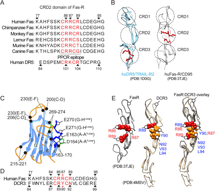
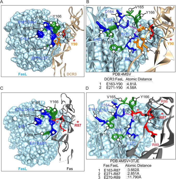
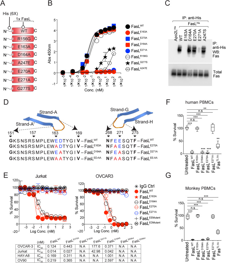
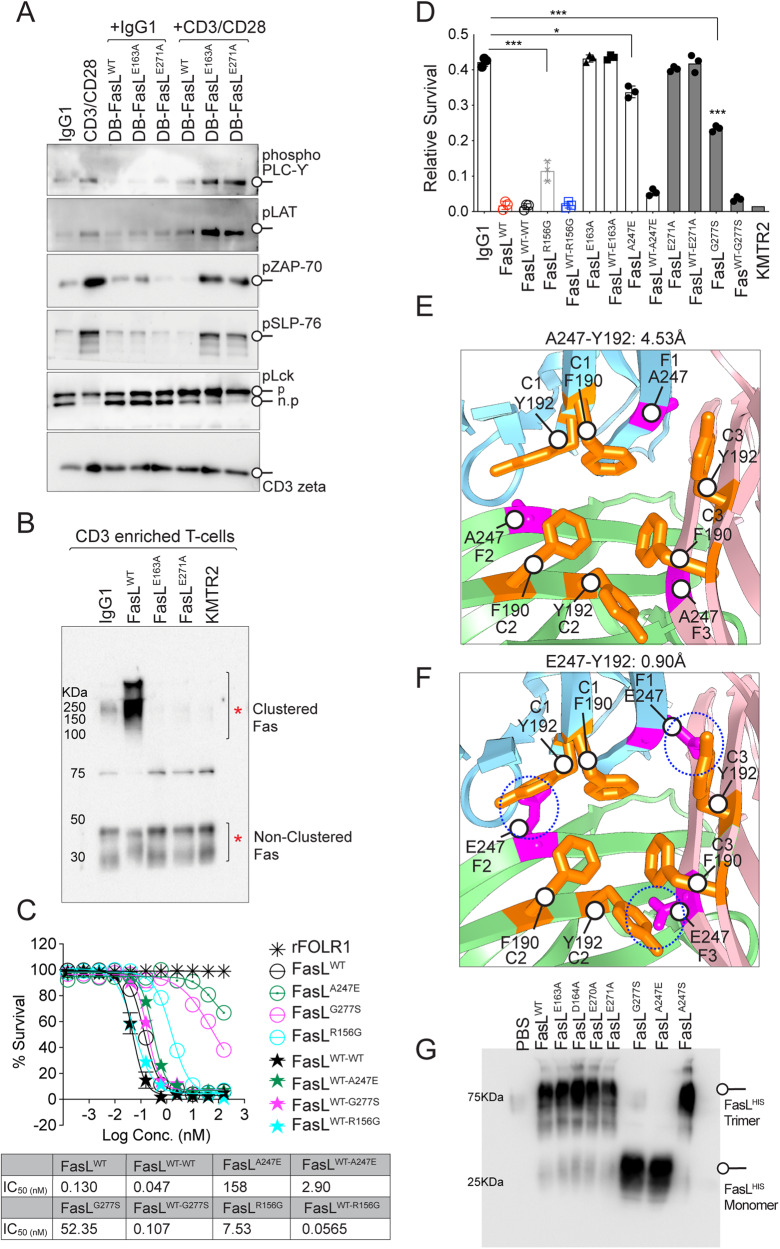
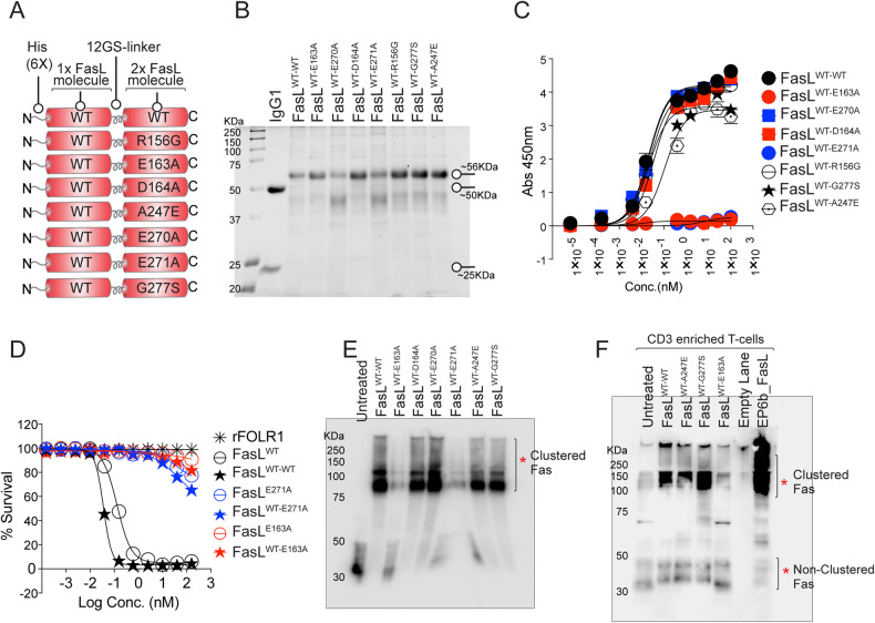
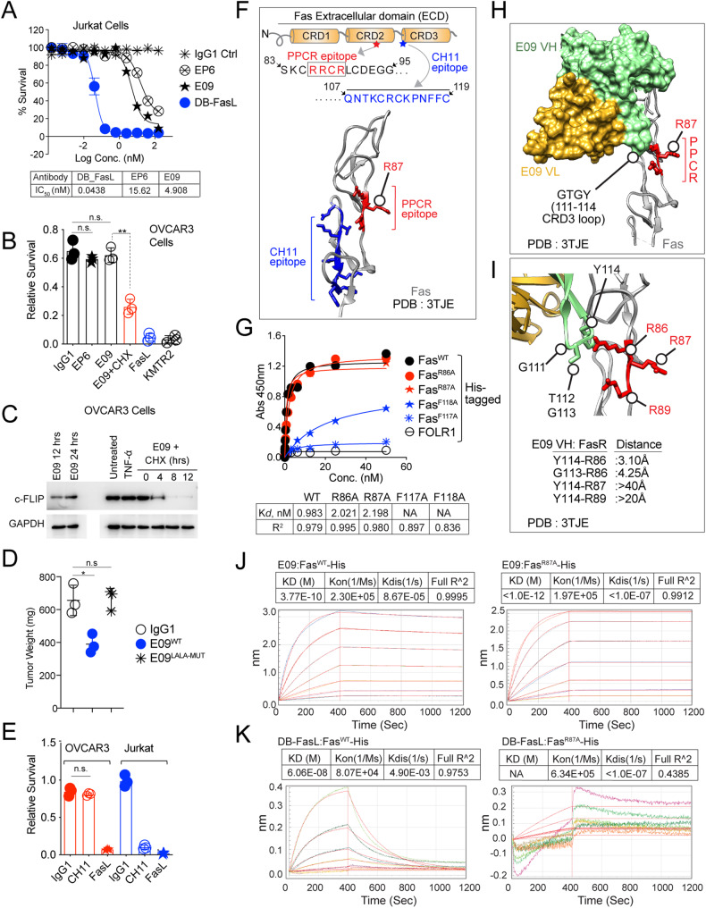
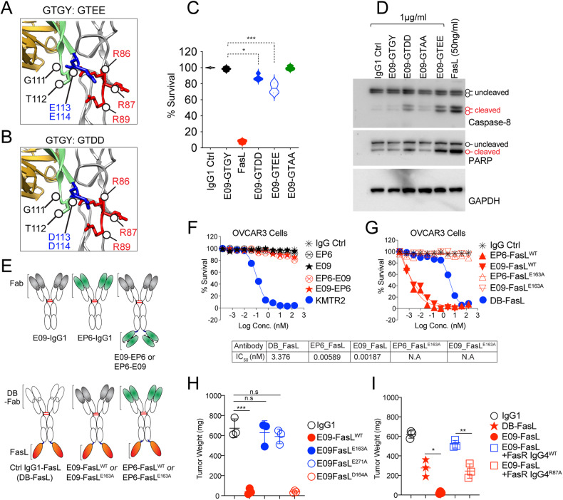
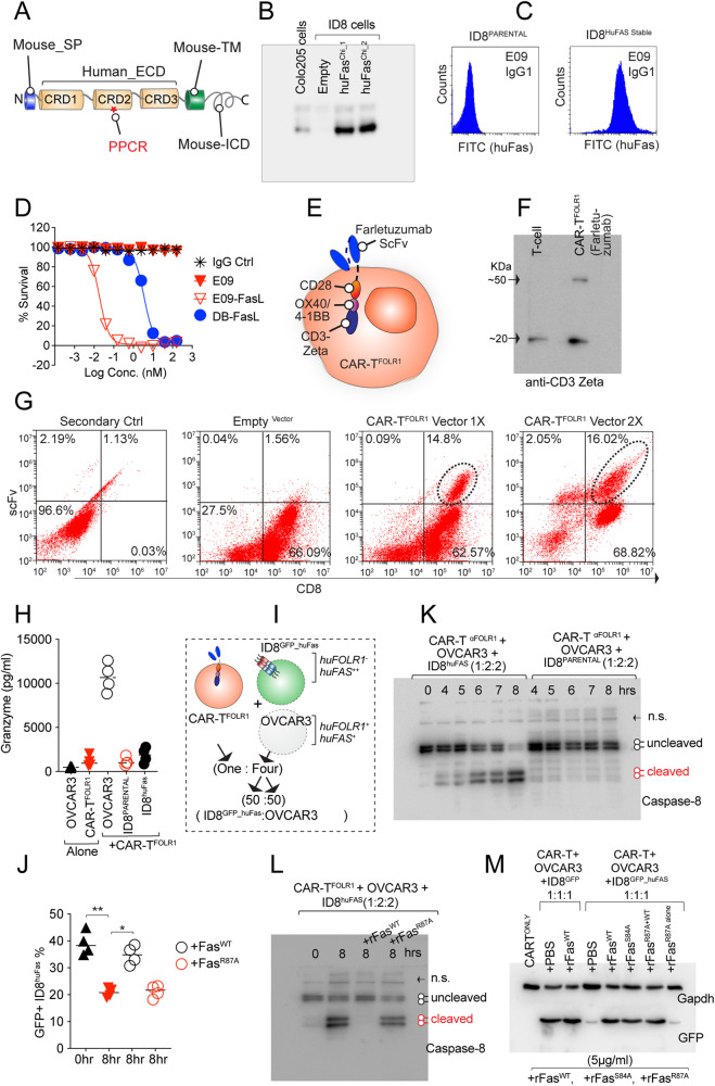
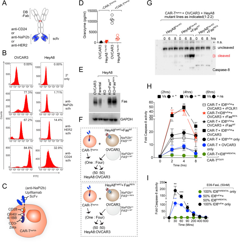
Receptor clustering is the most critical step to activate extrinsic apoptosis by death receptors belonging to the TNF superfamily. Although clinically unsuccessful, using agonist antibodies, the death receptors-5 remains extensively studied from a cancer therapeutics perspective. However, despite its regulatory role and elevated function in ovarian and other solid tumors, another tumor-enriched death receptor called Fas (CD95) remained undervalued in cancer immunotherapy until recently, when its role in off-target tumor killing by CAR-T therapies was imperative. By comprehensively analyzing structure studies in the context of the binding epitope of FasL and various preclinical Fas agonist antibodies, we characterize a highly significant patch of positively charged residue epitope (PPCR) in its cysteine-rich domain 2 of Fas. PPCR engagement is indispensable for superior Fas agonist signaling and CAR-T bystander function in ovarian tumor models. A single-point mutation in FasL or Fas that interferes with the PPCR engagement inhibited apoptotic signaling in tumor cells and T cells. Furthermore, considering that clinical and immunological features of the autoimmune lymphoproliferative syndrome (ALPS) are directly attributed to homozygous mutations in FasL, we reveal differential mechanistic details of FasL/Fas clustering at the PPCR interface compared to described ALPS mutations. As Fas-mediated bystander killing remains vital to the success of CAR-T therapies in tumors, our findings highlight the therapeutic analytical design for potentially effective Fas-targeting strategies using death agonism to improve cancer immunotherapy in ovarian and other solid tumors.


Conflict of interest statement

The authors declare no competing interests.

Figures

Fig. 1
Fig. 1. PPCR remains conserved in Fas and DR5.
A Alignment of Fas PPCR region sequences from dog to humans. B Ribbon depiction of PPCR in DR5 and Fas. The three CRD′s are marked with dotted circles. C Jelly-like structure of FasL colored blue (beta-sheets) and orange (connecting loops). D Alignment of Fas PPCR region with the corresponding sequence in DCR3. E Overlay of DCR3 (gold, PDB:4MSV) and Fas (dark gray, PDB:3TJE). Positively charged residues are shown in red spheres. The critical R87 is replaced with Y90 in DCR3.
Fig. 2
Fig. 2. FasL forms salt bridges with Fas R87 residue.
A A space-filling representation of FasL surface (sky blue) at the interface of PPCR corresponding region of DCR3 (gold color shown as ribbon structure). Blue and green surfaces in FasL highlight the negatively charged and hydrophobic residues, respectively. Fas PPCR corresponding residues of DCR3 are shown in red and orange sticks. B Zoomed interface of FasL and DCR3 at PPCR from (A). Y90 (orange stick near red asterisk) of DCR3 acts like a “key” to insert itself into hydrophobically stabilized “lock” of negatively charged residues (E163, E271). Additional hydrophobic residues of DCR3 at its Fas PPCR corresponding region are shown in orange. C Same as (A), except Fas (Gray ribbon) is overlayed instead of DCR3. Fas PPCR residues are shown in red sticks. D Zoomed interface of FasL and Fas at PPCR. Instead of tyrosine (Y90) in DCR3, R87 (red stick near red asterisk) of Fas form significantly close salt-bridges (3.66 and 2.85 Å) with FasL negatively charged residues E163 and E271. The guanidine side chains of R86 and R89 are oriented away from the FasL PPCR interface motif.
Fig. 3
Fig. 3. A single-point mutation in PPCR Interacting FasL Residues abolishes its function.
A Schematic of various His-tagged FasL mutants described in the figure. B Binding analysis of His-tagged FasL to 96-well-immobilized Fc-tagged Fas by ELISA. C OVCAR3 cells were treated with indicated His-tagged FasL, followed by immunoprecipitation using anti-His antibody and immunoblotting using anti-Fas antibody. Total Fas levels were equal in all samples. D Schematic of FasL A-A′ and G-H beta loops with corresponding residues either WT or mutated (Red) in the loops are shown. Negatively charged residues are blue. E Cell-killing assay of OVCAR3 and Jurkat cells 36 h after treatment of indicated FasL mutants. IC50 values are shown at the bottom. F, G Cell survival assay of human and murine PBMC-derived and CD3-positive T cells 48 h after treatment of indicated FasL mutants. Error bars in (B), (F) and (G) represent SEM (n = 3+).
Fig. 4
Fig. 4. Differential Fas trimerization and activation profile by ALPS and PPCR mutations.
A CD3-enriched human PBMC-derived T cells were treated with either IgG1 alone or CD3/CD28 agonist alone or indicated Fas-ligand mutants (DB-FasL) alone or together for 60 min (as indicated), followed by immunoblotting of indicated T-cell scaffold proteins and kinases. B CD3-enriched human PBMC-derived T cells were treated as indicated on top (DB-FasL mutants) for 1 h, followed by BS3 crosslinker treatment to capture the membrane-clustered Fas. Lysates were run in a non-reducing and partially denaturing gel and immunoblotted for Fas. C Jurkat cell killing assay of indicated FasL APLS mutants. rFOLR1 is a negative control. IC50 values are shown. D Same as (C), except human PBMC-derived CD3-enriched pan-T cells were used after activation with CD3/CD28 antibodies. E Ribbon diagram showing zoomed FasL trimer interface in sky blue (1), green (2), and pink (3) colors (PDB: 4MSV). Hydrophobic residues Y192 and F190 (colored orange and represented with sticks) on the C beta-strand of three FasL monomers (C1, C2, and C3) are shown. A247, with a short sidechain, is on the F beta-strands of three FasL monomers (F1, F2, and F3) and is colored pink and represented with sticks. F Same as (E), except the ALPS mutant E247 (substituted with A247 of the F beta-strands of three FasL monomers) with sidechain potentially sterically clashing with Y192 (on the C beta-strands of other FasL monomers) is shown in pink sticks. G Indicated FasL mutants on top were added to Jurkat cells in culture wells. After 30 min, soup and total lysates were pelleted together, followed by SDS-PAGE on non-reducing and partially denaturing gel and immunoblotting with anti-His antibody to detect recombinant FasL trimer formation on cells. Error bars in (D) represent SEM (n = 3+).
Fig. 5
Fig. 5. Haploinsufficiency of key ALPS mutants does not entirely block the FasLWT function.
A Schematic of genetic construction of various N-terminal His-tagged FasL mutants generated in tandem right next to FasLWT. B CHO cells expressing various tandem FasL mutants (as in A) were purified using HisTrap Ni Sepharose and were run on reducing gel along with an IgG1 for size confirmation. C Binding of indicated His-tagged tandem FasL mutants (as indicated on the right) to 96-well-immobilized Fc-tagged Fas by ELISA. D Cell survival assay of Jurkat cells after treatment with indicated His-tagged tandem FasL mutants. E Jurkat cells were treated with indicated His-tagged tandem FasL mutants on top for 1 hr, followed by low-dose BS3 treatment on ice (15 min) to capture the membrane-clustered Fas. Lysates were run in a non-reducing and partially denaturing gel, followed by Fas immunoblotting. F CD3-enriched human PBMC-derived T cells were treated with indicated His-tagged tandem FasL mutants followed by Fas clustering as described in (E).
Fig. 6
Fig. 6. Preclinical Fas agonist antibodies are ineffective in tumor cells and do not engage Fas PPCR.
A Jurkat cell killing assay of indicated Fas antibodies and dabigatran Fc-conjugated FasL, DB-FasL (see Supplementary Fig. S4A and Fig. 7 for DB-FasL design) (representative of 3). B Cell survival assay of OVCAR3 cells treated with indicated Fas agonist ± cFLIP inhibitor. DR5 agonist KMTR2 and FasL are positive control (n = 3). C Immunoblotting analysis of total cFLIP in the presence of cycloheximide ± E09 IgG1 and other controls. D 1 × 106–2 × 106 OVCAR3 tumor cells were injected subcutaneously in Athymic Nude animals with Matrigel in PBS. When tumors appeared on animals (3–4 weeks), animals were i.p. injected with indicated Fas agonists (4–6 doses, 50 mg), followed by tumor extraction and weight measurements. The same E09 IgG1 antibody was tested ± ADCC effector function (L234, L235 CH2 or L234A, L235A CH2). E Cell Survival assay of OVCAR3 and Jurkat cells treated CH11 IgM antibody, and FasL (n = 3). As a negative control, instead of IgM, IgG1 available in our lab was used. F Top: schematic of Fas ECD showing CH11 antibody epitope location in the context of PPCR. Bottom: ribbon depiction of CH11 antibody epitope (blue sticks) and PPCR (red sticks). G Binding of increasing concentrations of CH11 IgG1 against His-tagged indicated Fas PPCR mutants and F117A and F118A (CH11 epitope) mutants in a 96-well-immobilized ELISA assay. H A surface model interface of E09-Fas antibody against Fas PPCR (PDB:3TJE). VH (green) and VL (gold) are shown as space-filling models, while Fas (Gray) is in a ribbon structure. PPCR residues are shown as red sticks. I Same as (H) except for the E09 VH CDR3 loop with GTGY residues (green) and PPCR (red sticks) is focused with shown atomic distances. J The binding kinetics of immobilized His-tagged FasWT and FasR87A against the E09 antibody was measured using biolayer interferometry (BLI). K The binding kinetics of immobilized His-tagged FasWT and FasR87A against DB-FasL (see Supplementary Fig. S4A and Fig. 7 for DB-FasL design) were measured using BLI. Error bars in (A), (B), (D), and (E) represent SEM (n = 3).
Fig. 7
Fig. 7. Negative charge substitution in CRD3 of E09 VH partly restores its Fas agonist activity in tumor cells.
A, B The zoomed-in structure of Fas ECD region in complex with E09 IgG1. The interface of the E09 Fab against Fas PPCR is highlighted (PDB:3TJE). VH (green), VL (gold), and Fas (gray) are as depicted as ribbons. WT E09 contains GTGY (green sticks) in the CDR3 loop (see Supplementary Fig. 6I). GTGY residues of the CRD3 loop were mutated to GTEE or GTDD at 111–114 positions for the experiments described in (C) and (D). C 0.5 µg/ml WT E09 IgG1 (GTGY) or E09 IgG1 (GTEE or GTDD or GTAA) at 111–114 positions of E09 VH CRD3 along with other controls were added onto OVCAR3 in cell survival analysis post 36 h (n = 3+). D Same as (C), except indicated molecules (FasL at 20-fold lower concentration) were added for 10 hrs, followed by immunoblotting for caspase-8 and PARP. E Schematic showing genetic construction of generation of Fas agonist (E09, EP6) bispecific antibodies and antibody ligand bispecific combinations. F A 96-well plate OVCAR3 cell killing assay of indicated E09, EP6 Fas agonist monospecific antibodies, and E09-EP6 combination bispecific antibodies. KMTR2 was used as a positive control of killing. G Same as (F), except E09-FasL and EP6-FasL bispecific combinations were used with FasLWT or FasLE163A. IC50 values are shown (n = 3). H 1 × 106–1.5 × 106 OVCAR3 tumor cells were injected subcutaneously in NOD.Cg-PrkdcscidIl2rgtm1Wjl/SzJ animals with Matrigel in PBS. When tumors appeared on animals (3–4 weeks), animals were i.p. injected with indicated E09-FasL bispecific combinations either with FasLWT or FasLE163A or FasLD164A or FasLE271A (n = 3+). I Same as (H), except 4-fold recombinant Fas-IgG4WT and Fas-IgG4R87A proteins were co-injected in indicated cases along with E09-FasL (n = 3+). Error bars in (H) and (I) represent SEM (n = 3+).
Fig. 8
Fig. 8. Fas PPCR R87 is critical for CAR-T bystander function.
A Cartoon schematic of chimeric Fas with human ectodomain and murine TM, ICD, and signal peptide. B Immunoblotting confirmation of murine ID8 cell clones expressing huFas chimeric clones 1 and 2. C E09 IgG1 Flow cytometry confirmation of murine ID8 cells expressing huFas ectodomain. D Confirmation of cytotoxic sensitivity of chimeric huFas stable murine ID8 cells against Fas agonist reagents. E Cartoon schematic of anti-FOLR1 (Farletuzumab) expressing CAR-T (CAR-TFOLR1). F Immunoblotting confirmation of CAR-TFOLR1 construct expression in T cells using CD3 zeta antibody. G Flow cytometry using goat anti-human Fab specific antibody after two rounds of CAR-T construction lentiviral infections (represents n = 3). H CAR-TFOLR1 cells were co-cultured either alone or with OVCAR3 cells or ID8Parental cells, or ID8huFas stable cells at an effector/target (E/T) ratio 5:1 for 4 h followed by granzyme B measurement using ELISA (n = 4). I Schematic of CAR-TFOLR1 cells co-cultured with the 50:50 mix of GFP+ ID8huFas stable cells and OVCAR3. In additional sets, cultured plates were precoated either with recombinant rFasWT or rFasR87A (5 μg/ml). J %GFP flow cytometry signal as an indicator of FOLR1 antigen negative but GFP positive ID8huFas stable cell lysis from the experiment described in I (n = 3). K Same as (J) and (I), except in one additional set, a 50:50 mix of ID8Parental (huFas-) cells and OVCAR3 were used for indicated times. Immunoblotting analyzed the total cell lysates for cleaved caspase-8 levels. L Same as (K), except additional controls were included via precoating the culture plates with rFasWT or rFasR87A (5 μg/ml) (n = 3). M Same as (K) and (L), except GFP levels (as an indicator of ID8huFas stable cell lysis) were analyzed using immunoblotting. Error bars in (J) represent SEM (n = 3+).
Fig. 9
Fig. 9. CAR-T-targeting antigen-positive cells next to antigen-negative cells in heterogenous cultures are crucial for optimal caspase-8 activation.
A Schematic of anti-CD24, anti-NaPi2b, and anti-HER2 scFvs (in blue) conjugated to idarucizumab-Fc (DB-Fc) used for flow cytometry in (B). B Flow cytometry analysis of OVCAR3 and Hey-A8 cells using a list of scFv described in (A). C Cartoon schematic of anti-NaPi2b (Upifitamab) expressing CAR-T (CAR-TNaPi2b). D OVCAR3 cells or Hey-A8 FasKO cells were co-cultured either alone or with CAR-TNaPi2b cells at 5:1 ratio for 4 h followed by granzyme B measurement using ELISA (n = 3+). E Immunoblotting confirmation Fas expression in Hey-A8PARENTAL, Hey-A8FasKO, and Hey-A8FasKO transfected with FasWT and FasR87A. OVCAR3 control lysates were used side-by-side. F Schematic of CAR-TNaPi2b T cells co-cultured with the 50:50 mix of Hey-A8FasKO overexpressing exogenous FasWT (top) or overexpressing exogenous FasR87A (bottom) and OVCAR3 cells. G Same as (F), except total lysates of indicated combinations were immunoblotted for caspase-8 cleavage. H In one set, CAR-TFOLR1 cells were co-cultured either with huFOLR1 antigen+ OVCAR3 alone (blue circle) or huFOLR1 antigen- ID8PARENTAL alone (green circle) or huFOLR1 antigen- ID8huFas stable (black circle) alone. In the second set, CAR-TFOLR1 cells were co-cultured with a 50:50 mix of huFOLR1 antigen+ OVCAR3+ huFOLR1 antigen- ID8huFas stable in the presence of indicated precoated recombinant proteins [rFasWT or rFasR87A or rFOLR1 (5 μg/ml)]. After 2, 4, and 8 hr, all sets were analyzed for caspase-8 assays. I E09-FasL bispecific antibody was added for indicated times either to the 50:50 mix of GFP+ ID8huFas stable and ID8 regular cells or 100% GFP+ ID8huFas stable alone cells followed by caspase-8 activity analysis. Error bars in (H) and (I) represent SD (n = 3+).

References

    1. Ashkenazi A. Directing cancer cells to self-destruct with pro-apoptotic receptor agonists. Nat Rev Drug Discov. 2008;7:1001–12. - PubMed
    1. Annibaldi A, Walczak H. Death receptors and their ligands in inflammatory disease and cancer. Cold Spring Harb Perspect Biol. 2020;12:a036384. - PMC - PubMed
    1. Shivange G, Mondal T, Lyerly E, Bhatnagar S, Landen CN, Reddy S, et al. A patch of positively charged residues regulates the efficacy of clinical DR5 antibodies in solid tumors. Cell Rep. 2021;37:109953. - PMC - PubMed
    1. Du G, Zhao L, Zheng Y, Belfetmi A, Cai T, Xu B, et al. Autoinhibitory structure of preligand association state implicates a new strategy to attain effective DR5 receptor activation. Cell Res. 2023;33:131–46. - PMC - PubMed
    1. Adams C, Totpal K, Lawrence D, Marsters S, Pitti R, Yee S, et al. Structural and functional analysis of the interaction between the agonistic monoclonal antibody Apomab and the proapoptotic receptor DR5. Cell Death Differ. 2008;15:751–61. - PubMed

Publication types