. 2023 Dec 5:261:115854.
doi: 10.1016/j.ejmech.2023.115854. Epub 2023 Oct 4.

Development of potent isoflavone-based formyl peptide receptor 1 (FPR1) antagonists and their effects in gastric cancer cell models

Affiliations

Development of potent isoflavone-based formyl peptide receptor 1 (FPR1) antagonists and their effects in gastric cancer cell models

Fabio Francavilla et al. Eur J Med Chem. .

Abstract

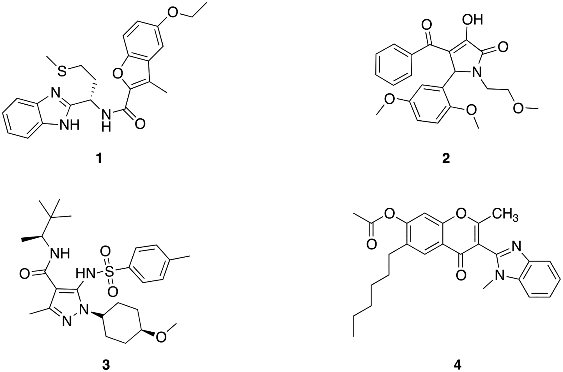
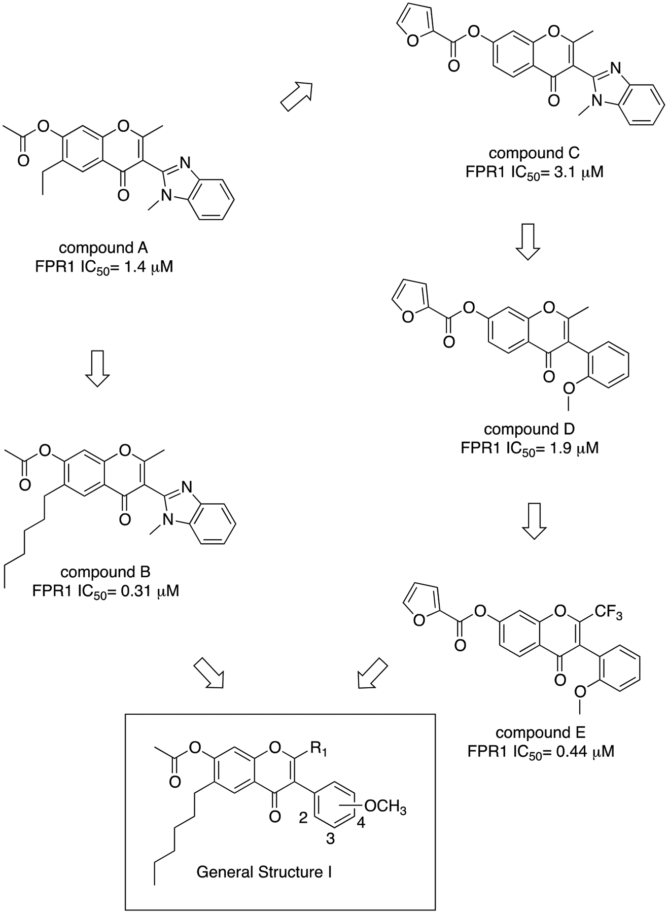
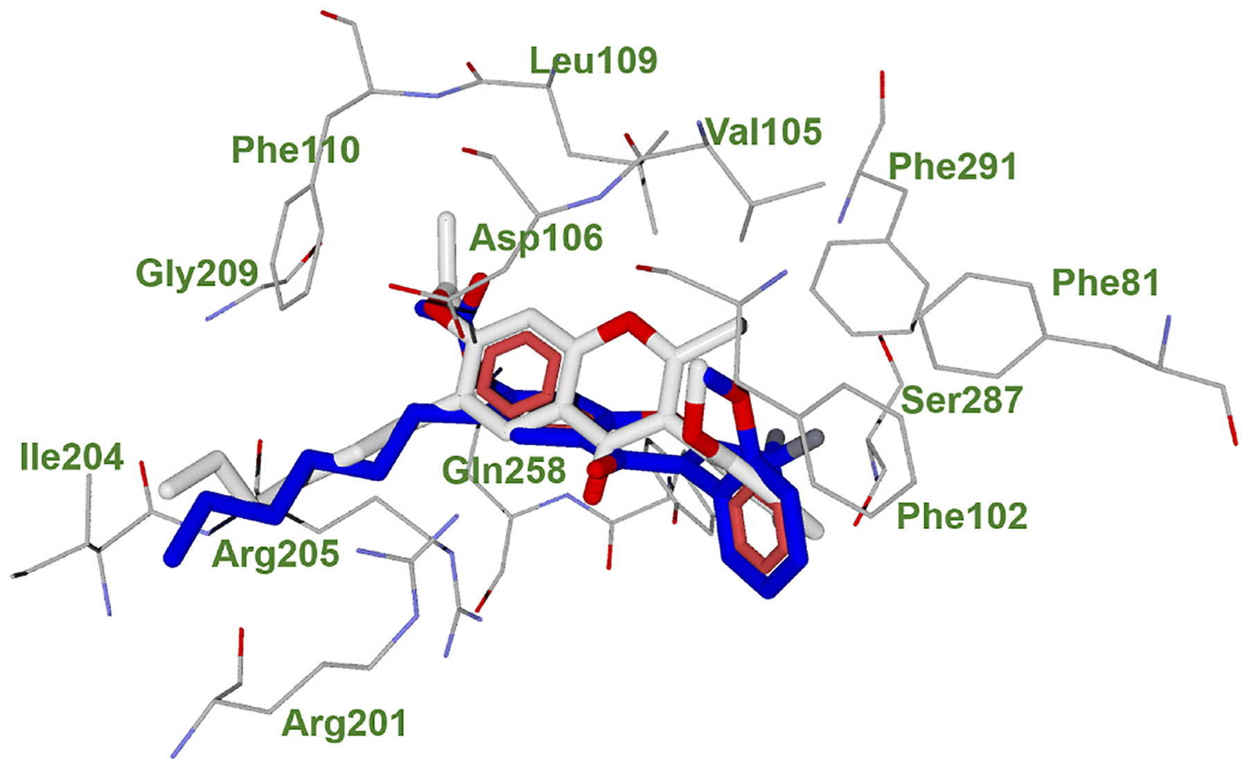
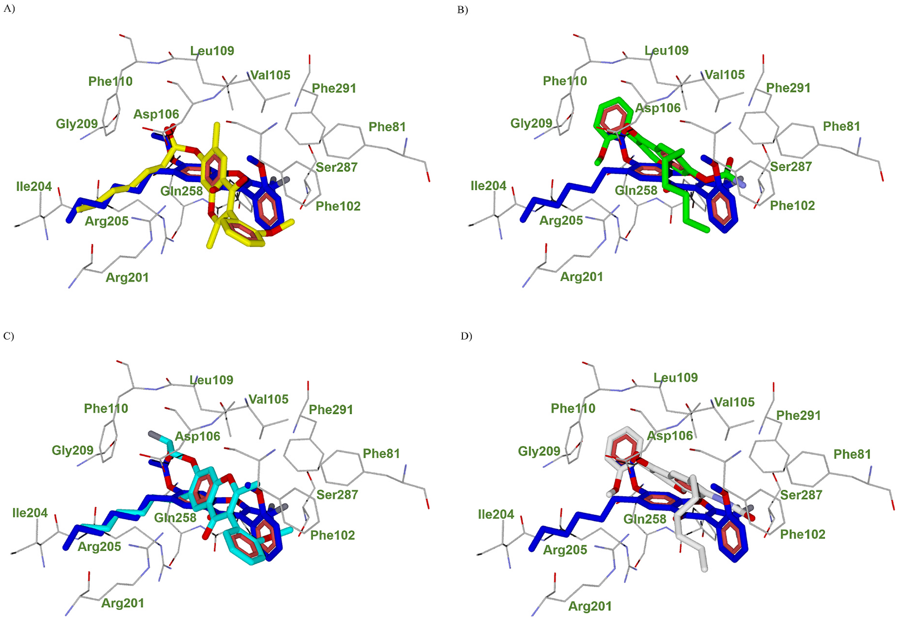
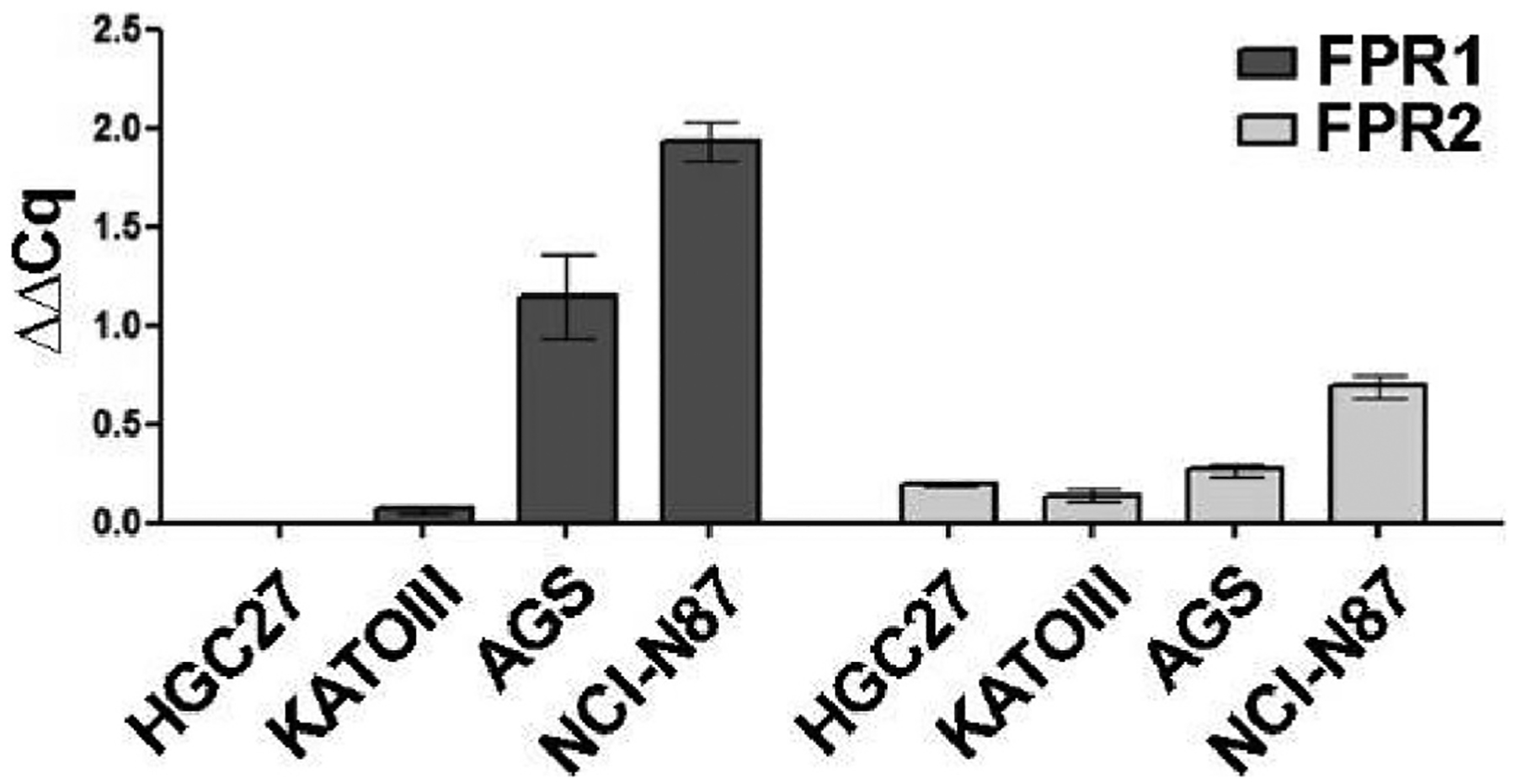
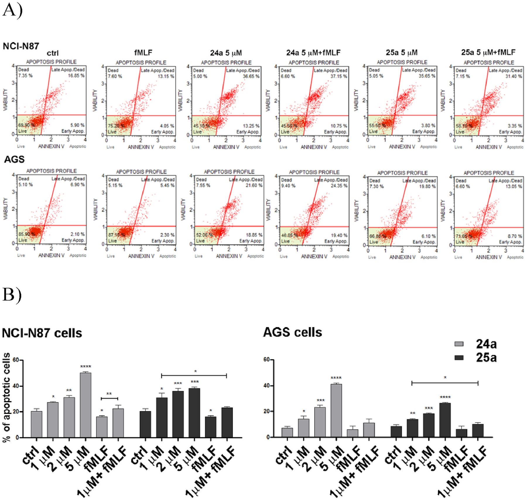
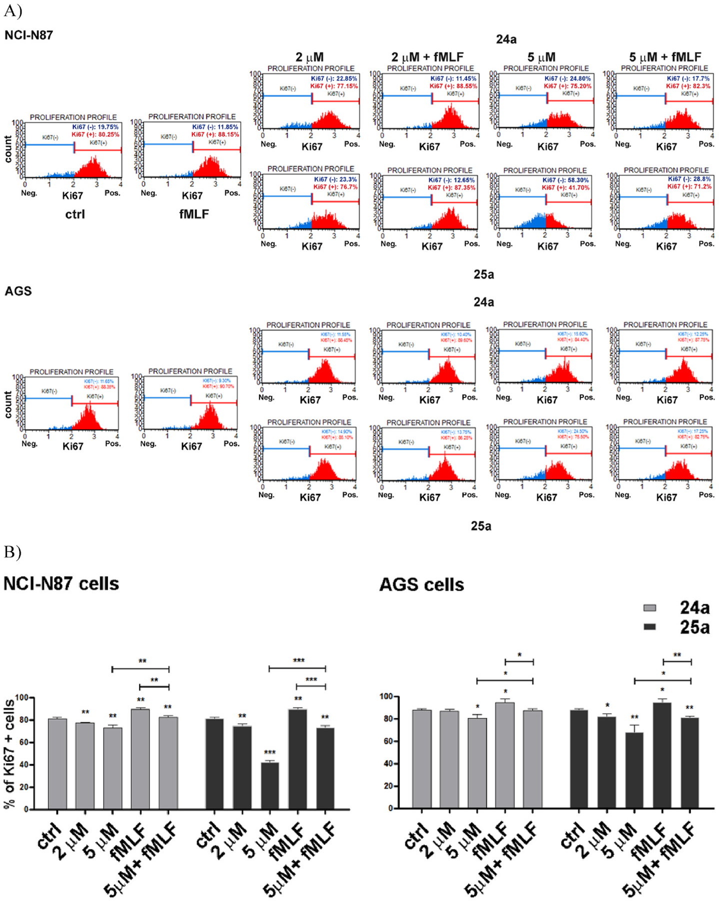
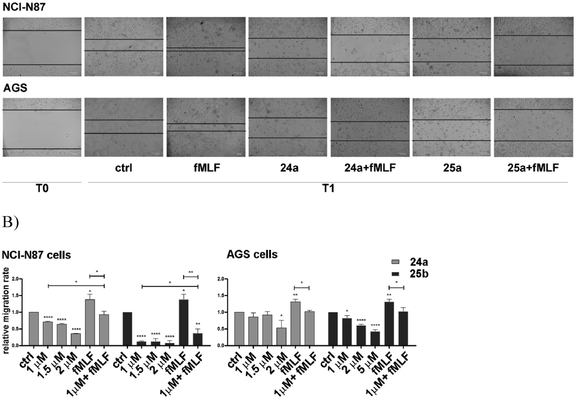
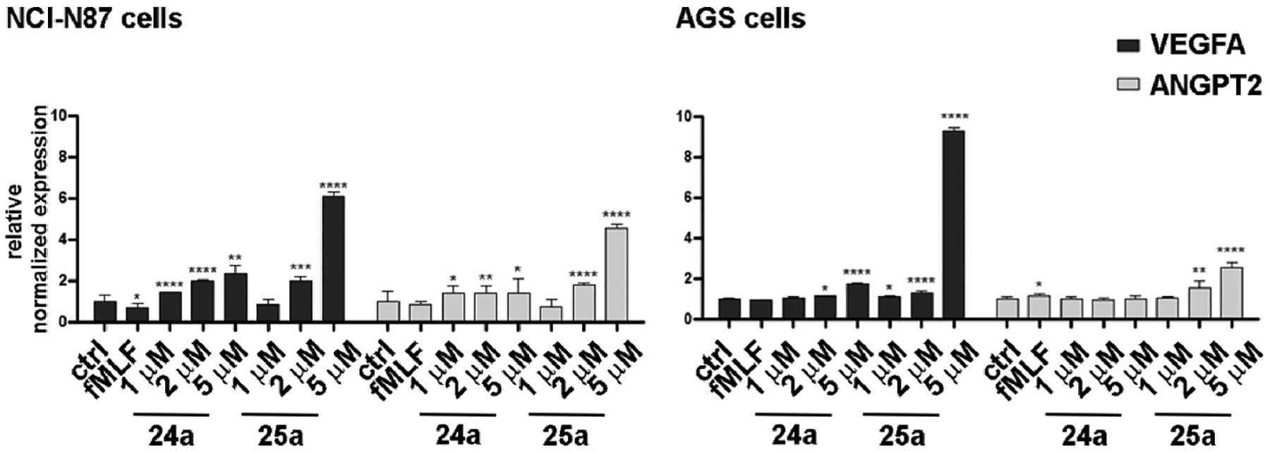
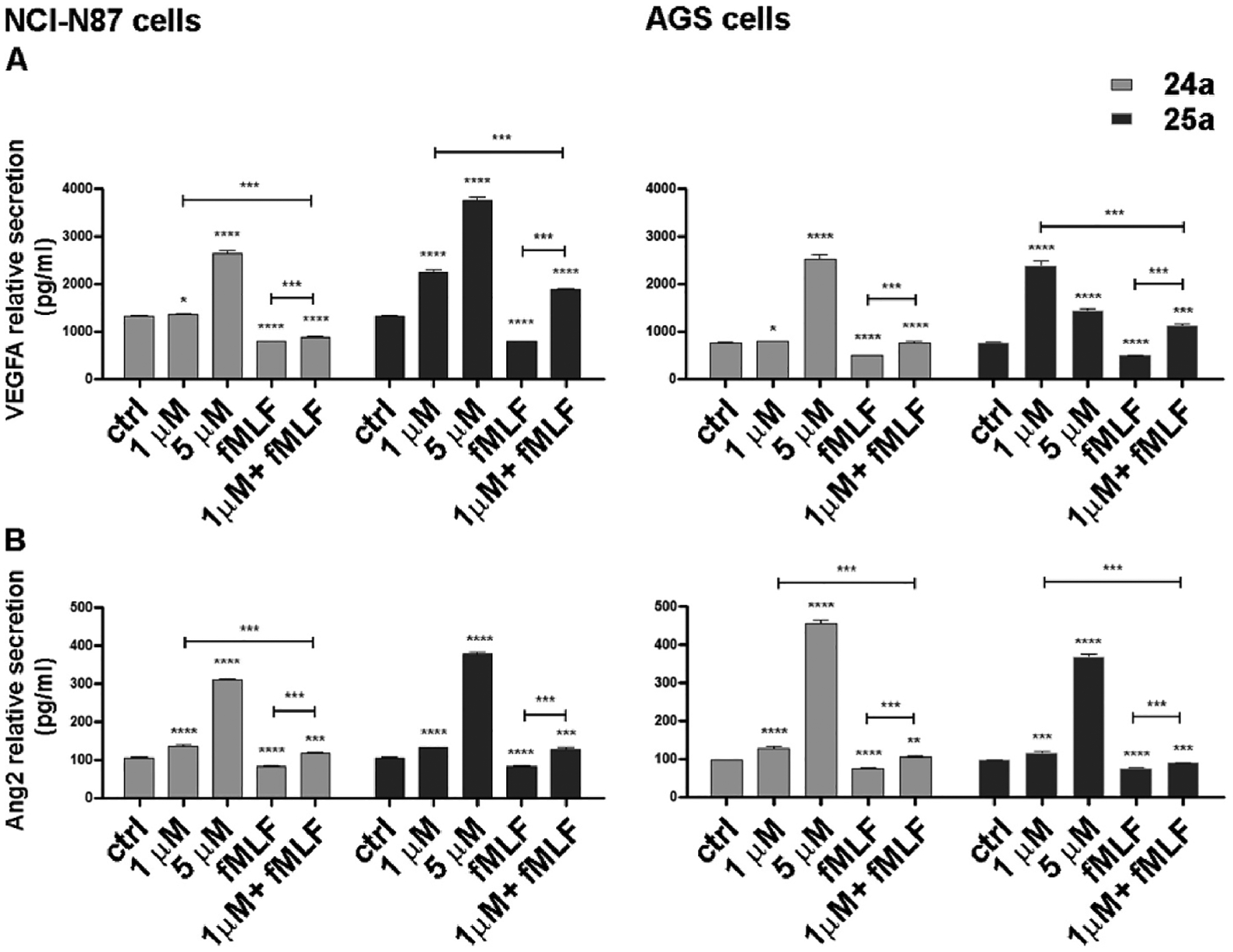
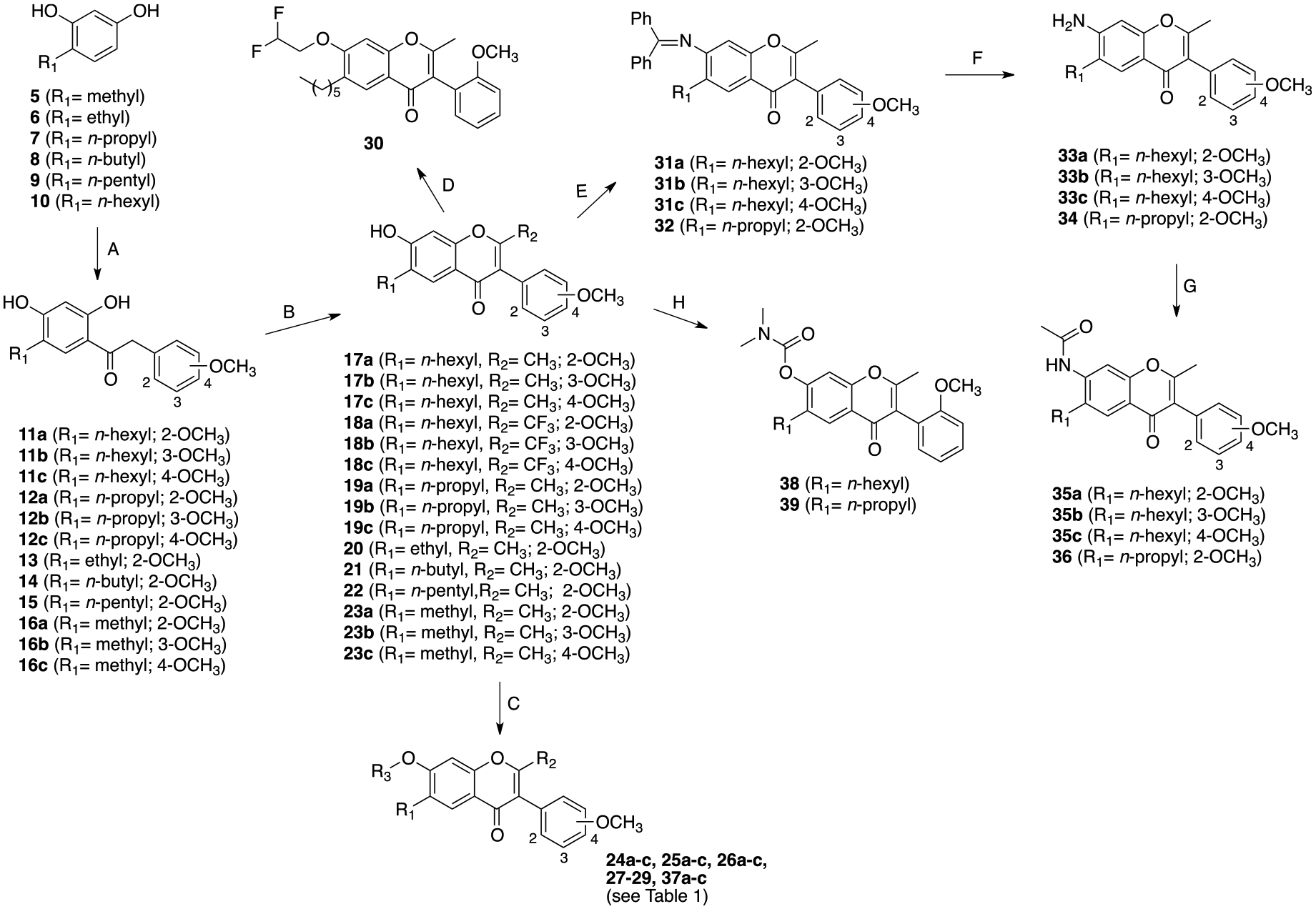
Formyl peptide receptor-1 (FPR1) is a G protein-coupled chemoattractant receptor that plays a crucial role in the trafficking of leukocytes into the sites of bacterial infection and inflammation. Recently, FPR1 was shown to be expressed in different types of tumor cells and could play a significant role in tumor growth and invasiveness. Starting from the previously reported FPR1 antagonist 4, we have designed a new series of 4H-chromen-2-one derivatives that exhibited a substantial increase in FPR1 antagonist potency. Docking studies identified the key interactions for antagonist activity. The most potent compounds in this series (24a and 25b) were selected to study the effects of the pharmacological blockade of FPR1 in NCl-N87 and AGS gastric cancer cells. Both compounds potently inhibited cell growth through a combined effect on cell proliferation and apoptosis and reduced cell migration, while inducing an increase in angiogenesis, thus suggesting that FPR1 could play a dual role as oncogene and onco-suppressor.

Keywords: 4H-chromen-2-one derivatives; Cell growth; Docking studies; FPR1; Gastric cancer.


Conflict of interest statement

Declaration of competing interest The authors declare that they have no known competing financial interests or personal relationships that could have appeared to influence the work reported in this paper.

Figures

Fig. 1.
Fig. 1.
Chemical structures of small-molecule FPR1 antagonists.
Fig. 2.
Fig. 2.
Graphical representation of the design of the new isoflavone FPR1 antagonists.
Fig. 3.
Fig. 3.
Superimposed docking poses of compounds 24a (grey) and 25a (blue). Residues within 3 Å from 25a are shown.
Fig. 4.
Fig. 4.
Pairwise superimposition of the docking poses of 37a (yellow) (panel A); 38 (green) (panel B); 30 (light blue) (panel C); 35a (grey) (panel D) on the docking pose of 25a (blue). Residues within 3 Å from 25a are shown.
Fig. 5.
Fig. 5.
FPR1 and FPR2 RNA expression level in GC cell lines. Real-time PCR experiments in HGC27, KATOIII, AGS and NCl–N87 cell lines with specific primers for FPR1 and FPR2 genes. The mRNA expression was normalized on the GAPDH housekeeping gene. Data were mean ± SD (n = 3).
Fig. 6.
Fig. 6.
Apoptotic profile of 24a, 25a in NCl–N87 and AGS cell lines. Muse Annexin V Cell Assay was assessed after 48 h of drug treatment with fMLF (10 nM) and 24a or 25a (1, 2, 5 μM) administrated alone or combining the lowest dose of 24a or 25a with fMLF. Representative flow cytometry charts (panel A). Four cell populations can be distinguished relative to the percentage of cells alive (bottom left quadrant), in early apoptosis (bottom right quadrant), in late apoptosis (top right quadrant), and dead (top left quadrant). Statistical charts (panel B). The results derived from three independent experiments were expressed as means ± SD and reported in the relative graphs. Statistical analysis was assessed by comparing the values obtained using single drug treatment to those of corresponding untreated cells and the combined treatments to those of the single treatments, *p < 0.05; **p < 0.001; ***p < 0.0002; ****p < 0.0001.
Fig. 7.
Fig. 7.
Effect of 24a and 25a on cell proliferation in NCl–N87 and AGS cell lines. Muse Ki67 Assay was assessed after 24 h of drug treatment with fMLF (10 nM) and 24a or 25a (2 or 5 μM) administrated alone or combining the higher dose of 24a or 25a with fMLF. Representative flow cytometry charts reporting percentage of Ki67 negative (blue) and positive (red) cells (panel A). Statistical charts reporting the results from three independent experiments and expressed as means ± SD (panel B). Statistical analysis was assessed by comparing the values obtained using single drug treatment to those of corresponding untreated cells and the combined treatments to those of the single treatments, *p < 0.05; **p < 0.001; ***p < 0.0001.
Fig. 8.
Fig. 8.
Effect of 24a and 25a on cell migration in NCl–N87 and AGS cell lines. Scratch assay assessed on cells treated with fMLF (10 nM) and 24a or 25a (1, 1.5, 2 μM) administrated alone or combining the lowest dose of 24a or 25a with fMLF. The cells were microscopically analyzed at the time of the scratch (T0) and after 24 h (T1). The relative migration rate was calculated by setting the percentage of migration of the control cells at time T1 equal to 1 and comparing the migration percentage of the cells after each drug treatment to this value. Representative original photographs (panel A) and quantitative analysis (panel B) of the cell-free scratch path ares. The experiments were performed in triplicate, and the mean values ± SD were plotted in the relative graph. Statistical analysis was assessed by comparing the values obtained using a single drug treatment to those of corresponding untreated cells and the combined treatments to those of the single treatments, *p < 0.05, **p < 0.001, ****p < 0.0001.
Fig. 9.
Fig. 9.
Effect of 24a and 25a on mRNA expression of VGFA and ANGPT2 in NCl–N87 and AGS cell lines. Real-time PCR experiments in NCl–N87 and AGS cells with specific primers for VEGFA and ANGPT2 genes. The mRNA expression was normalized on the GAPDH housekeeping gene. Expression analysis was performed after treatment with fMLF (10 nM) and 24a or 25a (1, 2, 5 μM) administrated alone or combining the lowest dose of 24a or 25a with fMLF. All expression values were calculated against the value of untreated PTX-sensitive cells, set equal to 1. Statistical analysis was assessed by comparing the values obtained using single drug treatment to those of corresponding untreated cells and the combined treatments to those of single treatments. The values of untreated PTX-resistant cells were compared with those of untreated PTX-sensitive ones. Data were mean ± SD (n = 3). *p < 0.05, **p < 0.001, ***p < 0.0002, ****p < 0.0001.
Fig. 10.
Fig. 10.
Effect of 24a and 25a on the secretion of VGFA (A) and Ang2 (B) in NCl–N87 and AGS cell lines. The ELISA assays were assessed on cells after 48 h of drug treatment with fMLF (10 nM) and 24a or 25a (1, 5 μM) administrated alone or combining the lowest dose of 24a or 25a with fMLF. The concentration of VEGFA (A) or Ang2 (B) was determined in the medium and normalized for the cell number. The values ± SD, obtained from three independent experiments expressed as pg/mL, were shown in the relative graphs. Statistical analysis was assessed by comparing the values obtained using a single drug treatment to those of corresponding untreated cells and the combined treatments to those of the single treatments. **p < 0.001; ***p < 0.0002; ****p < 0.0001.
Scheme 1.
Scheme 1.
Synthesis of Target Compoundsa aReagents and Conditions: (A) methoxyphenyl acetyl chlorides, AlCl3, anhydrous 1,2-dichlorobenzene, 70 °C, 2h, 10–57% yield; (B) acetic anhydride, anhydrous DMF, 115 °C, 2 h or trifluoroacetic anhydride, anhydrous pyridine, r.t, overnight, yield 10–85%; (C) acetic anhydride, anhydrous pyridine, r.t., 4 days, or heptanoyl chloride, DMAP, Et3N, anhydrous DMF, r.t., 2 h, 14–71% yield; (D) 2,2-difluoroethyl methanesulfonate, K2CO3, anhydrous DMF, 90 °C, 16 h, 32% yield; (E) i: trifluoromethanesulfonic anhydride, DMAP, pyridine, anhydrous CH2Cl2, r.t., 4 h; ii: benzophenone imine, Pd(OAc)2, Cs2CO3, BINAP, anhydrous THF, 80 °C, 22 h, 15–25%; (F) 2 M HCl, THF, r.t., 1 h; (G) acetyl chloride, Et3N, CH2Cl2, r.t., 5 h, 53–90% yield; (H) N,N-dimethylcarbamoylcarbonate, K2CO3, acetonitrile, reflux, 5 h, 35–42% yield.

Similar articles

Cited by

References

    1. Ye RD, Boulay F, Wang JM, Dahlgren C, Gerard C, Parmentier M, Serhan CN, Murphy PM, International union of basic and clinical pharmacology. LXXIII. Nomenclature for the formyl peptide receptor (FPR) family, Pharmacol. Rev 61 (2009) 119–161. - PMC - PubMed
    1. Liu M, Zhao J, Chen K, Bian X, Wang C, Shi Y, Wang J. Ming, G protein-coupled receptor FPR1 as a pharmacologic target in inflammation and human glioblastoma, Int. Immunopharm 14 (2012) 283–288. - PMC - PubMed
    1. He HQ, Ye RD, The formyl peptide receptors: diversity of ligands and mechanism for recognition, Molecules 22 (2017) E455. - PMC - PubMed
    1. Fu H, Karlsson J, Bylund J, Movitz C, Karlsson A, Dahlgren C, Ligand recognition and activation of formyl peptide receptors in neutrophils, J. Leukoc. Biol 79 (2006) 247–256. - PubMed
    1. Becker EL, Forouhar FA, Grunnet ML, Boulay F, Tardif M, Bormann BJ, Sodja D, Ye RD, Woska JR, Murphy PM, Broad immunocytochemical localization of the formylpeptide receptor in human organs, tissues, and cells, Cell Tissue Res. 292 (1998) 129–135. - PubMed

LinkOut - more resources