. 2024 Jan;166(1):178-190.e16.
doi: 10.1053/j.gastro.2023.09.050. Epub 2023 Oct 14.

A Circulating Panel of circRNA Biomarkers for the Noninvasive and Early Detection of Pancreatic Ductal Adenocarcinoma

Affiliations

A Circulating Panel of circRNA Biomarkers for the Noninvasive and Early Detection of Pancreatic Ductal Adenocarcinoma

Caiming Xu et al. Gastroenterology. 2024 Jan.

Abstract

Background & aims: Pancreatic ductal adenocarcinoma (PDAC) is one of the most fatal malignancies. Delayed manifestation of symptoms and lack of specific diagnostic markers lead patients being diagnosed with PDAC at advanced stages. This study aimed to develop a circular RNA (circRNA)-based biomarker panel to facilitate noninvasive and early detection of PDAC.

Methods: A systematic genome-wide discovery of circRNAs overexpressed in patients with PDAC was conducted. Subsequently, validation of the candidate markers in the primary tumors from patients with PDAC was performed, followed by their translation into a plasma-based liquid biopsy assay by analyzing 2 independent clinical cohorts of patients with PDAC and nondisease controls. The performance of the circRNA panel was assessed in conjunction with the plasma levels of cancer antigen 19-9 for the early detection of PDAC.

Results: Initially, a panel of 10 circRNA candidates was identified during the discovery phase. Subsequently, the panel was reduced to 5 circRNAs in the liquid biopsy-based assay, which robustly identified patients with PDAC and distinguished between early-stage (stage I/II) and late-stage (stage III/IV) disease. The areas under the curve of this diagnostic panel for the detection of early-stage PDAC were 0.83 and 0.81 in the training and validation cohorts, respectively. Moreover, when this panel was combined with cancer antigen 19-9 levels, the diagnostic performance for identifying patients with PDAC improved remarkably (area under the curve, 0.94) for patients in the validation cohort. Furthermore, the circRNA panel could also efficiently identify patients with PDAC (area under the curve, 0.85) who were otherwise deemed clinically cancer antigen 19-9-negative (<37 U/mL).

Conclusions: A circRNA-based biomarker panel with a robust noninvasive diagnostic potential for identifying patients with early-stage PDAC was developed.

Keywords: CA19-9; Circular RNA; Diagnostic Biomarker; Liquid-Biopsy Assay; Pancreatic Ductal Adenocarcinoma.


Conflict of interest statement

Conflicts of Interest: The authors declare that they have no competing interests.

Figures

Figure 1.
Figure 1.
Genome-wide discovery of circRNA candidates for PDAC diagnosis. Volcano plots illustrate the log2 fold change and the corresponding P-values for the differentially expressed circRNAs in two independent circRNA expression datasets: GSE79634 (A) and GSE69362 (B). A p < 0.01 was considered statistically significant. C. The heatmap depicts the top 10 overlapping upregulated circRNAs (sorted by p-value) among the PDAC patients (n=20) and matched adjacent normal (AN) tissues (n=20) in the discovery dataset (GSE79634). D. Receiver operating characteristic (ROC) curve analysis shows the performance of the 10-circRNA panel. Red lines indicate the specificity and sensitivity with 95% CIs for each circRNA under the best threshold; red points show the optimal threshold for specificity and sensitivity.
Figure 2.
Figure 2.
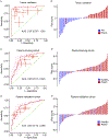
Validation and performance evaluation of the candidate circRNAs in tissue and plasma specimens from clinical cohorts. A. ROC curve analysis reveals the performance of the selected 7-circRNA panel in 32 pairs of PDAC and adjacent normal (AN) tissues. ROC curves are shown as 95% CIs. Red lines indicate the specificity and sensitivity with 95% CI for each circRNA under the best threshold. The red points depict the optimal threshold for specificity and sensitivity. B. The waterfall plot illustrates the risk probability distribution between matched PDAC and adjacent normal tissues. C. ROC curve analysis reveals the diagnostic potential of the final 5-circRNA panel in the training cohort. D. The waterfall plot illustrates the risk probability distribution between plasma samples of the PDAC patients (n= 70) and the non-disease controls (n= 35) in the training cohort. E. ROC curve analysis reveals the performance of the 5-circRNA panel in the validation cohort. F. The waterfall plot illustrates the risk probability distribution between PDAC patients (n= 88) and non-disease controls (n= 46) in the validation cohort.
Figure 3.
Figure 3.
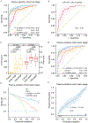
Performance evaluation of the developed circRNA panel to identify early-stage PDAC patients. A. The circRNA-based risk score was analyzed in the PDAC patients with early and advanced stages from the training cohort. B. ROC curve analysis demonstrates the 5-circRNA panel’s performance and the risk probability distribution between the plasma samples of the PDAC patients (n= 48) and non-disease controls (n= 35) in the training cohort. C. ROC curve analysis demonstrates the 5-circRNA panel’s performance and the risk probability distribution between the plasma samples of the PDAC patients (n = 63) and non-disease controls (n = 46) in the validation cohort. ROC curves are shown as 95% CI. Red lines indicate the specificity and sensitivity with 95% CI for each circRNA under the best threshold. Red points show the sensitivity and specificity with 95% CI for each circRNA under the best threshold. D. ROC curve analysis reveals the performance of the 5 circRNA biomarker-based risk score in different GI malignancies (esophageal squamous cell carcinoma, ESCC; colorectal cancer, CRC; gastric cancer, GC; hepatocellular cancer, HCC).
Figure 4.
Figure 4.
The combined performance of the circRNA panel and CA19–9 for the identification of PDAC patients. A. ROC curve analysis reveals the performance of the circRNA panel, CA19–9, and a combination of both for detecting all-stage PDAC patients in the validation cohort. B. ROC curve analysis reveals the performance of the circRNA panel in patients with CA19–9 levels under the cutoff value (37U/ml) from the validation cohort. C. The risk score levels of the circRNA panel and CA19–9 in non-disease controls, early-stage, and advanced-stage PDAC patients from the validation cohort. D. ROC curve analysis reveals the performance of the circRNA panel, CA19–9, and a combination of both in early-stage PDAC patients from the validation cohort. E. Decision curve Shows the net benefit curves for the circRNA panel alone and the combined CA19–9 and the circRNA in early-stage patients from the validation cohort. The X-axis indicates the threshold probability for PDAC diagnosis, and Y-axis indicates the net benefit. The dotted blue line indicates a combination of CA19–9 and the circRNA panel. F. Calibration curves of the combination of CA19–9 and the circRNA signature in early-stage PDAC patients from the validation cohort. The dashed blue line is the flexible calibration (loess) of CA19–9 combined with the circRNA signature. The dashed green line is the ideal line. The triangle sign indicates the grouped observations. The short blue line on the horizontal axis and the short black line represent the positive and negative cases, respectively.

References

    1. Siegel RL, Miller KD, Fuchs HE, Jemal A: Cancer statistics, 2022. CA Cancer J Clin 2022, 72(1):7–33. - PubMed
    1. Versteijne E, van Dam JL, Suker M, Janssen QP, Groothuis K, Akkermans-Vogelaar JM, Besselink MG, Bonsing BA, Buijsen J, Busch OR et al.: Neoadjuvant Chemoradiotherapy Versus Upfront Surgery for Resectable and Borderline Resectable Pancreatic Cancer: Long-Term Results of the Dutch Randomized PREOPANC Trial. J Clin Oncol 2022, 40(11):1220–1230. - PubMed
    1. Toft J, Hadden WJ, Laurence JM, Lam V, Yuen L, Janssen A, Pleass H: Imaging modalities in the diagnosis of pancreatic adenocarcinoma: A systematic review and meta-analysis of sensitivity, specificity and diagnostic accuracy. Eur J Radiol 2017, 92:17–23. - PubMed
    1. Capello M, Bantis LE, Scelo G, Zhao Y, Li P, Dhillon DS, Patel NJ, Kundnani DL, Wang H, Abbruzzese JL et al.: Sequential Validation of Blood-Based Protein Biomarker Candidates for Early-Stage Pancreatic Cancer. J Natl Cancer Inst 2017, 109(4). - PMC - PubMed
    1. Scara S, Bottoni P, Scatena R: CA 19–9: Biochemical and Clinical Aspects. Adv Exp Med Biol 2015, 867:247–260. - PubMed

Publication types

MeSH terms