. 2024 Mar 1;436(5):168313.
doi: 10.1016/j.jmb.2023.168313. Epub 2023 Oct 13.

Cyanobacteriochromes from Gloeobacterales Provide New Insight into the Diversification of Cyanobacterial Photoreceptors

Affiliations

Cyanobacteriochromes from Gloeobacterales Provide New Insight into the Diversification of Cyanobacterial Photoreceptors

Nathan C Rockwell et al. J Mol Biol. .

Abstract

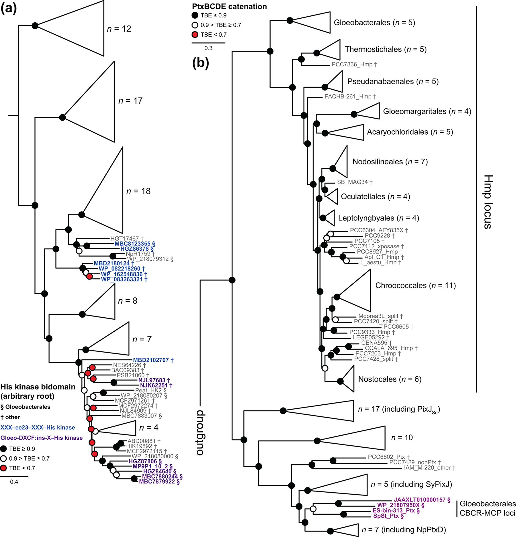
The phytochrome superfamily comprises three groups of photoreceptors sharing a conserved GAF (cGMP-specific phosphodiesterases, cyanobacterial adenylate cyclases, and formate hydrogen lyase transcription activator FhlA) domain that uses a covalently attached linear tetrapyrrole (bilin) chromophore to sense light. Knotted red/far-red phytochromes are widespread in both bacteria and eukaryotes, but cyanobacteria also contain knotless red/far-red phytochromes and cyanobacteriochromes (CBCRs). Unlike typical phytochromes, CBCRs require only the GAF domain for bilin binding, chromophore ligation, and full, reversible photoconversion. CBCRs can sense a wide range of wavelengths (ca. 330-750 nm) and can regulate phototaxis, second messenger metabolism, and optimization of the cyanobacterial light-harvesting apparatus. However, the origins of CBCRs are not well understood: we do not know when or why CBCRs evolved, or what selective advantages led to retention of early CBCRs in cyanobacterial genomes. In the current work, we use the increasing availability of genomes and metagenome-assembled-genomes from early-branching cyanobacteria to explore the origins of CBCRs. We reaffirm the earliest branches in CBCR evolution. We also show that early-branching cyanobacteria contain late-branching CBCRs, implicating early appearance of CBCRs during cyanobacterial evolution. Moreover, we show that early-branching CBCRs behave as integrators of light and pH, providing a potential unique function for early CBCRs that led to their retention and subsequent diversification. Our results thus provide new insight into the origins of these diverse cyanobacterial photoreceptors.

Keywords: chromatic acclimation; photosynthesis; phototaxis; phycocyanobilin; phytochrome.


Conflict of interest statement

Declaration of competing interest The authors declare that they have no known competing financial interests or personal relationships that could have appeared to influence the work reported in this paper.

Figures

Figure 1.
Figure 1.. Phylogenetic analysis of the CBCR domain.
(a) A maximum-likelihood phylogenetic tree is shown as a collapsed view for the CBCR domain, with a small group of GAF domains from knotless phytochromes as an outgroup. Root placement is between the outgroup and all CBCRs. This analysis provides good support for the ee23 clade (“earliest extant as of 2023”) as the earliest known branch in CBCR evolution, followed by the GGR clade. The tree was inferred as described in the Methods, using an alignment of 302 sequences and 156 characters. The complete tree is presented in Figs. S3–S6. Bold italic, lineages from which CBCRs were chosen for characterization in this study. (b) A detailed view is shown for the ee23 lineage. The complete ee23 and GGR clades are presented in context in Fig. S3. Bold, characterized CBCRs. ‡‡, CBCRs characterized in this study.
Figure 2.
Figure 2.. Characterization of the early-branching ee23 CBCR lineage.
(a) Absorption and difference spectra are presented for NIES2119_03185. The dashed line indicates zero absorbance. Blue, 15Z state; orange, 15E state; green, (15Z15E) photochemical difference spectrum. (b) Absorption and difference spectra are presented for WP_083263321, using the conventions of panel (a). (c) Absorption and difference spectra are presented for OSC10802_3032, using the conventions of panel (a). (d) Absorption and difference spectra are presented for MBF2026838, using the conventions of panel (a). (e) Absorption and difference spectra are presented for MBC8123355g2, using the conventions of panel (a). (f) Absorption and difference spectra are presented for HGZ86378, using the conventions of panel (a). (g) Normalized photochemical difference spectra are shown for NIES2119_03185 (dark blue), WP_083263321 (rose), and Oscil6304_4203 (grey; [40]). (h) Normalized photochemical difference spectra are shown for OSC10802_3032 (forest green), MBF2026838 (bronze), and HGZ86378 (aquamarine). Panels (g) and (h) were normalized by dividing ΔAbsorbance by the maximum observed value. All data taken at pH 7.5.
Figure 3.
Figure 3.. Characterization of knotless phytochromes mimicking ee23 CBCRs.
(a) The conserved phytochrome DIP motif [81, 82] is compared to the equivalent CϕP (Cys-hydrophobic-Pro) motif of the ee23 CBCRs and the EVFP motif of selected GGR CBCRs. The region around this motif and that around the canonical Cys are shown. A selection of knotless phytochromes are shown, including sequences used in the outgroup (Fig. S3) and characterized examples [–21]. bold, residues modified by site-directed mutagenesis. Light orange, conserved residues of knotless phytochromes; slate blue, conserved residues of ee23 CBCRs; light green, conserved residues of GGR CBCRs. (b) The domain structure of NpR4776 (Npun_R4776), a complex photoreceptor also containing red/green CBCR domains [28], is shown. Truncations expressing individual photosensory modules are indicated; NpR4776-GAF-PHY has been previously described [21]. (c) Absorption and difference spectra are presented for NpR4776g1, using the conventions of Fig. 2a. (d) Absorption and difference spectra are presented for the D86C variant of NpR4776g1 (NpR4776g1-PADCIP), using the conventions of Fig. 2a. (e) Normalized photochemical difference spectra are shown for wild-type NpR4776g1 (aquamarine), NpR4776g1-PADCIP (brick red), and the NpR4776-GAF-PHY bidomain [21]. (f) Absorption and difference spectra are presented for the isolated GAF domain of knotless phytochrome RfpA, RfpAg1, with G79E D80C substitutions (RfpAg1-PAECIP). Spectra are presented using the conventions of Fig. 2a. (g) Normalized photochemical difference spectra are shown for wild-type RfpAg1 (aquamarine; [21]), and RfpAg1-PAECIP (brick red). (h) Normalized absorption spectra are shown for wild-type OSC10802_3032 (coral) and RfpAg1-PAECIP (brick red) in the light-activated 15E state. Panels (e), (g), and (h) were normalized by dividing Absorbance or ΔAbsorbance by the maximum observed value.
Figure 4.
Figure 4.. Characterization of additional diversity in the GGR CBCR lineage.
(a) Absorption and difference spectra are presented for M595_1144, using the conventions of Fig. 2a. (b) Absorption and difference spectra are presented for Mic7113_1774g1, using the conventions of Fig. 2a. (c) Absorption and difference spectra are presented for Cri9333_0815, using the conventions of Fig. 2a. (d) Normalized photochemical difference spectra are shown for Mic7113_1774g1 (dark blue) and Cri9333_0815 (rose). Difference spectra were normalized by dividing by the maximum observed value. (e) Circular dichroism (CD) spectra are shown for RfpAg1-PAECIP, using the conventions of Fig. 2a. (f) CD spectra are shown for OSC10802_3032, using the conventions of Fig. 2a. (g) CD spectra are shown for HGZ86378, using the conventions of Fig. 2a. (h) CD spectra are shown for Mic7113_1774g1, using the conventions of Fig. 2a. All data taken at pH 7.5.
Figure 5.
Figure 5.. Titration of early-branching CBCRs in the 15Z dark-adapted state.
(a) Absorption spectra are shown for NIES2119_03185 after 10-fold dilution into buffers at pH 5 (red), pH 6 (orange), and pH 9 (aquamarine). (b) Absorption spectra are shown for OSC10802_3032 after 10-fold dilution into buffers at pH 7 (khaki), pH 8 (teal), pH 9 (aquamarine), and pH 10 (dark blue). (c) Absorption spectra are shown for MBF2026838 after 10-fold dilution into buffers at pH 6 (orange), pH 9 (aquamarine), and pH 10 (dark blue). (d) For estimation of pKa values, spectra were normalized on the aromatic amino acid band at ca. 280 nm, and chromophore absorbance was then plotted for different pH values (olive open circles, NIES2119_03185; brick red filled squares, OSC10802_3032). Data were fit under the assumption of a single titratable group using the procedure applied to RcaE [37]. Additional data are in Fig. S10, and pKa values are in Table 2. (e) Absorption spectra are shown for MBC8123355g2 after 10-fold dilution into buffers at pH 6 (orange), pH 7 (khaki), pH 9 (aquamarine), and pH 10 (dark blue). (f) Absorption spectra are shown for HGZ86378 after 10-fold dilution into buffers at pH 6 (orange), pH 9 (aquamarine), and pH 10 (dark blue). (g) Normalized absorption of MBC8123355g2 (mahogany open circles) and HGZ86378 (purple filled squares) was analyzed as in panel (d). (h) Absorption spectra are shown for RfpAg1-PAECIP after 10-fold dilution into buffers at pH 5 (red), pH 6 (orange), pH 9 (aquamarine), and pH 10 (dark blue). (i) Absorption spectra are shown for Mic7113_1774g1 after 10-fold dilution into buffers at pH 7 (khaki), pH 8 (teal), pH 9 (aquamarine), and pH 10 (dark blue). (j) Absorption spectra are shown for Cri9333_0815 after 10-fold dilution into buffers at pH 7 (khaki) and pH 10 (dark blue), with concentration-corrected data from pH 7.5 shown for comparison (dashed grey). (k) Normalized absorption of Mic7113_1774g1 (mauve filled circles) and Cri9333_0815 (forest green open squares) was analyzed as in panel (d). (l) Absorption spectra are shown for the red/green CBCR AnPixJg2 [27] after 10-fold dilution into buffers at pH 6 (orange), pH 7 (khaki), pH 8 (teal), pH 9 (aquamarine), and pH 10 (dark blue).
Figure 6.
Figure 6.. Integration of light and pH sensing in early-branching CBCRs.
(a) Normalized photochemical difference spectra are shown for NIES2119_03185 at pH 5.5 (red), pH 7.5 (dashed grey; see Fig. 2a) and pH 10 (dark blue). (b) Normalized photochemical difference spectra are shown for OSC10802_3032 at pH 8 (teal), pH 9 (aquamarine), and pH 10 (dark blue). (c) Normalized photochemical difference spectra are shown for MBF2026838 at pH 6 (orange), pH 7.75 (moss green), pH 8.5 (blue-green), and pH 10 (dark blue). (d) Normalized photochemical difference spectra are shown for MBC8123355g2 at pH 6 (orange), pH 7.75 (moss green), pH 9 (aquamarine), and pH 10 (dark blue). (e) Normalized photochemical difference spectra are shown for MBC8123355g2 (mauve), OSC10802_3032 (forest green), and MBF2026838 (bronze) at pH 6. (f) Normalized photochemical difference spectra are shown for the same proteins at pH 9. (g) Normalized photochemical difference spectra are shown for Mic7113_1774g1 at pH 8 (teal) and pH 10 (dark blue). (h) Normalized photochemical difference spectra are shown for Cri9333_0815 at pH 7 (khaki), pH 8 (teal), and pH 10 (dark blue). (i) Normalized photochemical difference spectra are shown for RcaE at pH 7 (khaki), pH 9 (aquamarine), and pH 10 (dark blue). (j) Normalized photochemical difference spectra are shown for Mic7113_1774g1 (dark blue), Cri9333_0815 (rose), and RcaE (khaki) at pH 10. (k) Normalized photochemical difference spectra are shown for HGZ86378 at pH 6.5 (brown), pH 9 (aquamarine), and pH 10 (dark blue). (l) Normalized photochemical difference spectra are shown for AnPixJg2 at pH 6 (orange), pH 9 (aquamarine), and pH 10 (dark blue). For panels (a)-(d), (g)-(i), and (k)-(l), difference spectra were normalized by dividing the difference spectrum by the peak absorbance measured for the long-wavelength chromophore band at that pH (Fig. 4), yielding the ΔAbsorbance as a fraction of the total to visualize efficiency of photoconversion. For panels (e), (f), and (j), difference spectra were instead divided by the maximum observed value to facilitate comparison of lineshapes.
Figure 7.
Figure 7.. A conserved lineage of DXCF CBCRs in Gloeobacterales.
(a) The number of full-length proteins containing one or more phytochrome and/or CBCR photoreceptor is plotted against assembly size in Mb for diverse cyanobacterial genomes and MAGs (filled blue diamonds) and for those from Prochlorococcaceae ([70]; open red circles) and Gloeobacter spp. (Fig. S7; open violet squares). (b) The photoreceptor density (calculated as phytochrome and/or CBCR open reading frames per Mb, or ORFs/Mb) was calculated for genomes and MAGs from panel (a) and is plotted by lineage. Open blue circles, Gloeobacter spp. (defined as in Fig. S7); filled violet circles, all other Gloeobacterales; open aquamarine triangles, Thermostichales; open green rectangles, Pseudanabaenales; filled dark blue diamonds, Gloeomargaritales; filled brick red squares, more derived cyanobacteria. Prochlorococcaceae lack phytochromes and CBCRs entirely and are omitted. (c) The tandem index (calculated as the average number of phytochrome or CBCR photosensors in a single open reading frame) is plotted as in panel (b). (d) The Gloeo-DXCF CBCR lineage is shown in detail (see Fig. 1a and Fig. S6). Bold, CBCRs characterized in this study. ‡, CBCRs from Thermostichales. (e) Absorption and difference spectra are presented for MP9P1_10_2A, using the conventions of Fig. 2a. (f) Absorption and difference spectra are presented for NJL97683g1, using the conventions of Fig. 2a. (g) Absorption and difference spectra are presented for WP_218079506g3, using the conventions of Fig. 2a.
Figure 8.
Figure 8.. Conserved XRG CBCRs in Gloeobacterales.
(a) A subset of the sequence alignment used to infer the CBCR phylogeny is shown. Second Cys residues of DXCF [25, 51, 53, 94, 136], hybrid [41], ins-Cys [17], and ins-X CBCRs are shown, as is the canonical Cys typically used for chromophore attachment [16, 22]. Sequences are shown for selected red/green, hybrid, ins-Cys, and ins-X CBCRs (see Fig. S6 for phylogenetic placement). (b) Absorption and difference spectra are presented for MP9P1_10_2B, using the conventions of Fig. 2a. (c) Absorption and difference spectra are presented for NJL97683g2, using the conventions of Fig. 2a. (d) Absorption and difference spectra are presented for WP_027842869, using the conventions of Fig. 2a. (e) Absorption and difference spectra are presented for WP_06508324, using the conventions of Fig. 2a.
Figure 9.
Figure 9.. Characterization of pH responses in CBCR lineages from Gloeobacterales.
(a) Absorption spectra are shown for MP9P1_10_2A in the 15Z state after 10-fold dilution into buffers at pH 6 (orange), pH 7 (khaki), pH 8 (teal), pH 9 (aquamarine), and pH 10 (dark blue). (b) Normalized photochemical difference spectra are shown for MP9P1_10_2A after 10-fold dilution into buffers at pH 6 (orange), pH 7 (khaki), pH 8 (teal), pH 9 (aquamarine), and pH 10 (dark blue). (c) Absorption spectra are shown for MP9P1_10_2A in the 15E state after 10-fold dilution into buffers at pH 6 (orange), pH 7 (khaki), pH 8 (teal), pH 9 (aquamarine), and pH 10 (dark blue). (d) Absorbance at 568 nm for MP9P1_10_2A in the 15E state (panel (c)) was normalized by peak absorption on the UV band and plotted as a function of pH. Values were fit to a model describing a single titrating group [37] as described in the Methods, yielding an estimated pKa of 8.5 ± 0.2 (Table 2). (e) Absorption spectra are shown for WP_218079506g3 in the 15Z state after 10-fold dilution into buffers at pH 6 (orange), pH 7 (khaki), pH 8 (teal), pH 9 (aquamarine), and pH 10 (dark blue). (f) Normalized photochemical difference spectra are shown for MP9P1_10_2A after 10-fold dilution into buffers at pH 6 (orange), pH 7 (khaki), pH 8 (teal), pH 9 (aquamarine), and pH 10 (dark blue). (g) Absorption spectra are shown for MP9P1_10_2B in the 15Z state after 10-fold dilution into buffers at pH 6 (orange), pH 7 (khaki), pH 8 (teal), pH 9 (aquamarine), and pH 10 (dark blue). (h) Normalized photochemical difference spectra are shown for MP9P1_10_2A after 10-fold dilution into buffers at pH 6 (orange), pH 7 (khaki), pH 8 (teal), pH 9 (aquamarine), and pH 10 (dark blue). All difference spectra were normalized as in Fig. 5.
Figure 10.
Figure 10.. Phylogenetic analysis of signaling domains from Gloeobacterales CBCRs.
(a) A maximum-likelihood phylogenetic tree is shown as a collapsed view for selected His kinase bidomains. Root placement is arbitrary, and His kinases associated with ee23 CBCRs (blue) or with Gloeo-DXCF:ins-X tandem CBCR pairs (purple) are shown in detail. (b) A maximum-likelihood phylogenetic tree is shown as a collapsed view for a catenation of candidate HmpBCDE/PtxBCDE sequences, using only the MCP region of HmpD/PtxD proteins. PtxBCDE candidates associated with Gloeo-DXCF CBCRs are highlighted in purple, and the clade of candidate Hmp loci is indicated. Sequences from Gloeobacterales (§) and all other cyanobacteria (†) are indicated.
Figure 11.
Figure 11.. Early-branching CBCR lineages and cyanobacterial light-harvesting pigments.
(a) Approximate absorption spectra are shown for cells of Synechocystis sp. PCC 6803 ([112]; solid cyan), Nostoc punctiforme grown under green light ([36]; solid brown), and Chlorobium tepidum ([118]; dashed dark green), for intracytoplasmic membranes of Rhodopseudomonas palustris ([117]; dashed purple), and for the anoxygenic, homodimeric reaction center of Heliobacterium modesticaldum ([114]; dashed grey), with wavelength ranges and perceived colors indicated underneath. (b) Approximate peak wavelengths are shown for major cyanobacterial light-harvesting pigments [112, 113]: chlorophyll a (Chl a; 440 and 680 nm), allophycocyanin (APC; 650 nm), phycocyanin (PC; 620 nm); phycoerythrin (PE; 564 nm), and phycoerythrocyanin (PEC; 568 nm). (c) Cartoon red/far-red photocycles are shown for representative cyanobacterial phytochromes using PCB (Fig. S1c) or biliverdin IXα (BV). (d) Cartoon photocycles are shown for selected CBCRs from the ee23 and GGR lineages.

Similar articles

Cited by

References

    1. Rockwell NC, Su YS, Lagarias JC. Phytochrome structure and signaling mechanisms. Annu Rev Plant Biol. 2006;57:837–58. - PMC - PubMed
    1. Franklin KA, Quail PH. Phytochrome functions in Arabidopsis development. J Exp Bot. 2010;61:11–24. - PMC - PubMed
    1. Casal JJ. Photoreceptor signaling networks in plant responses to shade. Annu Rev Plant Biol. 2013;64:403–27. - PubMed
    1. Wagner JR, Brunzelle JS, Forest KT, Vierstra RD. A light-sensing knot revealed by the structure of the chromophore binding domain of phytochrome. Nature. 2005;438:325–31. - PubMed
    1. Essen LO, Mailliet J, Hughes J. The structure of a complete phytochrome sensory module in the Pr ground state. Proc Natl Acad Sci USA. 2008;105:14709–14. - PMC - PubMed

Publication types

MeSH terms

Associated data