

In situ photo-crosslinked hydrogel promotes oral mucosal wound healing through sustained delivery of ginsenoside Rg1
- PMID: 37840668
- PMCID: PMC10569426
- DOI: 10.3389/fbioe.2023.1252574
In situ photo-crosslinked hydrogel promotes oral mucosal wound healing through sustained delivery of ginsenoside Rg1
Abstract
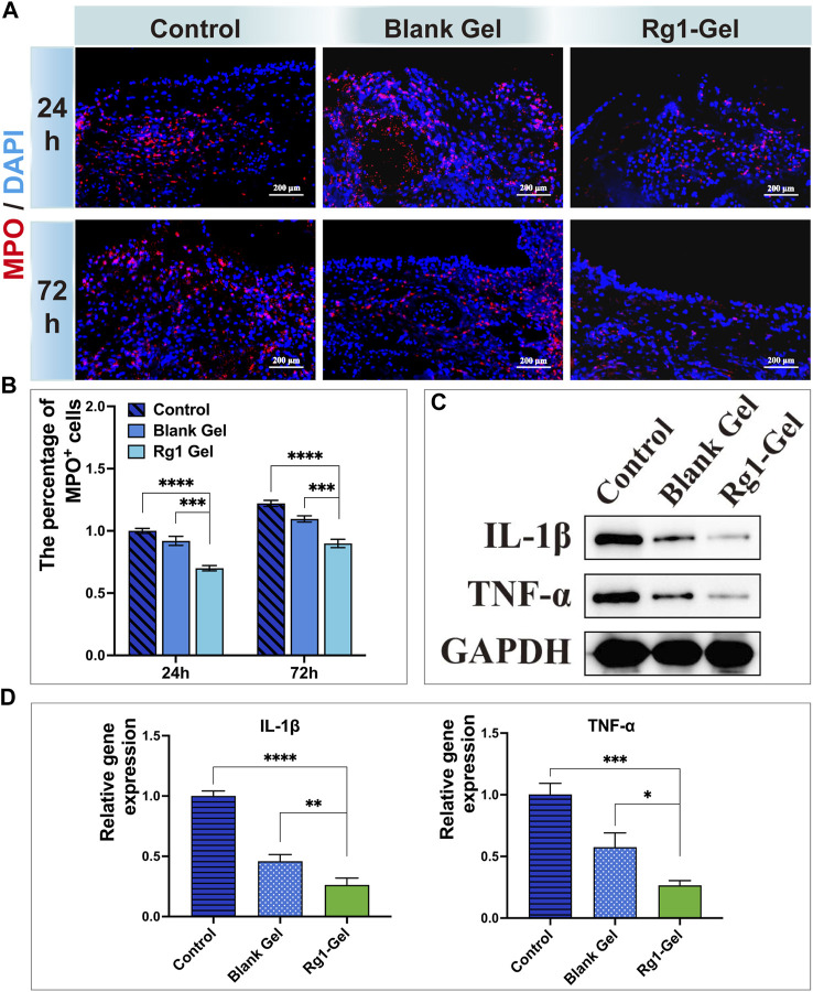
Oral mucosal wounds exhibit an increased susceptibility to inflammation as a consequence of their direct exposure to a diverse range of microorganisms. This causes pain, slow healing, and other complications that interfere with patients' daily activities like eating and speaking. Consequently, patients experience a significant decline in their overall quality of life. Therefore, the pursuit of novel treatment approaches is of great importance. In this study, ginsenoside Rg1, a natural active substance extracted from ginseng root, was chosen as a therapeutic agent. It was encapsulated in a screened photo-crosslinked hydrogel scaffold for the treatment of mucosal defects in the rat palate. The results demonstrated that Rg1-hydrogel possessed excellent physical and chemical properties, and that oral mucosa wounds treated with Rg1-hydrogel exhibited the greatest healing performance, as evidenced by more pronounced wound re-epithelialization, increased collagen deposition, and decreased inflammatory infiltration. Subsequent investigations in molecular biology confirmed that Rg1-hydrogel stimulated the secretion of repair-related factors and inhibited the secretion of inflammatory factors. This study demonstrated that the hydrogel containing ginsenoside Rg1 significantly promotes oral mucosal tissue healing in vivo. Based on the findings, it can be inferred that the Rg1-hydrogel has promising prospects for the therapeutic management of oral mucosal wounds.
Keywords: antiinflammation; ginsenoside Rg1; hydrogel; oral mucosa wound; tissue repair; visible-light crosslinking.
Copyright © 2023 Xu, Zhang, Ren, Zhang, Zhou, Lan and Guo.
Conflict of interest statement
The authors declare that the research was conducted in the absence of any commercial or financial relationships that could be construed as a potential conflict of interest.
Figures


References
-
- Alolga R. N., Nuer-Allornuvor G. F., Kuugbee E. D., Yin X., Ma G. (2020). Ginsenoside Rg1 and the control of inflammation implications for the therapy of type 2 diabetes: A review of scientific findings and call for further research. Pharmacol. Res. 152, 104630. 10.1016/j.phrs.2020.104630 - DOI - PubMed
-
- Atia G. A. N., Shalaby H. K., Ali N. G., Morsy S. M., Ghobashy M. M., Attia H. A. N., et al. (2023). New challenges and prospective applications of three-dimensional bioactive polymeric hydrogels in oral and craniofacial tissue engineering: A narrative review. Pharm. (Basel) 16 (5), 702. 10.3390/ph16050702 - DOI - PMC - PubMed
LinkOut - more resources
Full Text Sources