. 2023 Sep 28:16:1244133.
doi: 10.3389/fnmol.2023.1244133. eCollection 2023.

A quest for the stereo-electronic requirements for selective agonism for the neurotrophin receptors TrkA and TrkB in 17-spirocyclic-dehydroepiandrosterone derivatives

Affiliations

A quest for the stereo-electronic requirements for selective agonism for the neurotrophin receptors TrkA and TrkB in 17-spirocyclic-dehydroepiandrosterone derivatives

Daniele Narducci et al. Front Mol Neurosci. .

Abstract

Introduction: The neurotrophin system plays a pivotal role in the development, morphology, and survival of the nervous system, and its dysregulation has been manifested in numerous neurodegenerative and neuroinflammatory diseases. Neurotrophins NGF and BDNF are major growth factors that prevent neuronal death and synaptic loss through binding with high affinity to their specific tropomyosin-related kinase receptors namely, TrkA and TrkB, respectively. The poor pharmacokinetic properties prohibit the use of neurotrophins as therapeutic agents. Our group has previously synthesized BNN27, a prototype small molecule based on dehydroepiandrosterone, mimicking NGF through the activation of the TrkA receptor.

Methods: To obtain a better understanding of the stereo-electronic requirements for selective activation of TrkA and TrkB receptors, 27 new dehydroepiandrosterone derivatives bearing a C17-spiro-dihydropyran or cyclobutyl moiety were synthesized. The new compounds were evaluated for their ability (a) to selectively activate the TrkA receptor and its downstream signaling kinases Akt and Erk1/2 in PC12 cells, protecting these cells from serum deprivation-induced cell death, and (b) to induce phosphorylation of TrkB and to promote cell survival under serum deprivation conditions in NIH3T3 cells stable transfected with the TrkB receptor and primary cortical astrocytes. In addition the metabolic stability and CYP-mediated reaction was assessed.

Results: Among the novel derivatives, six were able to selectively protect PC12 cells through interaction with the TrkA receptor and five more to selectively protect TrkB-expressing cells via interaction with the TrkB receptor. In particular, compound ENT-A025 strongly induces TrkA and Erk1/2 phosphorylation, comparable to NGF, and can protect PC12 cells against serum deprivation-induced cell death. Furthermore, ENT-A065, ENT-A066, ENT-A068, ENT-A069, and ENT-A070 showed promising pro-survival effects in the PC12 cell line. Concerning TrkB agonists, ENT-A009 and ENT-A055 were able to induce phosphorylation of TrkB and reduce cell death levels in NIH3T3-TrkB cells. In addition, ENT-A076, ENT-A087, and ENT-A088 possessed antiapoptotic activity in NIH-3T3-TrkB cells exclusively mediated through the TrkB receptor. The metabolic stability and CYP-mediated reaction phenotyping of the potent analogs did not reveal any major liabilities.

Discussion: We have identified small molecule selective agonists of TrkA and TrkB receptors as promising lead neurotrophin mimetics for the development of potential therapeutics against neurodegenerative conditions.

Keywords: DHEA derivatives; TrkA; TrkB; neurodegeneration; neuroprotection; neurotrophin mimetics; receptor agonists; signaling.


Conflict of interest statement

The authors declare that the research was conducted in the absence of any commercial or financial relationships that could be construed as a potential conflict of interest.

Figures

Figure 1
Figure 1
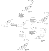
Conceptual representation of the design of the compounds of the present study.
Figure 2
Figure 2
Synthesis of ENT-A002.
Figure 3
Figure 3
Synthesis of the key intermediate 6 and compounds ENT-A008, ENT-A025, ENT-A034, and ENT-A035.
Figure 4
Figure 4
Synthesis of compounds ENT-A026, ENT-A036, ENT-A056, ENT-A075, and ENT-A033 and structure of LM22B-10.
Figure 5
Figure 5
Synthesis of the 1,2,3-triazolyl-substituted derivatives ENT-A037, ENT-A046, ENT-A047, and ENT-A055.
Figure 6
Figure 6
Synthesis of the unsaturated derivatives ENT-A007, ENT-A009, ENT-A065, ENT-A066, and ENT-A068.
Figure 7
Figure 7
Synthesis of C5′-alkyl-substituted derivatives ENT-A070 and ENT-A069.
Figure 8
Figure 8
Synthesis of the key intermediate 13 and final compounds ENT-A076, ENT-A077, ENT-A079, ENT-A080, ENT-A087, and ENT-A088.
Figure 9
Figure 9
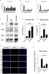
(A–C) Screening and identification of compounds for their ability to activate TrkA and its downstream signaling kinases as well as to promote PC12 cell survival. (A) Quantification of CellTox assay on PC12 cells starved from serum for 4 h and treated with compound (500 nM) or NGF (100 ng/mL) for 24 h, n = 3–6 independent experiments. (B, C) Quantification of phosphorylation of TrkA downstream signaling kinases Akt and ERK1/2 on PC12 cells treated with NGF (100 ng/mL) or compound (500 nM) for 30 min, n = 2–3 independent experiments for AKT, n = 3–4 independent experiments for ERK1/2. (D–H) ENT-A025 strongly activates TrkA, its downstream signaling kinase ERK1/2, and protects PC12 cells from cell death. (D–F) Western Blot and quantification of TrkA and ERK1/2 phosphorylation on PC12 cells treated with ENT-A025 (500 nM) or NGF (100 ng/mL). Data are shown as ±SEM, ***p < 0.001; Student's t-test against negative control, n = 4 independent experiments. (G, H) CellTox assay and quantification of PC12 cells starved from serum and treated with ENT-A025 (500 nM) or NGF (100 ng/mL). Data are shown as ±SEM, **p < 0.01; One-way ANOVA, multiple comparisons, Turkey's test correction, n = 5 independent experiments.
Figure 10
Figure 10
ENT-A065, ENT-A066, ENT-A068, ENT-A069, and ENT-A070 protect PC12 cells from serum deprivation-induced death. (A) PC12 cells were starved of serum and treated with each compound (1 μM) or NGF (100 ng/mL). CellTox dye for dead cells (green) and Hoechst for the total number of cells (blue) were used. (B) Quantification of CellTox+ cells showed that compounds ENT-A065, ENT-A066, ENT-A068, ENT-A069, and ENT-A070 promote cell survival as well as NGF does under stress conditions. Data are the average for 3 independent experiments, presented as mean ± SEM. Statistical analyses were performed by unpaired t-test; *p < 0.05.
Figure 11
Figure 11
CellTox, Cell Toxicity Assay in the NIH-3T3 TrkB-expressing cell line. Quantification and representative images of NIH-3T3 cells, untreated and treated with BDNF (500 ng/mL) or compounds ENT-A007, ENT-A009, ENT-A046, and ENT-A055 (1 μM) for 24 h, after 24 h of serum starvation, n = 3–5 independent experiments, error bars represent SEM. *p < 0.05, **p < 0.01, ***p < 0.001.
Figure 12
Figure 12
Phosphorylation assay in primary astrocytes. Quantification and representative images [(top), phospho-TrkB, and β-Actin] for the Western Blot Assay, n = 3 independent experiments. Cells were treated with BDNF (500 ng/mL) or compounds (1 μM) for 20 min, after 6 h of serum starvation. Error bars represent SEM. *p < 0.05.
Figure 13
Figure 13
ENT-A076, ENT-A087, and ENT-A088 protect NIH3T3 TrkB stable transfected cells from serum deprivation-induced death. (A) NIH3T3 TrkB stable transfected cells were starved of serum and treated with each compound (1 μM) or BDNF (500 ng/mL). CellTox dye for dead cells (green) and Hoechst for the total number of cells (blue) were used. (B) Quantification of CellTox+ cells showed that compounds ENT-A076, ENT-A087, and ENT-A088 promote cell survival as well as BDNF under stress conditions. Data are the average for 3 independent experiments, presented as mean ± SEM. Statistical analyses were performed by unpaired t-test; *p < 0.05; **p < 0.01.
Figure 14
Figure 14
TrkB receptor is involved in the cell-protective effects of compounds ENT-A076, ENT-A087, and ENT-A088. (A) NIH3T3 naive cells were starved of serum and treated with each compound (1 μM) or BDNF (500 ng/mL). CellTox dye for dead cells (green) and Hoechst for the total number of cells (blue) were used. (B) Quantification of CellTox+ cells showed that neither BDNF nor compound administration was not capable of saving cells from death. Thus, we can assume that the cell survival effect is exclusively mediated through the activation of the TrkB receptor. Data are the average for 3 independent experiments, presented as mean ± SEM. Statistical analysis was performed by unpaired t-test.
Figure 15
Figure 15
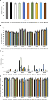
(A) Microsomal stability of test compounds at 1 μM upon incubation with pooled human liver microsomes (60 min). (B) Enzyme (catalytic) activity of the CYP1A2, CYP2A6, CYP2C9, CYP2C19, CYP2D6, and CYP3A4 isoenzymes upon the administration of test compounds at 1 μM (60 min). RFU, relative fluorescence units. Reaction Not Disturbed, reaction without test compounds. (C) CYP450 (%) inhibition of CYP1A2, CYP2A6, CYP2C9, CYP2C19, CYP2D6, and CYP3A4 isoenzymes upon the administration of test compounds at 1 μM (60 min). (D) CYP450 (%) metabolic activity of CYP1A2, CYP2A6, CYP2C9, CYP2C19, CYP2D6, and CYP3A4 isoenzymes upon the administration of test compounds at 1 μM (60 min).

References

    1. Alexaki V. I., Fodelianaki G., Neuwirth A., Mund C., Kourgiantaki A., Ieronimaki E., et al. . (2018). DHEA inhibits acute microglia-mediated inflammation through activation of the TrkA-Akt1/2-CREB-Jmjd3 pathway. Mol. Psychiat. 23, 1410–1420, 10.1038/mp.2017.167 - DOI - PubMed
    1. Allen S. J., Dawbarn D. (2006). Clinical relevance of the neurotrophins and their receptors. Clin. Sci. 110, 175–191. 10.1042/CS20050161 - DOI - PubMed
    1. Baeza-Raja B., Li P., Le Moan N., Sachs B. D., Schachtrup C., Davalos D., et al. . (2012). P75 neurotrophin receptor regulates glucose homeostasis and insulin sensitivity. PNAS 109, 5838–5843. 10.1073/pnas.1103638109 - DOI - PMC - PubMed
    1. Bai L., Pang P. T., Woo N. H. (2005). The yin and yang of neurotrophin action. Nat. Rev. Neurosci. 6, 603–614. 10.1038/nrn1726 - DOI - PubMed
    1. Barter Z. E., Bayliss M. K., Beaune P. H., Boobis A. R., Carlie D. J., Edwards R. J., et al. . (2007). Scaling factors for the extrapolation of in vivo metabolic drug clearance from in vitro data: reaching a consensus on values of human microsomal protein and hepatocellularity per gram of liver. Curr. Drug. Meta. 8 33–45. 10.2174/138920007779315053 - DOI - PubMed