Case Reports
. 2023 Oct 4;18(12):4384-4388.
doi: 10.1016/j.radcr.2023.09.014. eCollection 2023 Dec.

Giant serpentine aneurysm: Neuroradiological and neurosurgical management in a left-handed patient

Affiliations
Case Reports

Giant serpentine aneurysm: Neuroradiological and neurosurgical management in a left-handed patient

Andrea Romano et al. Radiol Case Rep. .

Abstract

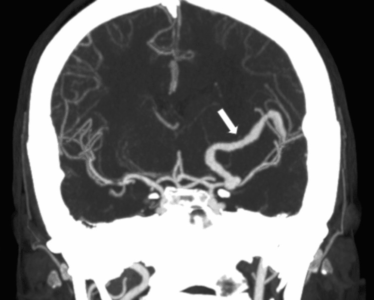
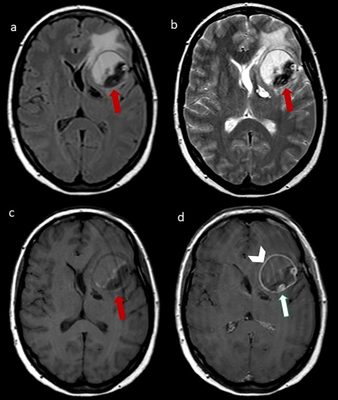
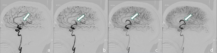
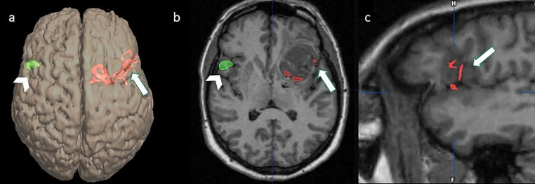
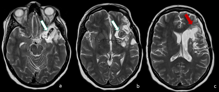
Giant serpentine aneurysms are rare huge and partially thrombosed aneurysms, with an eccentric tortuous intra-aneurysmal vascular channel. Surgical treatment is often necessary due to the great mass effect. We describe a case of a left-handed woman with a giant serpentine aneurysm of the left middle cerebral artery whose management was complex. The challenge was to exclude the aneurysm from circulation, reduce the mass effect, and, mostly, preserve the language function. Since the patient was left-handed the language dominance needed to be assessed; functional MRI (fMRI) and Wada test (WT) showed a right dominance. Surgical treatment was performed, as a complication, the patient developed left fronto-basal ischemia with a slight paresis of the right hand but without any language deficit. Our case shows the importance of a multidisciplinary team in patient management, with a pivotal role of neuroradiological functional tests in presurgical planning.

Keywords: Functional MRI; Language dominance; Serpentine aneurysm; Wada test.


Figures

Fig. 1
Fig. 1
Maximum intensity projection (MIP) coronal reconstruction of a contrast-enhanced CT showing a serpentine eccentric patent vessel (arrow).
Fig. 2
Fig. 2
(A) Axial FLAIR, (B) axial T2- weighted image, (C) T1- weighted image, (D) T1- weighted postcontrastographic images showing a well-defined mass in the left frontal lobe characterized by inhomogeneous signal intensity on T1 and T2 weighted images, compatible with different stages of organizing hematoma, surrounded by perilesional edema. After contrast medium administration, an intralesional “serpentine” eccentric patent vessel (arrow) and a linear enhancement of the mass walls are evident (arrowhead).
Fig. 3
Fig. 3
(A–D): Different phases of digital subtraction angiography (DSA) showing a partially thrombosed giant serpentine aneurysm arising from the M1 segment of the left middle cerebral artery (arrow) with distal small arterial branches reaching the fronto-basal region. These dynamic angiograms showed the slow flow inside the patent component of aneurysm with high vascular enhancement evident in the latest stage of acquisitions (D).
Fig. 4
Fig. 4
(A) Volume rendering (VR) reconstruction of volumetric T1 MPRAGE, (B) axial T1 MPRAGE, and (C) sagittal T1 MPRAGE images co-registered with DSA images and fMRI images showing vessels arising from the distal segment of the serpentine aneurysm reached the left fronto-basal cortex (A–C, arrow) and the activation of the right fronto-basal region during the task of verb generation (Broca's area) (A, B, arrowhead), suggestive of right dominance.
Fig. 5
Fig. 5
(A–C) follow-up axial T2-weighted images 10 months after the surgical treatment documented significant volumetric reduction of the residual aneurysm (A, B, white arrow) and malacic ischemic damage in the fronto-basal region (C, red arrow).

Similar articles

Cited by

  • A comprehensive analysis and systematic review of giant serpentine aneurysms.
    Atallah O, Hammoud Z, Aljeradat B, Almealawy YF, Badary A, Elqader JA, Sanker V, Farooq M, Muthana A, Alrubaye SN, Dwebi A, Chaurasia B, Azab MA. Atallah O, et al. Ann Med Surg (Lond). 2025 Feb 26;87(3):1458-1466. doi: 10.1097/MS9.0000000000003000. eCollection 2025 Mar. Ann Med Surg (Lond). 2025. PMID: 40213197 Free PMC article. Review.

References

    1. Xu K, Yu T, Guo Y, Yu J. Study and therapeutic progress on intracranial serpentine aneurysms. Int J Med Sci. 2016;13:432–439. - PMC - PubMed
    1. Day AL, Gaposchkin CG, Yu CJ, Rivet DJ, Dacey RG., Jr. Spontaneous fusiform middle cerebral artery aneurysms: characteristics and a proposed mechanism of formation. J Neurosurg. 2003;99:228–240. - PubMed
    1. Terao H, Muraoka I. Giant aneurysm of the middle cerebral artery containing an important blood channel. Case report. J Neurosurg. 1972;37:352–356. doi: 10.3171/jns.1972.37.3.0352. - DOI - PubMed
    1. Lan J, Fu Z, Zhang J, Ma C, Cao CJ, Zhao WY, et al. Giant serpentine aneurysm of the middle cerebral artery. World Neurosurg. 2018;117:109–114. - PubMed
    1. Verny C, Marc G, Pasco A, Dubas F. Middle cerebral artery dissection gives arise to giant serpentine aneurysm. Cerebrovasc Dis. 2008;25:283–285. - PubMed

Publication types

LinkOut - more resources