

The thalamus and basal ganglia are smaller in children with epilepsy after perinatal stroke
- PMID: 37840930
- PMCID: PMC10568465
- DOI: 10.3389/fneur.2023.1252472
The thalamus and basal ganglia are smaller in children with epilepsy after perinatal stroke
Abstract
Background: Epilepsy is one of the most serious consequences of perinatal stroke. Epilepsy itself has been proposed as a risk factor for impaired cognitive, language, and behavioral functioning. It is still unclear which children develop epilepsy after perinatal stroke. The current study aimed to evaluate the volume of the thalamus and the basal ganglia in children after perinatal stroke in relation to poststroke epilepsy.
Methods: The follow-up study included 29 children with perinatal arterial ischemic stroke (AIS), 33 children with presumed periventricular venous infarction (PVI), and 46 age- and sex-matched healthy controls. Magnetic resonance imaging was performed in children between the ages of 4 and 18 years, and volumetric analysis by segmentation was used to evaluate the size of the thalamus, caudate nucleus, putamen, globus pallidus, hippocampus, amygdala, and nucleus accumbens.
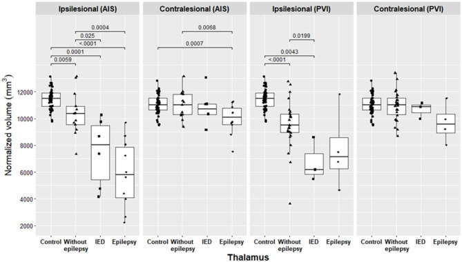
Results: During a median follow-up time of 12.8 years [interquartile range (IQR): 10.8-17.3] in the AIS group and 12.5 years (IQR: 9.3-14.8) in the PVI group (p = 0.32), epilepsy developed in 10 children (34.5%) with AIS and in 4 (12.1%) children with PVI, p = 0.036 [odds ratio (OR) = 3.8, 95%, confidence interval (CI): 1.04-14]. Epilepsy and interictal epileptiform discharges (IEDs) without clinical seizures were more often expressed in children with AIS (n = 16, 55%) than in children with PVI (n = 7, 21.2%), p = 0.0057 (OR = 3.8 95% CI: 1.04-14). In the AIS group, the ipsilesional and contralesional thalamus, ipsilesional caudate nucleus, and nucleus accumbens were significantly smaller in children with epilepsy compared to children without epilepsy. In the PVI group, the ipsilesional thalamus, caudate nucleus, and nucleus accumbens were smaller in the pooled group of epilepsy plus IED alone compared to children without epilepsy.
Conclusion: In children with AIS, epilepsy or IED occurred more often compared to children with PVI. Both patients with AIS and PVI with severe damage to the basal ganglia and the thalamus have a higher risk of developing poststroke epilepsy and should be monitored more closely throughout childhood to initiate timely antiseizure medication and rehabilitation.
Keywords: basal ganglia; epilepsy; interictal epileptiform discharges; ischemic perinatal stroke; thalamus.
Copyright © 2023 Vaher, Ilves, Ilves, Laugesaar, Männamaa, Loorits, Kool and Ilves.
Conflict of interest statement
The authors declare that the research was conducted in the absence of any commercial or financial relationships that could be construed as a potential conflict of interest.
Figures


Similar articles
-
Ipsilesional volume loss of basal ganglia and thalamus is associated with poor hand function after ischemic perinatal stroke.BMC Neurol. 2022 Jan 12;22(1):23. doi: 10.1186/s12883-022-02550-3. BMC Neurol. 2022. PMID: 35022000 Free PMC article.
-
Bihemispheric developmental alterations in basal ganglia volumes following unilateral perinatal stroke.Neuroimage Clin. 2022;35:103143. doi: 10.1016/j.nicl.2022.103143. Epub 2022 Aug 4. Neuroimage Clin. 2022. PMID: 36002972 Free PMC article.
-
Long-term neurodevelopmental outcome after perinatal arterial ischemic stroke and periventricular venous infarction.Eur J Paediatr Neurol. 2018 Nov;22(6):1006-1015. doi: 10.1016/j.ejpn.2018.07.005. Epub 2018 Jul 21. Eur J Paediatr Neurol. 2018. PMID: 30249407
-
The basal ganglia and apraxia.Brain. 1996 Feb;119 ( Pt 1):319-40. doi: 10.1093/brain/119.1.319. Brain. 1996. PMID: 8624692 Review.
-
Poststroke epilepsy: current perspectives on diagnosis and treatment.Neuropsychiatr Dis Treat. 2018 Dec 24;15:95-103. doi: 10.2147/NDT.S169579. eCollection 2019. Neuropsychiatr Dis Treat. 2018. PMID: 30636875 Free PMC article. Review.
Cited by
-
Brain growth until adolescence after a neonatal focal injury: sex related differences beyond lesion effect.Front Neurosci. 2024 Aug 23;18:1405381. doi: 10.3389/fnins.2024.1405381. eCollection 2024. Front Neurosci. 2024. PMID: 39247049 Free PMC article.
References
-
- Raju TNK Nelson KB Ferriero D Lynch JK The The NICHD-NINDS Perinatal Stroke Workshop Participants . Ischemic Perinatal Stroke: Summary of a Workshop Sponsored by the National Institute of Child Health and Human Development and the National Institute of Neurological Disorders and Stroke. Pediatrics. (2007) 120:609–16. 10.1542/peds.2007-0336 - DOI - PubMed
LinkOut - more resources
Full Text Sources