

Extracting decision-making features from the unstructured eye movements of clinicians on glaucoma OCT reports and developing AI models to classify expertise
- PMID: 37841006
- PMCID: PMC10571140
- DOI: 10.3389/fmed.2023.1251183
Extracting decision-making features from the unstructured eye movements of clinicians on glaucoma OCT reports and developing AI models to classify expertise
Abstract
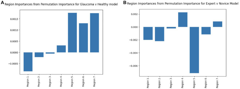
This study aimed to investigate the eye movement patterns of ophthalmologists with varying expertise levels during the assessment of optical coherence tomography (OCT) reports for glaucoma detection. Objectives included evaluating eye gaze metrics and patterns as a function of ophthalmic education, deriving novel features from eye-tracking, and developing binary classification models for disease detection and expertise differentiation. Thirteen ophthalmology residents, fellows, and clinicians specializing in glaucoma participated in the study. Junior residents had less than 1 year of experience, while senior residents had 2-3 years of experience. The expert group consisted of fellows and faculty with over 3 to 30+ years of experience. Each participant was presented with a set of 20 Topcon OCT reports (10 healthy and 10 glaucomatous) and was asked to determine the presence or absence of glaucoma and rate their confidence of diagnosis. The eye movements of each participant were recorded as they diagnosed the reports using a Pupil Labs Core eye tracker. Expert ophthalmologists exhibited more refined and focused eye fixations, particularly on specific regions of the OCT reports, such as the retinal nerve fiber layer (RNFL) probability map and circumpapillary RNFL b-scan. The binary classification models developed using the derived features demonstrated high accuracy up to 94.0% in differentiating between expert and novice clinicians. The derived features and trained binary classification models hold promise for improving the accuracy of glaucoma detection and distinguishing between expert and novice ophthalmologists. These findings have implications for enhancing ophthalmic education and for the development of effective diagnostic tools.
Keywords: eye-tracking; fixations; glaucoma; neural networks; optical coherence tomography; unsupervised clustering.
Copyright © 2023 Akerman, Choudhary, Liebmann, Cioffi, Chen and Thakoor.
Conflict of interest statement
KAT is receiving funding support from Topcon Healthcare, Inc., for a study whose topic does not overlap with the topic of this study. The remaining authors declare that the research was conducted in the absence of any commercial or financial relationships that could be construed as a potential conflict of interest.
Figures


Similar articles
-
Retinal nerve fiber layer imaging with spectral-domain optical coherence tomography: analysis of the retinal nerve fiber layer map for glaucoma detection.Ophthalmology. 2010 Sep;117(9):1684-91. doi: 10.1016/j.ophtha.2010.01.026. Epub 2010 Jul 21. Ophthalmology. 2010. PMID: 20663563
-
Glaucoma detection in Latino population through OCT's RNFL thickness map using transfer learning.Int Ophthalmol. 2021 Nov;41(11):3727-3741. doi: 10.1007/s10792-021-01931-w. Epub 2021 Jul 1. Int Ophthalmol. 2021. PMID: 34212255
-
Predicting Clinician Fixations on Glaucoma OCT Reports via CNN-Based Saliency Prediction Methods.IEEE Open J Eng Med Biol. 2024 Feb 20;5:191-197. doi: 10.1109/OJEMB.2024.3367492. eCollection 2024. IEEE Open J Eng Med Biol. 2024. PMID: 38606397 Free PMC article.
-
[Aiming for zero blindness].Nippon Ganka Gakkai Zasshi. 2015 Mar;119(3):168-93; discussion 194. Nippon Ganka Gakkai Zasshi. 2015. PMID: 25854109 Review. Japanese.
-
Detecting glaucoma with only OCT: Implications for the clinic, research, screening, and AI development.Prog Retin Eye Res. 2022 Sep;90:101052. doi: 10.1016/j.preteyeres.2022.101052. Epub 2022 Feb 23. Prog Retin Eye Res. 2022. PMID: 35216894 Review.
Cited by
-
A Practical Guide to Evaluating Artificial Intelligence Imaging Models in Scientific Literature.Ophthalmol Sci. 2025 Jun 9;5(6):100847. doi: 10.1016/j.xops.2025.100847. eCollection 2025 Nov-Dec. Ophthalmol Sci. 2025. PMID: 40778360 Free PMC article.
-
The Effect of Experience on Visual Search Patterns in Retinal Imaging Analysis.Ophthalmic Surg Lasers Imaging Retina. 2025 Jun;56(6):336-344. doi: 10.3928/23258160-20250228-03. Epub 2025 Mar 1. Ophthalmic Surg Lasers Imaging Retina. 2025. PMID: 40163634 Free PMC article.
-
The Use of Machine Learning in Eye Tracking Studies in Medical Imaging: A Review.IEEE J Biomed Health Inform. 2024 Jun;28(6):3597-3612. doi: 10.1109/JBHI.2024.3371893. Epub 2024 Jun 6. IEEE J Biomed Health Inform. 2024. PMID: 38421842 Free PMC article. Review.
References
LinkOut - more resources
Full Text Sources