

Developing a trans-multisynaptic tracer to map the neural circuit of recovered sciatic nerve after treatment with nerve growth factor
- PMID: 37841085
- PMCID: PMC10570716
- DOI: 10.1016/j.ibneur.2023.09.011
Developing a trans-multisynaptic tracer to map the neural circuit of recovered sciatic nerve after treatment with nerve growth factor
Abstract
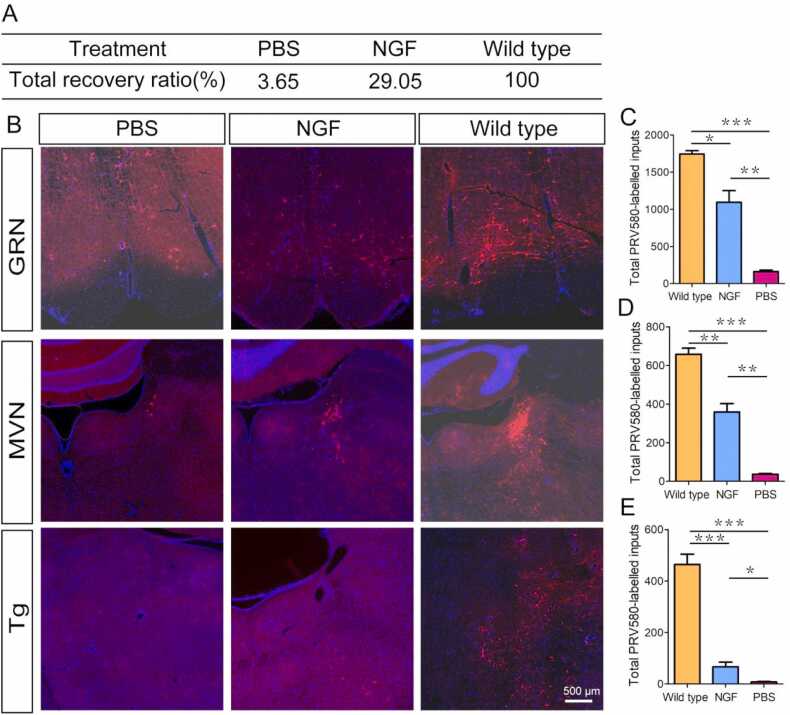
Nerve growth factor (NGF) has been shown to support the survival and differentiation of neurons. In this study, we first developed a retrograde trans-multisynaptic tracer PRV580 expressing the mCherry fluorescent protein based on pseudorabies virus Bartha strain to map the neural circuit of sciatic nerve. Secondly, the newly developed PRV580 was used to map the neural circuit of the recovering sciatic nerve upon treatment with NGF. Our results showed that red signals from PRV580 were observed in various brain regions. Among these regions, many areas of the pyramidal system and the extra-pyramidal system had been mapped, accounting for as much as 56.8 % of the total inputs. Furthermore, we found that NGF could significantly increase the ratio of total input (29.05 %) compared to PBS (3.65 %), indicating that NGF indeed can aid in the repair of injured sciatic nerve. These findings indicated that NGF has therapeutic ability for the treatment of peripheral nerve injuries and virus-based tracers can be used to monitor the recovery.
Keywords: Nerve growth factor; Neural circuit; Retrograde trans-multisynaptic tracer; Sciatic nerve.
© 2023 The Authors.
Conflict of interest statement
The authors declare no competing financial interests.
Figures


Similar articles
-
Development of Cre-dependent retrograde trans-multisynaptic tracer based on pseudorabies virus bartha strain.Mol Brain. 2025 Apr 14;18(1):33. doi: 10.1186/s13041-025-01204-y. Mol Brain. 2025. PMID: 40229811 Free PMC article.
-
Optimization of the Fluorescent Protein Expression Level Based on Pseudorabies Virus Bartha Strain for Neural Circuit Tracing.Front Neuroanat. 2019 Jun 21;13:63. doi: 10.3389/fnana.2019.00063. eCollection 2019. Front Neuroanat. 2019. PMID: 31281245 Free PMC article.
-
Gene Expression Profiling with Cre-Conditional Pseudorabies Virus Reveals a Subset of Midbrain Neurons That Participate in Reward Circuitry.J Neurosci. 2017 Apr 12;37(15):4128-4144. doi: 10.1523/JNEUROSCI.3193-16.2017. Epub 2017 Mar 10. J Neurosci. 2017. PMID: 28283558 Free PMC article.
-
Single injection of a novel nerve growth factor coacervate improves structural and functional regeneration after sciatic nerve injury in adult rats.Exp Neurol. 2017 Feb;288:1-10. doi: 10.1016/j.expneurol.2016.10.015. Epub 2016 Oct 28. Exp Neurol. 2017. PMID: 27983992
-
Use of pseudorabies virus to delineate multisynaptic circuits in brain: opportunities and limitations.J Neurosci Methods. 2000 Nov 15;103(1):51-61. doi: 10.1016/s0165-0270(00)00295-8. J Neurosci Methods. 2000. PMID: 11074095 Review.
Cited by
-
Therapeutic Potential of Vitamin B Complex in Peripheral Nerve Injury Recovery: An Experimental Rat Model Study.Medicina (Kaunas). 2024 Sep 23;60(9):1556. doi: 10.3390/medicina60091556. Medicina (Kaunas). 2024. PMID: 39336597 Free PMC article.
References
-
- Cao X., Shoichet M.S. Defining the concentration gradient of nerve growth factor for guided neurite outgrowth. Neuroscience. 2001;103:831–840. - PubMed
-
- Collins J.J., Lin C.E., Berthoud H.R., Papka R.E. Vagal afferents from the uterus and cervix provide direct connections to the brainstem. Cell Tissue Res. 1999;295:43–54. - PubMed
-
- Derby A., Engleman V.W., Frierdich G.E., Neises G., Rapp S.R., Roufa D.G. Nerve growth factor facilitates regeneration across nerve gaps: morphological and behavioral studies in rat sciatic nerve. Exp. Neurol. 1993;119:176–191. - PubMed
LinkOut - more resources
Full Text Sources