. 2023 Sep 26;8(40):37391-37401.
doi: 10.1021/acsomega.3c05343. eCollection 2023 Oct 10.

Binding of Stimuli-Responsive Ruthenium Aqua Complexes with 9-Ethylguanine

Affiliations

Binding of Stimuli-Responsive Ruthenium Aqua Complexes with 9-Ethylguanine

Atsuki Maeda et al. ACS Omega. .

Abstract

Stimuli-responsive ruthenium complexes proximal- and distal-[Ru(C10tpy)(C10pyqu) OH2]2+ (proximal-1 and distal-1; C10tpy = 4'-decyloxy-2,2':6',2″-terpyridine and C10pyqu = 2-[2'-(6'-decyloxy)-pyridyl]quinoline) were experimentally studied for adduct formation with a model DNA base. At 303 K, proximal-1 exhibited 1:1 adduct formation with 9-ethylguanine (9-EtG) to yield proximal-[Ru(C10tpy)(C10pyqu)(9-EtG)]2+ (proximal-RuEtG). Rotation of the guanine ligand on the ruthenium center was sterically hindered by the presence of an adjacent quinoline moiety at 303 K. Results from 1H NMR measurements indicated that photoirradiation of a proximal-RuEtG solution caused photoisomerization to distal-RuEtG, whereas heating of proximal-RuEtG caused ligand substitution to proximal-1. The distal isomer of the aqua complex, distal-1, was observed to slowly revert to proximal-1 at 303 K. In the presence of 9-EtG, distal-1 underwent thermal back-isomerization to proximal-1 and adduct formation to distal-RuEtG. Kinetic analysis of 1H NMR measurements showed that adduct formation between proximal-1 and 9-EtG was 8-fold faster than that between distal-1 and 9-EtG. This difference may be attributed to intramolecular hydrogen bonding and steric repulsion between the aqua ligand and the pendant moiety of the bidentate ligand..


Conflict of interest statement

The authors declare no competing financial interest.

Figures

Scheme 1
Scheme 1. Photoisomerization and Thermal Back-Isomerization between proximal-1 and distal-1
Scheme 2
Scheme 2. Top: Synthetic Route (4 Steps) of C10pyqu Starting from 2-Acetylpyridine,
Middle: 2-step syntheses of C10pyqu in this work starting from 6-bromo-2-acetylpyridine. Bottom: synthetic route for Ru complexes. Adapted in part with permission from M. Hirahara (2016). Visible-light-induced morphological changes of giant vesicles by photoisomerization of a ruthenium aqua complex. Chem. Eur. J.2016, 22, 2590–2594. Copyright 2016 Wiley-VCH GmbH.
Figure 1
Figure 1
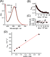
(A) Absorption spectral changes of proximal-1 (29 μM) in the presence of 9-EtG (570 μM, 20 equiv) at 303 K. Solvent: H2O/CH3OH/2,2,2-trifluoroethanol (TFE) = 70:50:2, v/v/v. (B) Absorbance changes at 550 nm (black dot) during the reaction and fitting curve (solid line) based on the pseudo-first-order reaction model. (C) Residual plots for the fitting curve. (D) Concentration dependency of the observed rate constants (kobs) for the ligand substitution reaction of proximal-1 with 9-EtG at 303 K.
Figure 2
Figure 2
ESI-MS spectra of [Ru(C10tpy)(C10pq)(9-EtG)]2+. Conditions: proximal-1 (0.1 mM) and 9-ethylguanine (2 mM) in mixed solvents (H2O/methanol/TFE = 70/50/2, v/v/v). The sample solution was left at room temperature in the dark for 24 h to complete the ligand substitution reaction. Peaks at 381.20 and 739.40 m/z correspond to the ionized species of [(9-EtG)2 + Na]+ and [(9-EtG)4 + Na]+, respectively. Inset: observed spectra at 516 m/z and calculated isotope pattern of [Ru(C10tpy)(C10pq)(9-EtG)]2+.
Figure 3
Figure 3
1H NMR spectra of (A) proximal-1 (B) 10 min after mixing proximal-1 (1 mM) with 9-EtG (5 mM) and (C) proximal-1 and 9-EtG after incubation for 1 day. Peaks associated with proximal-1 at 6.64 ppm and proximal-RuEtG at 6.93 ppm are colored gray and red, respectively. In (C), peaks corresponding to aromatic protons of C10tpy of proximal-RuEtG are highlighted with yellow. Solvent: D2O/CD3OD/2,2,2-trifluoroethanol (TFE) = 70:50:2, v/v/v. The singlet at 7.8 ppm corresponds to the C–H proton of the free 9-EtG.
Figure 4
Figure 4
Kinetic traces of proximal-1 (1 mM) during the substitution reaction with 5 mM 9-EtG at 303 K. Red: proximal-RuEtG; black: proximal-1. The concentrations of the complexes were calculated by integrating peaks at 6.92 ppm (proximal-RuEtG) and 6.63–6.70 ppm (proximal-1 and proximal-RuEtG).
Figure 5
Figure 5
(A) Absorption spectral changes of proximal- and distal-1 (28.3 μM) in the presence of 9-EtG (566 μM, 20 equiv) at 303 K. (B) Kinetic profile (black dot) during the reaction and fitting curve (red line) based on the double exponential curve. (C) Residual plots for the fitting curve. Solvent: D2O/CD3OD/TFE = 70:50:2, v/v/v.
Figure 6
Figure 6
1H NMR spectral changes during the substitution reaction of proximal-1 and distal-1 with 9-EtG in the dark at 303 K. (A) Proximal-1 and distal-1 in the photostationary state. (B) Proximal-1 and distal-1 (1 mM) after 15 min of mixing with 9-EtG (5 mM). (C) Proximal-1 and distal-1 after 4 h of mixing with 9-EtG. (D) Proximal-RuEtG. Solvent: D2O/CD3OD/TFE = 70:50:2, v/v/v.
Figure 7
Figure 7
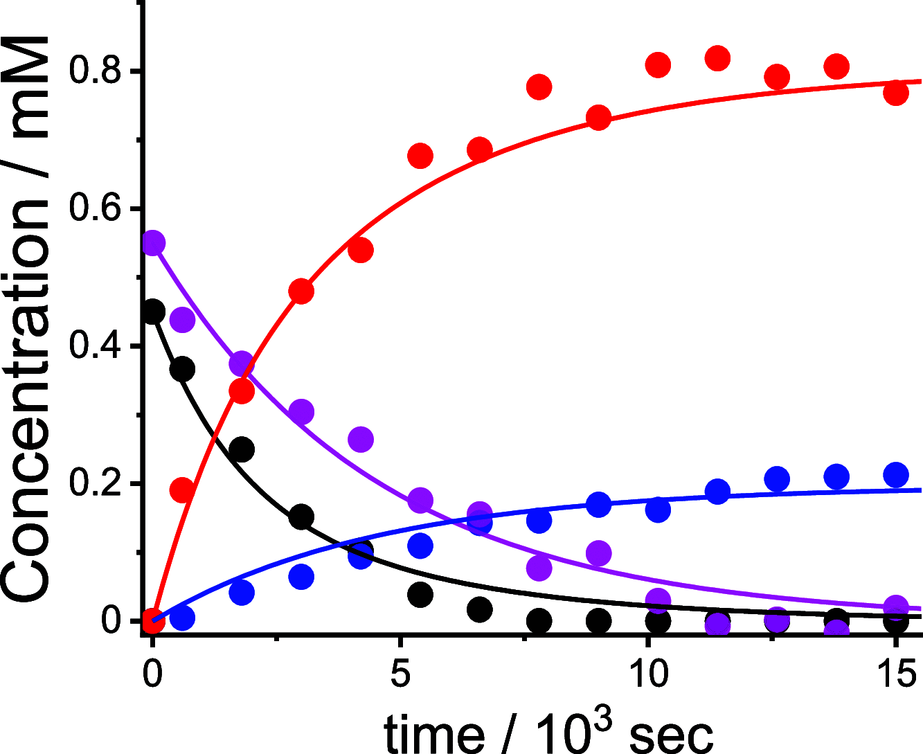
Kinetic traces of proximal-1 and distal-1 (total concentration: 1 mM) during the substitution reaction with 9-EtG (5 mM) at 303 K. Red: proximal-RuEtG; blue: distal-RuEtG; magenta: distal-1; black: proximal-1. The concentration of each complex was calculated based on the integration of peaks at 8.90 ppm (distal-RuEtG), 8.80 ppm (proximal-1), 6.92 ppm (distal-1 and proximal-RuEtG), and 6.63–6.70 ppm (proximal-1, proximal-RuEtG and distal-RuEtG). The solid lines are simulated curves according to first-order kinetics based on ligand substitution (proximal-1 to proximal-RuEtG, distal-1 to distal-RuEtG) and thermal isomerization (distal-1 to proximal-1) reactions.
Scheme 3
Scheme 3. Binding Reactions with 9-EtG for the Stimuli-Responsive Ruthenium Complexes proximal-1 and distal-1
The insets show steric repulsion between the aqua ligand and quinoline moiety in proximal-1 and intramolecular hydrogen bonding between the aqua ligand and alkoxy unit in distal-1.
Figure 8
Figure 8
1H NMR spectra of (A) proximal-RuEtG, (B) proximal- and distal-RuEtG, and (C) proximal-RuEtG after visible light irradiation for 16 h. Representative peaks of distal-RuEtG are highlighted as yellow. Solvent: D2O/CD3OD/TFE = 70:50:2, v/v/v.

References

    1. Bonnet S.; Limburg B.; Meeldijk J. D.; Klein Gebbink R. J. M.; Killian J. A. Ruthenium-Decorated Lipid Vesicles: Light-Induced Release of [Ru(terpy)(bpy)(OH2)]2+ and Thermal Back Coordination. J. Am. Chem. Soc. 2011, 133 (2), 252–261. 10.1021/ja105025m. - DOI - PubMed
    1. Wang J.; Feringa B. L. Dynamic Control of Chiral Space in a Catalytic Asymmetric Reaction Using a Molecular Motor. Science 2011, 331 (6023), 1429–1432. 10.1126/science.1199844. - DOI - PubMed
    1. Eisenreich F.; Kathan M.; Dallmann A.; Ihrig S. P.; Schwaar T.; Schmidt B. M.; Hecht S. A photoswitchable catalyst system for remote-controlled (co) polymerization in situ. Nat. Catal. 2018, 1 (7), 516–522. 10.1038/s41929-018-0091-8. - DOI
    1. Osorio-Planes L.; Rodríguez-Escrich C.; Pericàs M. A. Photoswitchable Thioureas for the External Manipulation of Catalytic Activity. Org. Lett. 2014, 16 (6), 1704–1707. 10.1021/ol500381c. - DOI - PubMed
    1. Blanco-Gómez A.; Cortón P.; Barravecchia L.; Neira I.; Pazos E.; Peinador C.; García M. D. Controlled binding of organic guests by stimuli-responsive macrocycles. Chem. Soc. Rev. 2020, 49 (12), 3834–3862. 10.1039/D0CS00109K. - DOI - PubMed