

High expression of P-selectin induces neutrophil extracellular traps via the PSGL-1/Syk/Ca2+/PAD4 pathway to exacerbate acute pancreatitis
- PMID: 37841279
- PMCID: PMC10568494
- DOI: 10.3389/fimmu.2023.1265344
High expression of P-selectin induces neutrophil extracellular traps via the PSGL-1/Syk/Ca2+/PAD4 pathway to exacerbate acute pancreatitis
Abstract
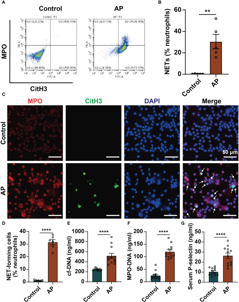
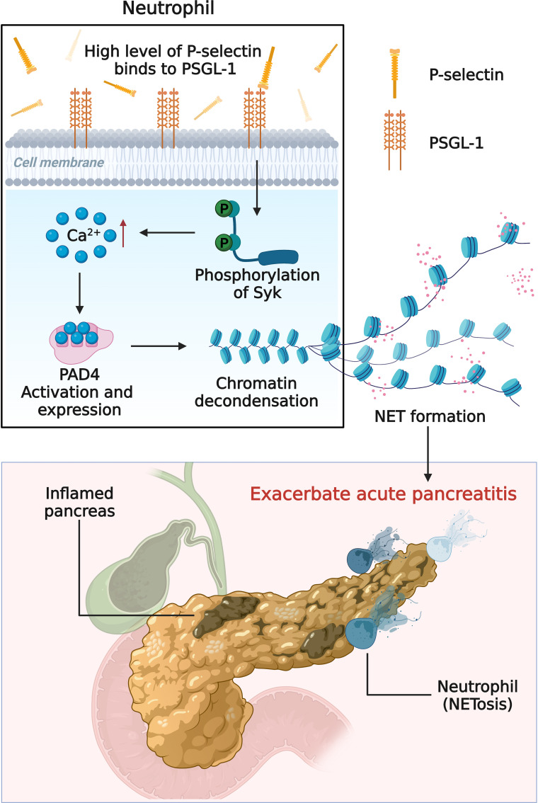
Background: Excessive neutrophil extracellular traps (NETs) is involved in the progression of acute pancreatitis (AP) but the mechanisms controlling NETs formation in AP are not fully understood. Therefore, our study sought to investigate the mechanism of the highly expressed P-selectin stimulating the formation of NETs in AP.
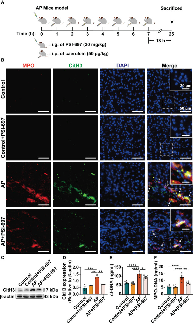
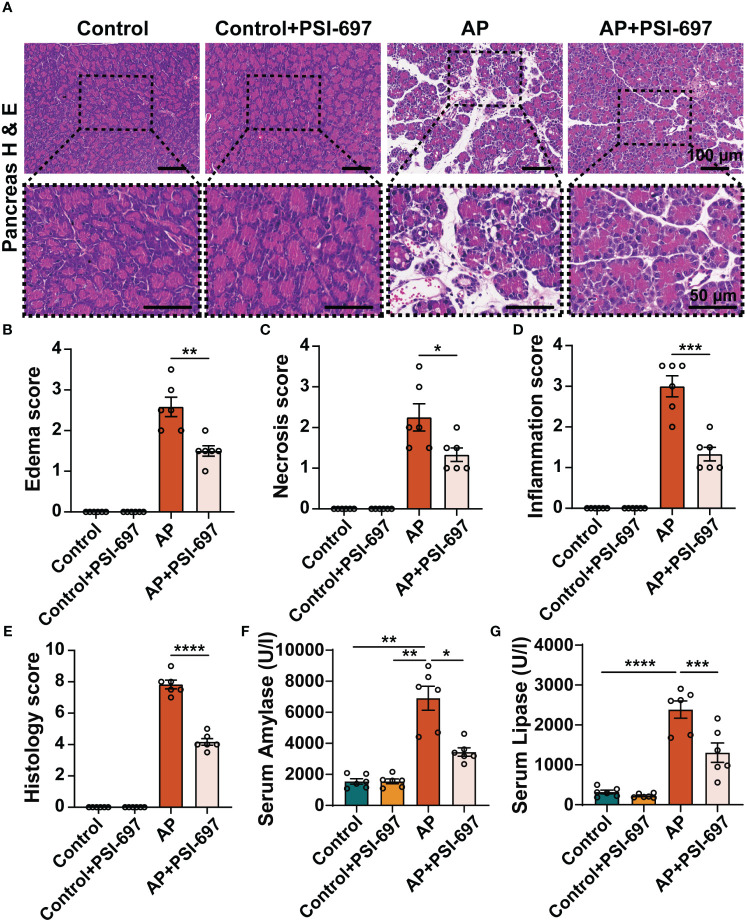
Methods: NETs formation was detected by flow cytometry, immunofluorescence staining, and cf-DNA and MPO-DNA complexes were measured as biomarkers of NETs formation. Neutrophils treated with P-selectin and pharmacological inhibitors were examined by western blot, immunofluorescence staining and flow cytometry. Mouse model of AP was established by caerulein and the effect of inhibiting P-selectin by PSI-697 on the level of NETs and PAD4 in pancreatic tissue was observed. The severity of AP was evaluated by histopathological score and the detection of serum amylase and lipase.
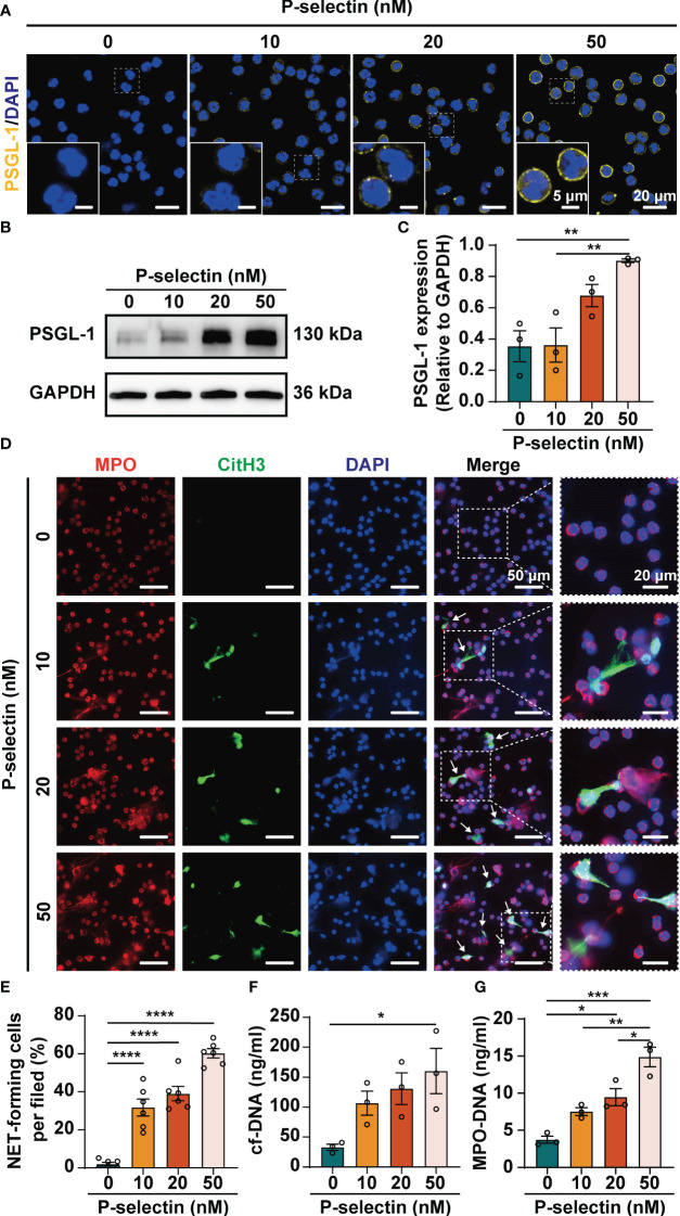
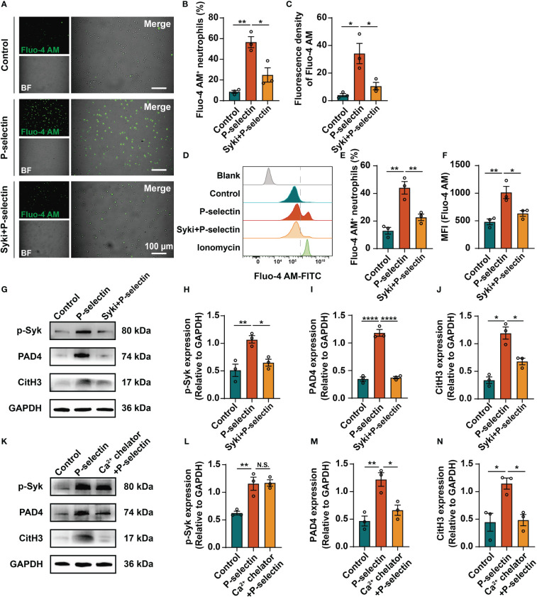
Results: Patients with AP had elevated levels of NETs and P-selectin compared with healthy volunteers. Stimulation of P-selectin up-regulated the expression of PSGL-1, increased the phosphorylation of Syk, mediated intracellular calcium signal and led to the activation and expression of PAD4, which modulated NETs formation in neutrophils. Pretreament with PSI-697 blunted NETs formation and PAD4 expression in the pancreatic tissue, and ameliorated the severity of AP in mice.
Conclusion: Taken together, these results suggest that P-selectin induces NETs through PSGL-1 and its downstream Syk/Ca2+/PAD4 signaling pathway, and that targeting this pathway might be a promising strategy for the treatment of AP.
Keywords: P-selectin; P-selectin glycoprotein ligand-1; acute pancreatitis; neutrophil extracellular traps; peptidylarginine deiminase 4.
Copyright © 2023 Xu, Shi, Ding, Xia, Luo, Lu, Zhang and Deng.
Conflict of interest statement
The authors declare that the research was conducted in the absence of any commercial or financial relationships that could be construed as a potential conflict of interest.
Figures

Similar articles
-
Protectin D1 decreases pancreatitis severity in mice by inhibiting neutrophil extracellular trap formation.Int Immunopharmacol. 2021 May;94:107486. doi: 10.1016/j.intimp.2021.107486. Epub 2021 Feb 24. Int Immunopharmacol. 2021. PMID: 33639566
-
Low P-Selectin Glycoprotein Ligand-1 Expression in Neutrophils Associates with Disease Activity and Deregulated NET Formation in Systemic Lupus Erythematosus.Int J Mol Sci. 2023 Mar 24;24(7):6144. doi: 10.3390/ijms24076144. Int J Mol Sci. 2023. PMID: 37047117 Free PMC article.
-
Neutrophil Extracellular Traps Induce Trypsin Activation, Inflammation, and Tissue Damage in Mice With Severe Acute Pancreatitis.Gastroenterology. 2015 Dec;149(7):1920-1931.e8. doi: 10.1053/j.gastro.2015.08.026. Epub 2015 Aug 22. Gastroenterology. 2015. PMID: 26302488
-
Roles, detection, and visualization of neutrophil extracellular traps in acute pancreatitis.Front Immunol. 2022 Aug 5;13:974821. doi: 10.3389/fimmu.2022.974821. eCollection 2022. Front Immunol. 2022. PMID: 36032164 Free PMC article. Review.
-
Recent advances in the role of neutrophils and neutrophil extracellular traps in acute pancreatitis.Clin Exp Med. 2023 Dec;23(8):4107-4122. doi: 10.1007/s10238-023-01180-4. Epub 2023 Sep 19. Clin Exp Med. 2023. PMID: 37725239 Review.
Cited by
-
P-selectin glycoprotein ligand-1 and cardiovascular diseases: from a general perspective to an HIV infection context.Front Cardiovasc Med. 2025 Feb 18;12:1521158. doi: 10.3389/fcvm.2025.1521158. eCollection 2025. Front Cardiovasc Med. 2025. PMID: 40041169 Free PMC article.
-
Mac-1 blockade impedes adhesion-dependent neutrophil extracellular trap formation and ameliorates lung injury in LPS-induced sepsis.Front Immunol. 2025 Mar 28;16:1548913. doi: 10.3389/fimmu.2025.1548913. eCollection 2025. Front Immunol. 2025. PMID: 40226627 Free PMC article.
-
Single-cell atlas of human gingiva unveils a NETs-related neutrophil subpopulation regulating periodontal immunity.J Adv Res. 2025 Jun;72:287-301. doi: 10.1016/j.jare.2024.07.028. Epub 2024 Jul 30. J Adv Res. 2025. PMID: 39084404 Free PMC article.
-
The role of P-selectin/PSGL-1 in regulating NETs as a novel mechanism in cerebral ischemic injury.Front Neurol. 2024 Jul 3;15:1442613. doi: 10.3389/fneur.2024.1442613. eCollection 2024. Front Neurol. 2024. PMID: 39022737 Free PMC article. Review.
-
Identification and analysis of neutrophil extracellular trap-related genes in periodontitis via bioinformatics and experimental verification.BMC Oral Health. 2025 Aug 6;25(1):1294. doi: 10.1186/s12903-025-06663-2. BMC Oral Health. 2025. PMID: 40770330 Free PMC article.
References
-
- Glasbrenner B, Adler G. Pathophysiology of acute pancreatitis. Hepato-gastroenterology (1993) 40:517–21. - PubMed
Publication types
MeSH terms
Substances
LinkOut - more resources
Full Text Sources
Medical
Research Materials
Miscellaneous