Review
. 2023 Aug 14;10(3):200-231.
doi: 10.3934/Neuroscience.2023017. eCollection 2023.

Depletion of dopamine in Parkinson's disease and relevant therapeutic options: A review of the literature

Affiliations
Review

Depletion of dopamine in Parkinson's disease and relevant therapeutic options: A review of the literature

Sairam Ramesh et al. AIMS Neurosci. .

Abstract

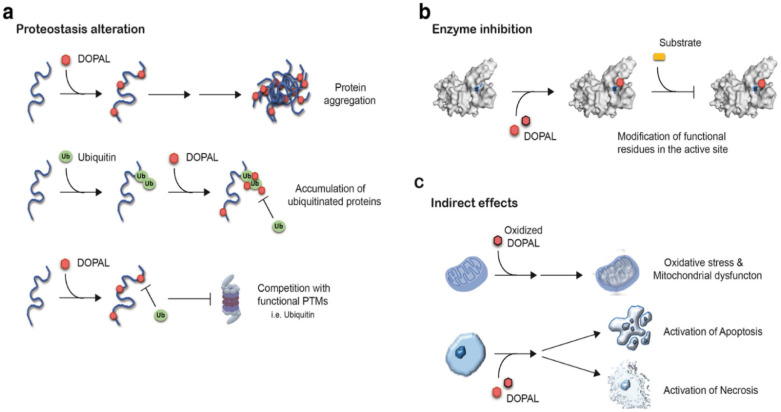
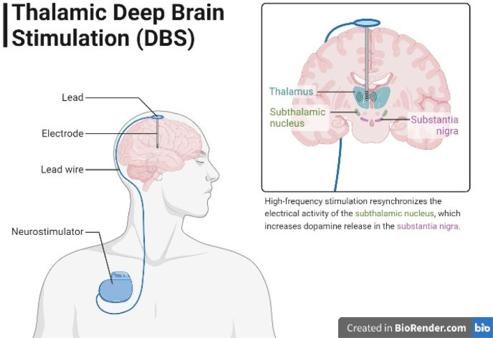
Parkinson's disease (PD) is a progressive neurodegenerative disorder that affects motor and cognition functions. The etiology of Parkinson's disease remains largely unknown, but genetic and environmental factors are believed to play a role. The neurotransmitter dopamine is implicated in regulating movement, motivation, memory, and other physiological processes. In individuals with Parkinson's disease, the loss of dopaminergic neurons leads to a reduction in dopamine levels, which causes motor impairment and may also contribute to the cognitive deficits observed in some patients. Therefore, it is important to understand the pathophysiology that leads to the loss of dopaminergic neurons, along with reliable biomarkers that may help distinguish PD from other conditions, monitor its progression, or indicate a positive response to a therapeutic intervention. Important advances in the treatment, etiology, and pathogenesis of Parkinson's disease have been made in the past 50 years. Therefore, this review tries to explain the different possible mechanisms behind the depletion of dopamine in PD patients such as alpha-synuclein abnormalities, mitochondrial dysfunction, and 3,4-dihydroxyphenylacetaldehyde (DOPAL) toxicity, along with the current therapies we have and the ones that are in development. The clinical aspect of Parkinson's disease such as the manifestation of both motor and non-motor symptoms, and the differential diagnosis with similar neurodegenerative disease are also discussed.

Keywords: Braak; COMT inhibitors; DOPAL neurotoxicity; L-DOPA; MAO inhibitors; Parkinson's disease; alpha-synuclein; deep brain stimulation; dopamine; therapies.


Conflict of interest statement

Conflicts of interest: The authors have no conflicts of interest to declare.

Figures

Figure 1.
Figure 1.. Diagram illustrating the direct and indirect pathways of the basal ganglia, which play a critical role in motor control. The direct pathway facilitates desired motor actions by reducing inhibition of the thalamus, promoting movement. In contrast, the indirect pathway suppresses unwanted movements by increasing thalamic inhibition. Together, they fine-tune motor movements, ensuring smooth and coordinated actions.
Figure 2.
Figure 2.. Braak staging is used to classify the degree of pathology in PD .
Figure 3.
Figure 3.. Illustrative representation of key traits of vulnerable neurons in PD. Concisely, neurons susceptible to Parkinson's disease (PD) exhibit key traits, and the disease is primarily driven by a malfunction in mitochondria and proteostasis. Mitochondrial dysfunction may result from the regulation of mitochondrial respiration by calcium and unidentified axonal bioenergetic factors, compounded by genetic and environmental influences (e.g., toxins). Proteostatic dysfunction arises from aSYN aggregation, promoted by oxidant stress, elevated cytosolic calcium, and DA quinones, as well as lysosomal dysfunction induced by increased mitophagy and oxidant damage to lysosomal proteins such as glucocerebrosidase. Solid lines represent firmly established connections in mammalian models, while dashed lines indicate mechanisms with strong but not definitive support .
Figure 4.
Figure 4.. Diagram showing the proposed functions of α-synuclein in controlling the cycling of presynaptic vesicles under varying levels of α-synuclein: (a) When α-synuclein levels are decreased, the reserve pool of vesicles is reduced, resulting in a higher number of readily available vesicles for release. This could lead to an augmentation in dopamine release. (b) In normal conditions, α-synuclein is believed to have a physiological role in regulating vesicle availability across different pools and in vesicle docking and fusion. (c) Conversely, elevated α-synuclein levels or mutations like E46K or A53T α-synuclein lead to a decrease in dopamine release. This may be due to their potential impact on a late stage in exocytosis or by reducing vesicle availability in the recycling pool through impaired vesicle endocytosis .
Figure 5.
Figure 5.. Neurotoxic molecular mechanisms associated with DOPAL (dihydroxyphenylacetaldehyde): DOPAL accumulation in dopaminergic neurons of the substantia nigra pars compacta (SNpc) triggers several neurotoxic processes: (a) Disruption of neuronal proteostasis, leading to protein aggregation, competition with functional post-translational modifications (PTMs) such as ubiquitination, SUMOylation, and acetylation, and buildup of ubiquitinated proteins. (b) Inhibition of enzymes within the neurons. (c) Indirect effects, including oxidative stress, mitochondrial dysfunction, and activation of necrotic and apoptotic pathways .
Figure 6.
Figure 6.. Molecular imaging of dopaminergic dysfunction in Parkinson's disease (PD) involves the use of PET (positron emission tomography) and SPECT (single-photon emission computed tomography) imaging techniques. In PD patients, these imaging studies reveal a reduction in VMAT2 (type 2 vesicular monoamine transporter) activity, DAT (dopamine transporter) availability, and DDC (dopa decarboxylase) activity when compared to healthy individuals used as controls. PET and SPECT imaging provides valuable insights into the molecular changes associated with PD, showcasing lower levels of VMAT2, DAT, and DDC functions, which are indicative of dopaminergic dysfunction in the disease .
Figure 7.
Figure 7.. Illustration depicting the concept of thalamic deep brain stimulation (DBS). A coronal section of a brain with the thalamus highlighted shows the placement of the DBS lead within the thalamic region. Electrical signals are represented in blue traveling from the implanted pulse generator to the thalamus, symbolizing the delivery of stimulation. This high-frequency stimulation synchronizes the electrical activity of the subthalamic nucleus, leading to an elevation in dopamine release within the substantia nigra. Thalamic DBS is a neurosurgical procedure used for treating various neurological disorders including Parkinson's disease.
Figure 8.
Figure 8.. The figure illustrates the mechanism of action of drugs used for Parkinson's disease management. The diagram showcases the key components involved in the process. For a detailed summary of these treatments, refer to Table 1.

References

    1. Parkinson J. An Essay on the Shaking Palsy. J Neuropsych Clin N. 2002;14(2):223–236. https://neuro.psychiatryonline.org/doi/full/10.1176/jnp.14.2.223 . - DOI - PubMed
    1. de Lau LML, Breteler MMB. Epidemiology of Parkinson's disease. Lancet Neurol. 2006;5:525–535. doi: 10.1016/S1474-4422(06)70471-9. - DOI - PubMed
    1. Poewe W, Seppi K, Tanner CM, et al. Parkinson disease. Nat Rev Dis Primers. 2017;3:17013. doi: 10.1038/nrdp.2017.13. - DOI - PubMed
    1. Kalia LV, Lang AE. Parkinson's disease. Lancet. 2015;386:896–912. doi: 10.1016/S0140-6736(14)61393-3. - DOI - PubMed
    1. Hernandez DG, Reed X, Singleton AB. Genetics in Parkinson disease: Mendelian versus non-Mendelian inheritance. J Neurochem. 2016;139:59–74. doi: 10.1111/jnc.13593. - DOI - PMC - PubMed

LinkOut - more resources