. 2023 Sep 28:10:1225796.
doi: 10.3389/fvets.2023.1225796. eCollection 2023.

Morphological, ultrastructural, genetic characteristics and remarkably low prevalence of macroscopic Sarcocystis species isolated from sheep and goats in Kurdistan region, Iraq

Affiliations

Morphological, ultrastructural, genetic characteristics and remarkably low prevalence of macroscopic Sarcocystis species isolated from sheep and goats in Kurdistan region, Iraq

Salh Nawshirwan et al. Front Vet Sci. .

Abstract

Introduction: Sarcocystis is a genus of cyst-forming parasites that infest both humans and livestock. Some parasites cause clinical and subclinical diseases in their hosts, resulting in economic losses.

Methods: Esophagus, diaphragm, and skeletal muscle from slaughtered sheep and goats were examined macroscopically, microscopically, and ultrastructurally and subjected to DNA analysis.

Results: We isolated macrocysts of S. gigantea and of S. caprafelis moulei from naturally infected sheep (Ovis aries) and goats (Capra hircus). The macrocyst wall thickness was 18.9 µm in sheep and 15.3 µm in goats, and consisted of an inner Periodic acid Schiff- (PAS) negative primary wall and an outer glycoconjugates containing i.e. PAS-positive secondary wall. The walls inner surface was compartmentalized and filled with bradyzoites. In S. gigantea the bradyzoites were approximently 12.3 x 2.6 µm in size, while in S. caprafelis moulei they were 13.9 x 4.4 µm. Ultrastructurally, both species have nearly identical morphology: cauliflower-like protrusions with numerous microtubules and often dendritic-like filaments, branching from the primary wall. The 18S rRNA gene in S. gigantea was 85.9% identical to that in S. medusiformis and 80.4% to the S. caprafelis moulei gene. The 28S rRNA gene in S. gigantea was 94.6% identical to that in S. medusiformis and 97.3% to the S. caprafelis moulei.

Conclusion: This study is the first to (i) detail the ultrastructure of the macrocyst wall of S. caprafelis moulei, (ii) identify S. medusiformis in Iraqi sheep, and (iii) compare the prevalence of macroscopic Sarcocystis at different time periods within the same region. A positive finding was the reduction of macroscopic sarcocystosis occurrences (0.01% in sheep and 0.02% in goats) compared to our previous data from 1992 (4.1%: sheep, 33.6%: goats).

Keywords: Sarcocystis gigantea; Sarcocystis medusiformis; Sarcocystis moulei; genetic analysis; goat; sheep.


Conflict of interest statement

The authors declare that the research was conducted in the absence of any commercial or financial relationships that could be construed as a potential conflict of interest.

Figures

Figure 1
Figure 1
Macroscopy and light microscopy of S. gigantea (A–C) and S. caprafelis moulei (D–F) macrocysts Gross appearance of macrocysts of S. gigantea in sheep (A) and S. caprafelis moulei in goat (D) in the heavily infected esophagi, scale bar in cm. Light microscopic appearance of an oval macrocyst of S. gigantea (B) and S. caprafelis moulei (E), both macrocysts have a PAS-positive outer secondary thick wall (SW), and a PAS-negative inner primary relatively thick wall (PW), HT = Host tissue. Higher magnification shows the primary and secondary walls of the macrocysts of S. gigantea (C) and S. caprafelis moulei (F). Between the primary and secondary walls is a relatively thick layer of degenerating host cells (DL). Beneath the primary wall are numerous compartments (Co) separated by tissue septa (S) and filled with banana-shaped bradyzoites (Br), (B) and (E) 5 μm paraffin sections, Tri-PAS staining; (C) and (F) 0.5 μm semithin sections, methylene blue staining.
Figure 2
Figure 2
Statistical variantion of macrocyst parameters between S. gigantea (sheep) and S. caprafelis moulei (goat). In all tests p < 0.05 was considered significant and marked with * p < 0.01 was assigned with **, p < 0.001 with ***, and **** p < 0.0001. ± = standard error of the mean. (A,B) macrocysts length and width: S. gigantea 6.7× 2.9 mm, S. caprafelis moulei 6.3 × 4.0 mm (n = 32 sheep and n = 70 goat; two-tailed Mann–Whitney test). (C) Inner primary wall thickness of macrocysts: S. gigantea 7.3 μm, S. caprafelis moulei 7.3 μm. (D) Outer secondary wall thickness of macrocysts: S. gigantea 11.6 μm, S. caprafelis moulei 8.0 μm (n = 25 sheep, n = 6 goat; two-tailed Mann–Whitney test). (E) Total wall thickness of macrocysts, i.e., primary and secondary cyst walls together: S. gigantea 18.9, S. caprafelis moulei 15.3 μm (n = 25 sheep, n = 6 goat; two-tailed Mann–Whitney test). (F) Area compartment of macrocysts: S. gigantea 1,065 μm 2 , S. caprafelis moulei 917 μm2 (n = 39 sheep, n = 96 goat; two-tailed Mann–Whitney test). (G) Bradyzoite length: S. gigantea 12.3, S. caprafelis moulei 13.9 μm (n = 49 sheep, n = 30 goat; two-tailed Mann–Whitney test). (H) Bradyzoite width: S. gigantea 2.6 μm, S. caprafelis moulei 4.4 μm (n = 49 sheep, n = 30 goat; two-tailed t-test). (I) width of macrocyst villar protrusions: S. gigantea 2.4 μm, S. caprafelis moulei 3.5 μm (n = 27 sheep, n = 23 goat; two-tailed Mann–Whitney test). (J) height of macrocyst villar protrusions: S. gigantea 3.1 μm, S. caprafelis moulei 3.4 μm (n = 27 sheep, n = 23 goat; two-tailed Mann–Whitney test). (K) Thickness of the degenerating layer: S. gigantea 4.5 μm, S. caprafelis moulei 4.9 μm (n = 40 sheep, n = 38 goat; two-tailed t-test), for more details see Supplementary material S2.
Figure 3
Figure 3
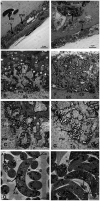
Electron micrographs of macrocysts in the esophagus of sheep (S. gigantea) (A–D) and goats (S. caprafelis moulei) (E–H). (A–C) and (E–G): showing the macrocyst wall of S. gigantea and S. caprafelis moulei, respectively. In both species, the degenerating host cells layer (DL) is located between the primary (PW) and secondary (SW) cyst walls and contains many vacuoles (V). In (E), the secondary cyst wall ist absent due to mechanical damage during tissue processing. (B,C) and (F,G): High magnifications of cauliflower-like protrusions (Pr) extended from the primary to the secondary cyst wall, and numerous long dendritic-like filaments (F) protruded from both surfaces of the protrusions and the primary wall of the macrocyst. In goats (G) the protrusions are more pronounced and the filaments are slightly longer and more numerous than in sheep (C). Within the protrusions, long microtubules (MT) extended longitudinally and transversely from the core of the free protrusions into the ground substance (GS). In both species, coarse and electron-dense granules (ED) of nearly spherical in shape were observed at the base and margin of the projections and in the ground substance. (D) and (H): Ultrastructure findings of the bradyzoites of S. gigantea and S. caprafelis moulei, respectively. They exhibited a bilayered pellicle (Pe), slightely thickened at the anterior end of the banana-shaped bradyzoites and numerous small rod-shaped micronemes (Mn) with rounded ends at the anterior part (Co = conoid). Beneath the micronemes are about 8–12 irregularly shaped rhoptries (Rh), as well as some elongated tubular mitochondria and a relatively small number of amylopectin granules (AG). The cell nucleus (N) is located at the posterior end of the cell body.

References

    1. Dubey JP, Carlero-Bernal R, Rosenthal BM, Speer CA, Fayer R. Sarcocystosis of animals and humans. 2nd ed. Boca Raton, FL: CRC Press; (2016).
    1. Heckeroth AR, Tenter AM. Development and validation of species-specific nested PCRs for diagnosis of acute sarcocystosis in sheep. Int J Parasitol. (1999) 29:1331–49. doi: 10.1016/s0020-7519(99)00111-3, PMID: - DOI - PubMed
    1. Prakas P, Bultkauslkas D, Rudaityte E, Kutkiene L, Srugosa A, Puraite I. Morphoogical and molecular characterization of Sarcocystis taeniata and Sarcocystis pilosa n. sp. from the sika deer (Cervus Nippon) in Lithuania. Parasitol Res. (2016) 115:3021–32. doi: 10.1007/s00436-016-5057-7, PMID: - DOI - PubMed
    1. Pipia AP, Varcasia A, Zidda A, Dessì G, Panzalis R, Tamponi C, et al. . Cross-sectional investigation on sheep sarcosporidiosis in Sardinia, Italy. Vet Parasitol. (2016) 3-4:13–7. doi: 10.1016/j.vprsr.2016.05.004, PMID: - DOI - PubMed
    1. Hoeve-Bakker BJA, Van der Giessen JWB, Franssen FFJ. Molecular identification targeting cox1 and 18S genes confirms the high prevalence of Sarcocystis spp. in cattle in the Netherlands. Int J Parasitol. (2019) 49:859–66. doi: 10.1016/j.ijpara.2019.05.008, PMID: - DOI - PubMed

LinkOut - more resources