[Preprint]. 2023 Sep 28:rs.3.rs-3179362.
doi: 10.21203/rs.3.rs-3179362/v1.

Neural Correlates of Positive and Negative Formal Thought Disorder in Individuals with Schizophrenia: An ENIGMA Schizophrenia Working Group Study

Affiliations

Neural Correlates of Positive and Negative Formal Thought Disorder in Individuals with Schizophrenia: An ENIGMA Schizophrenia Working Group Study

Thomas Nickl-Jockschat et al. Res Sq. .

Update in

Abstract

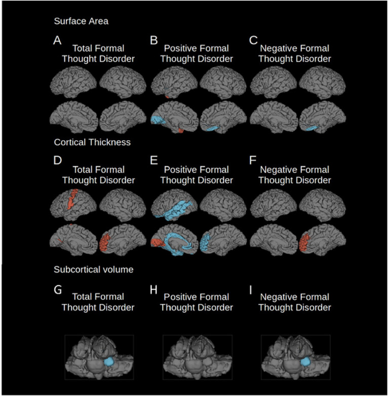
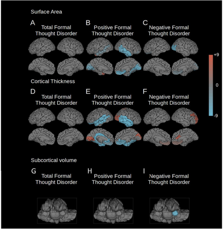
Formal thought disorder (FTD) is a key clinical factor in schizophrenia, but the neurobiological underpinnings remain unclear. In particular, relationship between FTD symptom dimensions and patterns of regional brain volume deficiencies in schizophrenia remain to be established in large cohorts. Even less is known about the cellular basis of FTD. Our study addresses these major obstacles based on a large multi-site cohort through the ENIGMA Schizophrenia Working Group (752 individuals with schizophrenia and 1256 controls), to unravel the neuroanatomy of positive, negative and total FTD in schizophrenia and their cellular bases. We used virtual histology tools to relate brain structural changes associated with FTD to cellular distributions in cortical regions. We identified distinct neural networks for positive and negative FTD. Both networks encompassed fronto-occipito-amygdalar brain regions, but negative FTD showed a relative sparing of orbitofrontal cortical thickness, while positive FTD also affected lateral temporal cortices. Virtual histology identified distinct transcriptomic fingerprints associated for both symptom dimensions. Negative FTD was linked to neuronal and astrocyte fingerprints, while positive FTD was also linked to microglial cell types. These findings relate different dimensions of FTD to distinct brain structural changes and their cellular underpinnings, improve our mechanistic understanding of these key psychotic symptoms.


Conflict of interest statement

Confiicts of Interest The authors declare that there is no conflict of interest.

Figures

Figure 1
Figure 1
Regions where cortical surface area or thickness were significantly associated with total, positive and negative FTD (FDR corrected p = 0.001). Left hemisphere is shown on the left of each group of images. Blue indicates regions of reduced surface area, cortical thickness or subcortical volumes, red indicates regions of relatively spared surface area, cortical thickness or subcortical volumes. A) surface area changes associated with total FTD; B) surface area changes associated with positive FTD; C) surface area changes associated with negative FTD; D) cortical thickness changes associated with total FTD; E) cortical thickness changes associated with positive FTD; F) cortical thickness changes associated with negative FTD; G) volume changes of subcortical structures associated with total FTD; H) volume changes of subcortical structures associated with positive FTD; I) volume changes of subcortical structures associated with negative FTD.
Figure 2
Figure 2
“Leave one out” analysis of FTD correlation. The regions associated with total, positive, or negative FTD (FDR corrected p = 0.001) of each of the 10 “leave one out” analyses were added together so that all positively affected regions are shown in red, and all negatively affected regions in blue. The intensity of the color represents the number of “leave one out” analyses where that region was affected. Leave one out analyses were conducted for the following parameters: A) surface area changes associated with total FTD; B) surface area changes associated with positive FTD; C) surface area changes associated with negative FTD; D) cortical thickness changes associated with total FTD; E) cortical thickness changes associated with positive FTD; F) cortical thickness changes associated with negative FTD; G) volume changes of subcortical structures associated with total FTD; H) volume changes of subcortical structures associated with positive FTD; I) volume changes of subcortical structures associated with negative FTD.

References

    1. Andreasen N. C., “Thought, language, and communication disorders. II. Diagnostic significance,” Arch. Gen. Psychiatry, vol. 36, no. 12, pp. 1325–1330, Nov. 1979, doi:10.1001/archpsyc.1979.01780120055007. - DOI - PubMed
    1. DeLisi L. E., “Speech disorder in schizophrenia: review of the literature and exploration of its relation to the uniquely human capacity for language,” Schizophr. Bull., vol. 27, no. 3, pp. 481–496, 2001, doi:10.1093/oxfordjournals.schbul.a006889. - DOI - PubMed
    1. Hartmann J. A. et al., “Declining transition rates to psychotic disorder in ‘ultra-high risk’ clients: Investigation of a dilution effect,” Schizophr. Res., vol. 170, no. 1, pp. 130–136, Jan. 2016, doi: 10.1016/j.schres.2015.11.026. - DOI - PubMed
    1. DeVylder J. E. et al., “Symptom trajectories and psychosis onset in a clinical high-risk cohort: the relevance of subthreshold thought disorder,” Schizophr. Res., vol. 159, no. 2–3, pp. 278–283, Nov. 2014, doi: 10.1016/j.schres.2014.08.008. - DOI - PMC - PubMed
    1. Armando M. et al., “Twelve-month psychosis-predictive value of the ultra-high risk criteria in children and adolescents,” Schizophr. Res., vol. 169, no. 1–3, pp. 186–192, Dec. 2015, doi: 10.1016/j.schres.2015.10.033. - DOI - PubMed

Publication types

LinkOut - more resources