Review
. 2023 Sep 28:14:1236550.
doi: 10.3389/fphar.2023.1236550. eCollection 2023.

A review of the pathophysiology and the role of ion channels on bronchial asthma

Affiliations
Review

A review of the pathophysiology and the role of ion channels on bronchial asthma

Indyra Alencar Duarte Figueiredo et al. Front Pharmacol. .

Abstract

Asthma is one of the main non-communicable chronic diseases and affects a huge portion of the population. It is a multifactorial disease, classified into several phenotypes, being the allergic the most frequent. The pathophysiological mechanism of asthma involves a Th2-type immune response, with high concentrations of allergen-specific immunoglobulin E, eosinophilia, hyperreactivity and airway remodeling. These mechanisms are orchestrated by intracellular signaling from effector cells, such as lymphocytes and eosinophils. Ion channels play a fundamental role in maintaining the inflammatory response on asthma. In particular, transient receptor potential (TRP), stock-operated Ca2+ channels (SOCs), Ca2+-activated K+ channels (IKCa and BKCa), calcium-activated chloride channel (TMEM16A), cystic fibrosis transmembrane conductance regulator (CFTR), piezo-type mechanosensitive ion channel component 1 (PIEZO1) and purinergic P2X receptor (P2X). The recognition of the participation of these channels in the pathological process of asthma is important, as they become pharmacological targets for the discovery of new drugs and/or pharmacological tools that effectively help the pharmacotherapeutic follow-up of this disease, as well as the more specific mechanisms involved in worsening asthma.

Keywords: CFTR; KCa channels; ORAI channels; P2X receptor; Piezo1 channel; TMEM16A channel; TRP channels; asthma.


Conflict of interest statement

The authors declare that the research was conducted in the absence of any commercial or financial relationships that could be construed as a potential conflict of interest.

Figures

FIGURE 1
FIGURE 1
Mainly asthma phenotypes and inflammatory response profile.
FIGURE 2
FIGURE 2
Inflammatory pathophysiology of allergic asthma. In the presence of an allergen, there is the release of cytokines from the epithelium, particularly IL-25 and IL-33, which induces co-stimulatory molecule of T lymphocytes in dendritic cells (DCs), which act to promote T cell survival and expansion. DCs are mobilized to local lymph nodes, where they present the antigenic peptide to naive CD4+ T cells, activating and converting them to a competent IL-4-producing state. These T cells migrate to B cell zones where they differentiate into follicular helper T cells (TFH) and move into the circulation to complete their maturation as helper T cells 2 (Th2). IL-4 secreting TFH cells in areas of B cells mediate IgE class switching in B lymphocytes, which attach to the surface of mast cells, basophils and eosinophils (not shown in figure), mediating the degranulation of these cells. IL-9 also stimulates mast cells. Th2 cells migrate to the airway epithelium and to the submucosa layer, where they secrete IL-5 and IL-13 to mediate the inflammatory response, with the accumulation of mast cells and eosinophils, airway remodeling and bronchial hyperreactivity.
FIGURE 3
FIGURE 3
TRPA and TRPV channel structure. ANK, ankyrin repeats (varies in number among members of subfamilies); coiled-coil domains; TRP box; CaM, calmodulin binding site; PDZ specific reason for binding to PDZ.
FIGURE 4
FIGURE 4
(A) STIM monomeric structure, including the PDB domain; the coiled-coil domains (CC1, CC2 and CC3); CAD/SOAR domain; sterile alpha motif (SAM) and EF-hand; (B) Structure of the Orai channel, containing four transmembrane domains (TM1-TM4) and the CAD/SOAR binding domain.
FIGURE 5
FIGURE 5
(A) IKCa structure, with 6 transmembrane segments and the calmodulin binding domain (CaM). (B) BKCa, with seven transmembrane segments and two Ca2+-sensitive domains that regulate K+ conductance.
FIGURE 6
FIGURE 6
Structure of a subunit of TMEM16A, with 10 transmembrane domains. Between α3-α7 there is the formation of the channel pore; Ca2+ binding sites is shown in blue circles.
FIGURE 7
FIGURE 7
CFTR structure with two canonical transmembrane domains (TMDs). NBD, cytosolic nucleotide-binding domain. R, regulatory domain.
FIGURE 8
FIGURE 8
PIEZO1 structure, with 38 transmembrane segments. THUs, transmembrane helical subunits; OH and IH, outer and inner transmembrane helices, respectively; CED, C-terminal domain.
FIGURE 9
FIGURE 9
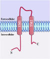
P2X structure with two transmembrane domain α-helices (TM1 and TM2), with TM2 forming the ion-conducting pore within the inner tunnel.
FIGURE 10
FIGURE 10
Ion channels as possible targets in allergic asthma.
FIGURE 11
FIGURE 11
Overview of ion channels in different airway cells associated with asthma pathophysiology.

References

    1. Adelman J. P. (2016). SK channels and calmodulin. Channels 10, 1–6. 10.1080/19336950.2015.1029688 - DOI - PMC - PubMed
    1. Aikawa T., Shimura S., Sasaki H., Ebina M., Takishima T. (1992). Marked goblet cell hyperplasia with mucus accumulation in the airways of patients who died of severe acute asthma attack. Chest 101, 916–921. 10.1378/chest.101.4.916 - DOI - PubMed
    1. Akuthota P., Wang H. B., Spencer L. A., Weller P. F. (2008). Immunoregulatory roles of eosinophils: A new look at a familiar cell. Clin. Exp. Allergy 38, 1254–1263. 10.1111/j.1365-2222.2008.03037.x - DOI - PMC - PubMed
    1. Al-Karagholi M. A. M., Gram C., Nielsen C. A. W., Ashina M. (2020). Targeting BKCa channels in migraine: Rationale and perspectives. CNS Drugs 34, 325–335. 10.1007/s40263-020-00706-8 - DOI - PubMed
    1. Aleksandrov L., Aleksandrov A. A., Chang X. B., Riordan J. R. (2002). The first nucleotide binding domain of cystic fibrosis transmembrane conductance regulator is a site of stable nucleotide interaction, whereas the second is a site of rapid turnover. J. Biol. Chem. 277, 15419–15425. 10.1074/jbc.M111713200 - DOI - PubMed