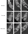
Prevalence of Radix Entomolaris in Mandibular Permanent Molars Analyzed by Cone-Beam CT in the Saudi Population of Ha'il Province
- PMID: 37841985
- PMCID: PMC10576614
- DOI: 10.7759/cureus.47034
Prevalence of Radix Entomolaris in Mandibular Permanent Molars Analyzed by Cone-Beam CT in the Saudi Population of Ha'il Province
Abstract
Background: The present study aimed to examine the prevalence of radix entomolaris (RE) in the mandibular permanent molar within a specific sub-population in Saudi Arabia.
Methods: A comprehensive analysis was conducted on 499 cone-beam computed tomography (CBCT) scans of a mandibular molar from a sample of Saudi patients aged between 18 and 65. The primary objective of this study was to investigate the anatomical characteristics of mandibular permanent molars, specifically focusing on the number of roots present. The chi-square test was employed to examine the relationship between various variables.
Results: In the case of mandibular first molars, it was observed that 95.3% of these molars exhibited a bifurcated root structure. In comparison, the remaining 4.7% displayed a triradicular configuration within the sample population under investigation. Although there were some variations, no significant differences in the number of roots were observed between males and females or left and right sides (p > 0.05). In the case of mandibular second molars, it was observed that 96.9% of them exhibited a bifurcated root structure, whereas 2.5% displayed a trifurcated root configuration, and a mere 0.6% possessed a single root. There were no statistically significant variations in the number of roots between males and females or left and right sides (p > 0.05).
Conclusions: The identification of RE was observed in the mandibular molars. Moreover, the discovered RE roots were predominantly found in the mandibular first molar, displaying a tendency for bilateral occurrence in both male and female individuals.
Keywords: anatomy; cone beam computed tomography; mandibular molars; radix entomolaris; saudi subpopulation.
Copyright © 2023, Almansour et al.
Conflict of interest statement
The authors have declared that no competing interests exist.
Figures
References
-
- The influence of missed canals on the prevalence of periapical lesions in endodontically treated teeth: a cross-sectional study. Baruwa AO, Martins JN, Meirinhos J, et al. J Endod. 2020;46:34–39. - PubMed
-
- Association between missed canals and apical periodontitis. Costa FF, Pacheco-Yanes J, Siqueira JF Jr, et al. Int Endod J. 2019;52:400–406. - PubMed
-
- An anatomic investigation of the mandibular first molar using micro-computed tomography. Harris SP, Bowles WR, Fok A, McClanahan SB. J Endod. 2013;39:1374–1378. - PubMed
-
- Missed anatomy: frequency and clinical impact. Cantatore G, Berutti E, Castellucci A. Endod Topics. 2009;15:3–31.
-
- Root canal anatomy of the human permanent teeth. Vertucci FJ. Oral Surg Oral Med Oral Pathol. 1984;58:589–599. - PubMed
LinkOut - more resources
Full Text Sources


