. 2023 Sep 25:11:1276333.
doi: 10.3389/fcell.2023.1276333. eCollection 2023.

Hyperactive KRAS/MAPK signaling disrupts normal lymphatic vessel architecture and function

Affiliations

Hyperactive KRAS/MAPK signaling disrupts normal lymphatic vessel architecture and function

Lorenzo M Fernandes et al. Front Cell Dev Biol. .

Abstract

Complex lymphatic anomalies (CLAs) are sporadically occurring diseases caused by the maldevelopment of lymphatic vessels. We and others recently reported that somatic activating mutations in KRAS can cause CLAs. However, the mechanisms by which activating KRAS mutations cause CLAs are poorly understood. Here, we show that KRASG12D expression in lymphatic endothelial cells (LECs) during embryonic development impairs the formation of lymphovenous valves and causes the enlargement of lymphatic vessels. We demonstrate that KRASG12D expression in primary human LECs induces cell spindling, proliferation, and migration. It also increases AKT and ERK1/2 phosphorylation and decreases the expression of genes that regulate the maturation of lymphatic vessels. We show that MEK1/2 inhibition with the FDA-approved drug trametinib suppresses KRASG12D-induced morphological changes, proliferation, and migration. Trametinib also decreases ERK1/2 phosphorylation and increases the expression of genes that regulate the maturation of lymphatic vessels. We also show that trametinib and Cre-mediated expression of a dominant-negative form of MEK1 (Map2k1 K97M) suppresses KRASG12D-induced lymphatic vessel hyperplasia in embryos. Last, we demonstrate that conditional knockout of wild-type Kras in LECs does not affect the formation or function of lymphatic vessels. Together, our data indicate that KRAS/MAPK signaling must be tightly regulated during embryonic development for the proper development of lymphatic vessels and further support the testing of MEK1/2 inhibitors for treating CLAs.

Keywords: Gorham-Stout disease; KRAS; complex lymphatic anomaly; lymphangiogenesis; lymphatic malformation; trametinib.


Conflict of interest statement

MD is the Director of Research for the Lymphatic Malformation Institute. The remaining authors declare that the research was conducted in the absence of any commercial or financial relationships that could be construed as a potential conflict of interest.

Figures

FIGURE 1
FIGURE 1
LEC KrasG12D embryos have enlarged jugular lymph sacs and malformed lymphovenous valves. (A). Schematics of the Lyve1-Cre and Kras LSL-G12D alleles. (B). Representative images of E14.5 LEC Ctrl and LEC KrasG12D embryos. The LEC KrasG12D embryo has edema. (C). Transverse sections of E14.5 embryos stained with an anti-Lyve1 antibody (brown) and hematoxylin (purple). (D). Jugular lymph sac area was significantly greater in LEC KrasG12D embryos (273391 ± 47694; n = 6 mice) than LEC Ctrl embryos (76949 ± 17566; n = 5 mice). (E). Coronal sections of E14.5 embryos stained with hematoxylin and eosin (H&E). The arrow points to a lymphovenous valve in a LEC Ctrl embryo. The arrow points to a cluster of cells in the lymphovenous valve region in a LEC KrasG12D embryo. Six LEC Ctrl and five LEC KrasG12D embryos were analyzed. (F). Coronal sections of E14.5 embryos stained with DAPI (blue) and antibodies against Lyve1 (yellow) and endomucin (red). The arrow points to a lymphovenous valve in a LEC Ctrl embryo. The arrow points to a cluster of Lyve1-positive cells in the lymphovenous valve region in a LEC KrasG12D embryo. Five LEC Ctrl and three LEC KrasG12D embryos were analyzed. (G). Immunostaining revealed that cell clusters in the jugular lymph sacs of LEC KrasG12D embryos contained Prox1 and Lyve-1 double-positive LECs. Six LEC KrasG12D embryos were analyzed. (H). Higher magnification image of cluster in panel (G). (I,J). Clusters also contained F4/80-positive cells (I) and CD45-positive cells (J). (K). Immunostaining revealed that clusters in LEC KrasG12D embryos contained Runx1-positive cells. (L–O). Higher magnification images of the separate channels in panel (K). Data are presented as mean ± SEM. **p < 0.01; unpaired Student’s t-tests. Scale bar in panel C = 200 µm. Scale bars in panels E, G, I, J, and K = 100 µm.
FIGURE 2
FIGURE 2
LEC KrasG12D embryos have abnormal dermal lymphatic vessels. (A,B). Back skin whole-mounts from E15.5 embryos stained for neuropilin-2. (C). LEC KrasG12D embryos (11.00 ± 1.732; n = 3 mice) had significantly fewer lymphatic vessel branch points than LEC Ctrl embryos (62.14 ± 8.382; n = 7 mice). (D). Lymphatic vessel diameter was significantly greater in LEC KrasG12D embryos (151.4 ± 20.05; n = 3 mice) than in LEC Ctrl embryos (43.30 ± 2.047; n = 7 mice). Data are presented as mean ± SEM. ** p < 0.01, **** p < 0.0001; unpaired Student’s t-tests. Scale bar = 250 µm.
FIGURE 3
FIGURE 3
KRASG12D induces cell morphological changes, proliferation, and migration. (A). Brightfield images of GFP-LECs and KRASG12D-LECs. The images were taken 72 h after treating primary human LECs with lentivirus particles that express GFP or KRASG12D. GFP-LECs exhibit a normal cobblestone morphology, whereas KRASG12D-LECs exhibit a spindle morphology. (B). Circularity index measurements for GFP-LECs and KRASG12D-LECs. (C). MTS viability assay results for GFP-LECs and KRASG12D-LECs. Viability was measured 72 h after treating cells with lentivirus particles that express GFP or KRASG12D. (D). Representative images of GFP-LECs and KRASG12D-LECs taken 0 and 14 h after scratching confluent monolayers of cells. (E). Graph showing scratch closure area 14 h after wounding. KRASG12D-LECs closed the scratched area significantly faster than GFP-LECs. Data are presented as mean ± SEM. **p < 0.01, ****p < 0.0001; unpaired Student’s t-tests. Scale bars = 300 µm.
FIGURE 4
FIGURE 4
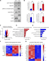
KRASG12D expression in LECs induces PI3K and MAPK signaling and changes in gene expression. (A). Western blot results for phospho-AKT, AKT, phospho-ERK1/2, ERK1/2, GFP, KRASG12D (mutation-specific antibody), and tubulin. Protein lysates were made 72 h after treating primary human LECs with GFP or KRASG12D expressing lentivirus particles. (B). Graphs of Western blot results. Phospho-AKT and phospho-ERK1/2 levels were significantly higher in KRASG12D-LECs than GFP-LECs. GFP-LECs specifically expressed GFP, and KRASG12D-LECs specifically expressed KRASG12D. (C). Volcano plot of RNA-Seq results comparing KRASG12D-LECs to GFP-LECs. Two thousand and nine genes were upregulated (red dots), and 1,396 genes were downregulated (blue dots) by KRASG12D (log2 fold-change ≥1 or ≤ −1; FDR <0.01). D,E. Select GO terms associated with genes upregulated by KRASG12D (D) and genes downregulated by KRASG12D (E). (F,G). Heatmaps of genes that regulate angio/lymphangiogenesis (F) and genes that control lymphatic valve development or lymphatic muscle cell recruitment (G). Data are presented as mean ± SEM. * p < 0.05, ** p < 0.01; unpaired Student’s t-tests.
FIGURE 5
FIGURE 5
Trametinib decreases KRASG12D-induced cell shape changes, proliferation, and migration. (A). Representative brightfield images of GFP-LECs and KRASG12D-LECs treated with DMSO or trametinib (10 nM) for 48 h. (B). Circularity index measurements for GFP-LECs and KRASG12D-LECs treated with DMSO or trametinib (10 nM). Trametinib significantly increased the circularity of KRASG12D-LECs. (C). MTS viability assay results for GFP-LECs and KRASG12D-LECs treated with DMSO or trametinib (10 nM) for 72 h. Trametinib decreased the proliferation of KRASG12D-LECs. (D). Representative images of GFP-LECs and KRASG12D-LECs taken 0 or 14 h after scratching confluent monolayers of cells. Cells were treated with DMSO or trametinib (10 nM) immediately after scratching. (E). Graph showing scratch closure area 14 h after wounding. Trametinib-treated KRASG12D-LECs closed the scratched area significantly slower than DMSO-treated KRASG12D-LECs. Data are presented as mean ± SEM. * p < 0.05, ** p < 0.01, **** p < 0.0001, ns = not significant; ANOVA Tukey’s multiple comparisons test. Scale bars = 300 µm.
FIGURE 6
FIGURE 6
Trametinib decreases MAPK signaling and increases the expression of lymphatic maturation genes. (A). Western blot analysis of phospho-AKT, AKT, phospho-ERK1/2, ERK1/2, GFP, KRASG12D (mutation-specific antibody), and tubulin. Protein lysates were made 16 h after treating GFP-LECs and KRASG12D-LECs with DMSO or trametinib (10 nM). (B). Graphs of Western blot results. Trametinib increased phospho-AKT levels and decreased phospho-ERK1/2 levels in KRASG12D-LECs. (C). Volcano plot of RNA-Seq data comparing trametinib-treated KRASG12D-LECs to DMSO-treated KRASG12D-LECs. RNA was isolated 16 h after treating cells with DMSO or trametinib (10 nM). Eight hundred forty-one genes were upregulated (red dots), and 424 genes were downregulated (blue dots) by trametinib (log2 fold-change ≥1 or ≤ −1; FDR <0.02). (D,E). Select GO terms associated with genes upregulated by trametinib (D) and genes downregulated by trametinib (E). (F,G). Heatmaps of genes that regulate angio/lymphangiogenesis (F) and genes that control lymphatic valve development or lymphatic muscle cell recruitment (G). Data are presented as mean ± SEM. * p < 0.05, ** p < 0.01, **** p < 0.0001, ns = not significant; ANOVA Tukey’s multiple comparisons test and Dunnett’s multiple comparisons test.
FIGURE 7
FIGURE 7
Trametinib partially suppresses KrasG12D-induced enlargement of lymphatic vessels. (A). Schematic showing when mice received vehicle or trametinib (1 mg/mL; p.o.; q.d.). (B). Lymphatic vessel branch points for vehicle-treated LEC Ctrl (90.50 ± 3.647; n = 5 mice), trametinib-treated LEC Ctrl (93.30 ± 9.332; n = 5 mice), vehicle-treated LEC KrasG12D (25.97 ± 3.578; n = 5 mice), and trametinib-treated LEC KrasG12D mice (23.50 ± 4.363; n = 6 mice). (C). Lymphatic vessel diameter measurements for vehicle-treated LEC Ctrl (46.24 ± 1.872; n = 5 mice), trametinib-treated LEC Ctrl (56.02 ± 4.381; n = 5 mice), vehicle-treated LEC KrasG12D (152.4 ± 14.14; n = 5 mice), and trametinib-treated LEC KrasG12D mice (85.42 ± 7.590; n = 6 mice). (D). Back skin whole-mounts from E14.5 embryos stained for neuropilin-2. Data are presented as mean ± SEM. * p < 0.05, *** p < 0.001, **** p < 0.0001, ns = not significant; ANOVA Tukey’s multiple comparisons test. Scale bar = 250 µm.
FIGURE 8
FIGURE 8
A dominant-negative form of MEK1 (Map2k1K97M) partially suppresses KrasG12D-induced enlargement of lymphatic vessels. (A). Schematics of the Lyve1-Cre, Kras LSL-G12D , and Tg LSL-Map2k1K97M alleles. (B). Lymphatic vessel branch points for LEC Ctrl (90.38 ± 9.783; n = 10 mice), LEC Map2k1K97M (101.8 ± 5.693; n = 13 mice), LEC KrasG12D (18.38 ± 2.151; n = 7 mice), and LEC Map2k1K97M;KrasG12D (32.33 ± 6.320; n = 4 mice). (C). Lymphatic vessel diameter measurements for LEC Ctrl (47.54 ± 2.628; n = 10 mice), LEC Map2k1K97M (37.05 ± 1.972; n = 13 mice), LEC KrasG12D (189.2 ± 6.192; n = 7 mice), and LEC Map2k1K97M;KrasG12D (100.4 ± 6.485; n = 4 mice). The Tg LSL-Map2k1K97M allele significantly decreased lymphatic vessel diameter in LEC KrasG12D embryos. (D). Back skin whole-mounts from E15.5 embryos stained for neuropilin-2. Data are presented as mean ± SEM. **** p < 0.0001, ns = not significant; ANOVA Tukey’s multiple comparisons test. Scale bar = 250 µm.
FIGURE 9
FIGURE 9
Conditional knockout of Kras in LECs does not impair the development or function of lymphatic vessels. (A). Schematics of the Lyve1-Cre and Kras loxp alleles. (B). RT-PCR results for Kras and Gapdh using cDNA generated from RNA isolated from pulmonary endothelial cells from LEC Ctrl and LEC ΔKras mice. (C). Ear skin whole-mount preparations from LEC Ctrl and LEC ΔKras mice stained for Lyve1. (D). The number of lymphatic vessel branch points was not significantly different between LEC Ctrl (21.68 ± 1.049; n = 6 mice) and LEC ΔKras mice (24.10 ± 0.9766; n = 5 mice). (E). The diameter of lymphatic vessels was not significantly different between LEC Ctrl (43.05 ± 1.669; n = 6 mice) and LEC ΔKras mice (47.47 ± 1.141; n = 5 mice). (F). Ear skin whole-mount preparations from LEC Ctrl and LEC ΔKras mice stained for Lyve1, CD31, and Vegfr3. (G). The number of lymphatic valves/mm vessel was not significantly different between LEC Ctrl (0.4812 ± 0.06282; n = 5 mice) and LEC ΔKras mice (0.5495 ± 0.02791; n = 6 mice). (H). Intradermally injected Evans blue dye was effectively transported from injection sites in LEC Ctrl (n = 4 mice) and LEC ΔKras mice (n = 4 mice). Data are presented as mean ± SEM. ns = not significant; unpaired Student’s t-tests. Scale bar in panel C = 250 µm. Scale bar in panel F = 100 µm.

Similar articles

Cited by

References

    1. Andreoti T. A., Berg S., Holm A., Angerer M., Oberlin M., Foeldi E., et al. (2023). Complex lymphatic anomalies: report on a patient registry using the latest diagnostic guidelines. Lymphat. Res. Biol. 21 (3), 230–243. 10.1089/lrb.2022.0041 - DOI - PubMed
    1. Barclay S. F., Inman K. W., Luks V. L., McIntyre J. B., Al-Ibraheemi A., Church A. J., et al. (2019). A somatic activating NRAS variant associated with kaposiform lymphangiomatosis. Genet. Med. 21 (7), 1517–1524. 10.1038/s41436-018-0390-0 - DOI - PMC - PubMed
    1. Boscolo E., Pastura P., Schrenk S., Goines J., Kang R., Pillis D., et al. (2022). NRAS(Q61R) mutation in human endothelial cells causes vascular malformations. Angiogenesis 25 (3), 331–342. 10.1007/s10456-022-09836-7 - DOI - PMC - PubMed
    1. Cha B., Geng X., Mahamud M. R., Chen L., Kim Y. (2020). YAP and TAZ maintain PROX1 expression in the developing lymphatic and lymphovenous valves in response to VEGF-C signaling. Development 147 (23), dev195453. 10.1242/dev.195453 - DOI - PMC - PubMed
    1. Cha B., Geng X., Mahamud M. R., Fu J., Mukherjee A., Kim Y., et al. (2016). Mechanotransduction activates canonical Wnt/β-catenin signaling to promote lymphatic vascular patterning and the development of lymphatic and lymphovenous valves. Genes. Dev. 30 (12), 1454–1469. 10.1101/gad.282400.116 - DOI - PMC - PubMed