. 2023 Sep 28:10:1144001.
doi: 10.3389/fmolb.2023.1144001. eCollection 2023.

VIDIIA Hunter diagnostic platform: a low-cost, smartphone connected, artificial intelligence-assisted COVID-19 rapid diagnostics approved for medical use in the UK

Affiliations

VIDIIA Hunter diagnostic platform: a low-cost, smartphone connected, artificial intelligence-assisted COVID-19 rapid diagnostics approved for medical use in the UK

Aurore C Poirier et al. Front Mol Biosci. .

Erratum in

Abstract

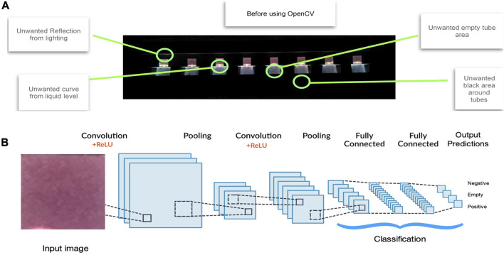
Introduction: Accurate and rapid diagnostics paired with effective tracking and tracing systems are key to halting the spread of infectious diseases, limiting the emergence of new variants and to monitor vaccine efficacy. The current gold standard test (RT-qPCR) for COVID-19 is highly accurate and sensitive, but is time-consuming, and requires expensive specialised, lab-based equipment. Methods: Herein, we report on the development of a SARS-CoV-2 (COVID-19) rapid and inexpensive diagnostic platform that relies on a reverse-transcription loop-mediated isothermal amplification (RT-LAMP) assay and a portable smart diagnostic device. Automated image acquisition and an Artificial Intelligence (AI) deep learning model embedded in the Virus Hunter 6 (VH6) device allow to remove any subjectivity in the interpretation of results. The VH6 device is also linked to a smartphone companion application that registers patients for swab collection and manages the entire process, thus ensuring tests are traced and data securely stored. Results: Our designed AI-implemented diagnostic platform recognises the nucleocapsid protein gene of SARS-CoV-2 with high analytical sensitivity and specificity. A total of 752 NHS patient samples, 367 confirmed positives for coronavirus disease (COVID-19) and 385 negatives, were used for the development and validation of the test and the AI-assisted platform. The smart diagnostic platform was then used to test 150 positive clinical samples covering a dynamic range of clinically meaningful viral loads and 250 negative samples. When compared to RT-qPCR, our AI-assisted diagnostics platform was shown to be reliable, highly specific (100%) and sensitive (98-100% depending on viral load) with a limit of detection of 1.4 copies of RNA per µL in 30 min. Using this data, our CE-IVD and MHRA approved test and associated diagnostic platform has been approved for medical use in the United Kingdom under the UK Health Security Agency's Medical Devices (Coronavirus Test Device Approvals, CTDA) Regulations 2022. Laboratory and in-silico data presented here also indicates that the VIDIIA diagnostic platform is able to detect the main variants of concern in the United Kingdom (September 2023). Discussion: This system could provide an efficient, time and cost-effective platform to diagnose SARS-CoV-2 and other infectious diseases in resource-limited settings.

Keywords: COVID-19; LAMP (loop mediated isothermal amplification); artificial intelligence; infectious diseases; rapid diagnostics.


Conflict of interest statement

Authors RRM, JC, and DR were employed by the company VIDIIA Ltd. Authors CC, MW, MaM, and MBu were employed by the company GB Electronics (UK) Ltd. VIDIIA Ltd is start-up company formed in 2020, in which the University of Surrey owns shares and has an Investor Director in place on the company Board of Directors. AP and RL are scientific advisors for VIDIIA. The remaining authors declare that the research was conducted in the absence of any commercial or financial relationships that could be construed as a potential conflict of interest.

Figures

FIGURE 1
FIGURE 1
Artificial Intelligence method. VIDIIA’s AI solution works in two stages: (A) Tube identification process. The tubes in the image are identified by using an edge detection method (OpenCV) that removes all the unwanted regions such as the empty tube areas, black areas around the tubes, reflection form lighting and curves from liquid levels that could mislead the Artificial Intelligence model. (B) Deep Learning process feature classification. By using over 10,000 images, and growing, as training data, our two-dimensional Deep Learning model (using convolutional neural network) classifies the input images into any of the specified categories: Negative, Positive and Empty. The overall training process of the Convolution Network may be summarized as follows. The filters and parameters/weights are first initialised with random values. The network then takes a training image as input, goes through the forward propagation step (convolution, ReLU and pooling operations along with forward propagation in the Fully Connected layer) and finds the output probabilities for each class. The total error at the output layer is calculated with the following formula: Total Error = ∑ ½ (target probability–output probability) 2. Backpropagation is used to calculate the gradients of the error with respect to all weights in the network and use gradient descent to update all filter values/weights and parameter values to minimize the output error. The weights are adjusted in proportion to their contribution to the total error. When the same image is input again, output probabilities might now be closer to the target vector. This means that the network has learnt to classify this particular image correctly by adjusting its weights/filters such that the output error is reduced. Parameters like number of filters, filter sizes, architecture of the network are all fixed before the training process is repeated with all images in the training set; until the model classifies the images correctly every time.
FIGURE 2
FIGURE 2
VIDIIA Hunter Diagnostics Platform process overview. (A) Using the VIDIIA companion app, the operator scans the unique identifiers attributed to nasal-pharyngeal swabs samples, that are collected in Virus transport media (VTM). (B) Samples are processed by full RNA extraction, using a commercial kit. (C) After preparing the RT-LAMP reactions and transferring the samples, according to VIDIIA’s instructions, the reaction tubes are inserted in the VH6 device. The VH6 device heats up the reaction tubes, for amplification and takes pictures throughout the amplification. At the end of the amplification, an artificial intelligence (deep-learning model) analyses the pictures and outputs the results. (D) The VIDIIA companion app connects to the device and retrieves the results from the VH6, displays them and uploads them to VIDIIA cloud. (E) VIDIIA users’ friendly online dashboard can then be used to consult and analyse data.
FIGURE 3
FIGURE 3
Artificial Intelligence results display. The VH6 software retrieves the images taken during the experiment and pass them through the inbuilt Artificial Intelligence (AI) model to obtain the predictions (Positive, Negative and Empty features); this information is then retrieved by the VIDIIA mobile app and displayed to the user showing the (A) input image, (B) output image with the probabilities of features (Positive, Negative and Empty) and the (C) “Positive features” graph created using the Predicted features of each tube calculated every minute until the end of the experiment.
FIGURE 4
FIGURE 4
Limit of detection of the VIDIIA Hunter diagnostic platform. The limit of detection of the VIDIIA Hunter was evaluated by testing different concentrations of the EDX SARS-CoV-2 Positive Run Control (Biorad, Watford, United Kingdom): 140, 14, 3, and 1.4 copies/µL. (A) Results obtained with a concentration of 3 copies of RNA/µL. A test was prepared with a negative control (NTC) in tube 1, 6 samples containing 3 copies of RNA/µL in tubes 2 to 7 and a positive control (PTC) in tube 8. After 30 min of amplification using the VIDIIA Hunter platform, the output image and positive features graph shows that 5 out of 6 samples containing 3 copies of RNA/uL are positives for SARS-CoV-2, the positive control is also positive and the negative control negative. The graph shows that the RNA started to be amplified between 15 and 20 min. (B) Results obtained with a concentration of 1.4 copies of RNA/µL. A test was prepared with a negative control (NTC) in tube 1, 6 samples containing 1.4 copies of RNA/uL in tubes 2 to 7 and a positive control (PTC) in tube 8. After 30 min of amplification using the VIDIIA Hunter platform, the output image and positive features graph shows that 5 out of 6 samples containing 1.4 copies of RNA/µL are positives for SARS-CoV-2, the positive control is also positive and the negative control negative. The graph shows that the RNA started to be amplified between 15 and 27 min.

References

    1. Alves P. A., de Oliveira E. G., Franco-Luiz A. P. M., Almeida L. T., Goncalves A. B., Borges I. A., et al. (2021). Optimization and clinical validation of colorimetric reverse transcription loop-mediated isothermal amplification, a fast, highly sensitive and specific COVID-19 molecular diagnostic tool that is robust to detect SARS-CoV-2 variants of concern. Front. Microbiol. 12, 713713. 10.3389/fmicb.2021.713713 - DOI - PMC - PubMed
    1. Aoki M. N., de Oliveira Coelho B., Goes L. G. B., Minoprio P., Durigon E. L., Morello L. G., et al. (2021). Colorimetric RT-LAMP SARS-CoV-2 diagnostic sensitivity relies on color interpretation and viral load. Sci. Rep. 11 (1), 9026. 10.1038/s41598-021-88506-y - DOI - PMC - PubMed
    1. Babiker A., Myers C. W., Hill C. E., Guarner J. (2020). SARS-CoV-2 testing. Am. J. Clin. Pathol. 153 (6), 706–708. 10.1093/ajcp/aqaa052 - DOI - PMC - PubMed
    1. Baek Y. H., Um J., Antigua K. J. C., Park J. H., Kim Y., Oh S., et al. (2020). Development of a reverse transcription-loop-mediated isothermal amplification as a rapid early-detection method for novel SARS-CoV-2. Emerg. Microbes Infect. 9 (1), 998–1007. 10.1080/22221751.2020.1756698 - DOI - PMC - PubMed
    1. Barnes L., Heithoff D. M., Mahan S. P., Fox G. N., Zambrano A., Choe J., et al. (2018). Smartphone-based pathogen diagnosis in urinary sepsis patients. EBioMedicine 36, 73–82. 10.1016/j.ebiom.2018.09.001 - DOI - PMC - PubMed