Review
. 2023 Dec 1;325(6):C1439-C1450.
doi: 10.1152/ajpcell.00349.2023. Epub 2023 Oct 16.

Exploring the role of urinary extracellular vesicles in kidney physiology, aging, and disease progression

Affiliations
Review

Exploring the role of urinary extracellular vesicles in kidney physiology, aging, and disease progression

Cristina Grange et al. Am J Physiol Cell Physiol. .

Abstract

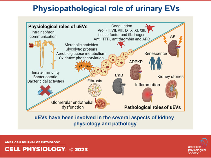
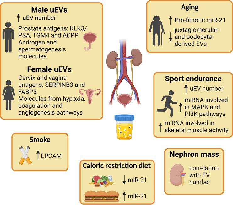
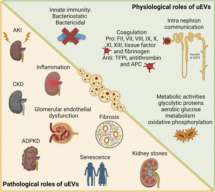
Extracellular vesicles (EVs), membranous vesicles present in all body fluids, are considered important messengers, carrying their information over long distance and modulating the gene expression profile of recipient cells. EVs collected in urine (uEVs) are mainly originated from the apical part of urogenital tract, following the urine flow. Moreover, bacterial-derived EVs are present within urine and may reflect the composition of microbiota. Consolidated evidence has established the involvement of uEVs in renal physiology, being responsible for glomerular and tubular cross talk and among different tubular segments. uEVs may also be involved in other physiological functions such as modulation of innate immunity, coagulation, or metabolic activities. Furthermore, it has been recently remonstrated that age, sex, endurance excise, and lifestyle may influence uEV composition and release, modifying their cargo. On the other hand, uEVs appear modulators of different urogenital pathological conditions, triggering disease progression. uEVs sustain fibrosis and inflammation processes, both involved in acute and chronic kidney diseases, aging, and stone formation. The molecular signature of uEVs collected from diseased patients can be of interest for understanding kidney physiopathology and for identifying diagnostic and prognostic biomarkers.

Keywords: EV release; exosomes; renal injury; renal tubular cells; urine.


Conflict of interest statement

C.G. and B.B. are members of the Task Force on Urinary Extracellular Vesicles of the ISEV community. None of the other authors has any conflicts of interest, financial or otherwise, to disclose.

Figures

None
Graphical abstract
Figure 1.
Figure 1.
Schematic representation of different EV subtypes and their origin. Different processes of EV generation are schematized, including EV released by budding of the plasma membrane (microvesicles, exopheres, and mitovesicles), by retraction fibers of migrating cells (migrasomes), or by membrane fusion of multivesicular bodies (exosomes). The intracellular steps of endosome maturation and multivesicular body formation and the release of small particles (exomeres and supermeres) are also shown. ILV, intraluminal vesicles; MVB, multivesicular body.
Figure 2.
Figure 2.
Physiological and exogenous factors that influence uEV cargo and release. Graphical representation of demographic parameters such as sex, nephron mass, and aging that affect uEV composition. Sport endurance, food intake, and smoke are some of the habits that modify uEV cargo. ACPP, prostatic acid phosphatase; EPCAM, epithelial cell adhesion molecule; FABP5, fatty acid-binding protein 5; KLK3/PSA, prostate specific antigen; MAPK, mitogen-activated protein kinase; PI3K, phosphoinositide 3-kinases; SERPINB3, serpin peptidase inhibitor member 3; TGM4, transglutaminase 4; uEV, urinary extracellular vesicle. Created with BioRender.com.
Figure 3.
Figure 3.
Physiological and pathological roles of uEVs. Graphical representation of different physiological and pathological conditions in which uEVs have been involved in the context of kidney pathophysiology. ADPKD, autosomal dominant polycystic kidney disease; AKI, acute kidney injury; APC, activated protein C; CKD, chronic kidney disease; TFPI, tissue factor pathway inhibitor; uEV, urinary extracellular vesicle. Created with BioRender.com.

References

    1. Erdbrügger U, Blijdorp CJ, Bijnsdorp IV, Borràs FE, Burger D, Bussolati B, , et al. Urinary extracellular vesicles: a position paper by the Urine Task Force of the International Society for Extracellular Vesicles. J Extracell Vesicles 10: e12093, 2021. doi: 10.1002/jev2.12093. - DOI - PMC - PubMed
    1. Wiklander OPB, Brennan MÁ, Lötvall J, Breakefield XO, El Andaloussi S. Advances in therapeutic applications of extracellular vesicles. Sci Transl Med 11: eaav8521, 2019. doi: 10.1126/scitranslmed.aav8521. - DOI - PMC - PubMed
    1. Van Niel G, Carter DRF, Clayton A, Lambert DW, Raposo G, Vader P. Challenges and directions in studying cell-cell communication by extracellular vesicles. Nat Rev Mol Cell Biol 23: 369–382, 2022. doi: 10.1038/s41580-022-00460-3. - DOI - PubMed
    1. Valcz G, Újvári B, Buzás EI, Krenács T, Spisák S, Kittel Á, Tulassay Z, Igaz P, Takács I, Molnár B. Small extracellular vesicle DNA-mediated horizontal gene transfer as a driving force for tumor evolution: facts and riddles. Front Oncol 12: 945376, 2022. doi: 10.3389/fonc.2022.945376. - DOI - PMC - PubMed
    1. Heidarzadeh M, Zarebkohan A, Rahbarghazi R, Sokullu E. Protein corona and exosomes: new challenges and prospects. Cell Commun Signal 21: 64, 2023. doi: 10.1186/s12964-023-01089-1. - DOI - PMC - PubMed

Publication types