. 2024 Feb 17;119(18):2902-2916.
doi: 10.1093/cvr/cvad154.

Prdm16 mutation determines sex-specific cardiac metabolism and identifies two novel cardiac metabolic regulators

Affiliations

Prdm16 mutation determines sex-specific cardiac metabolism and identifies two novel cardiac metabolic regulators

Jirko Kühnisch et al. Cardiovasc Res. .

Abstract

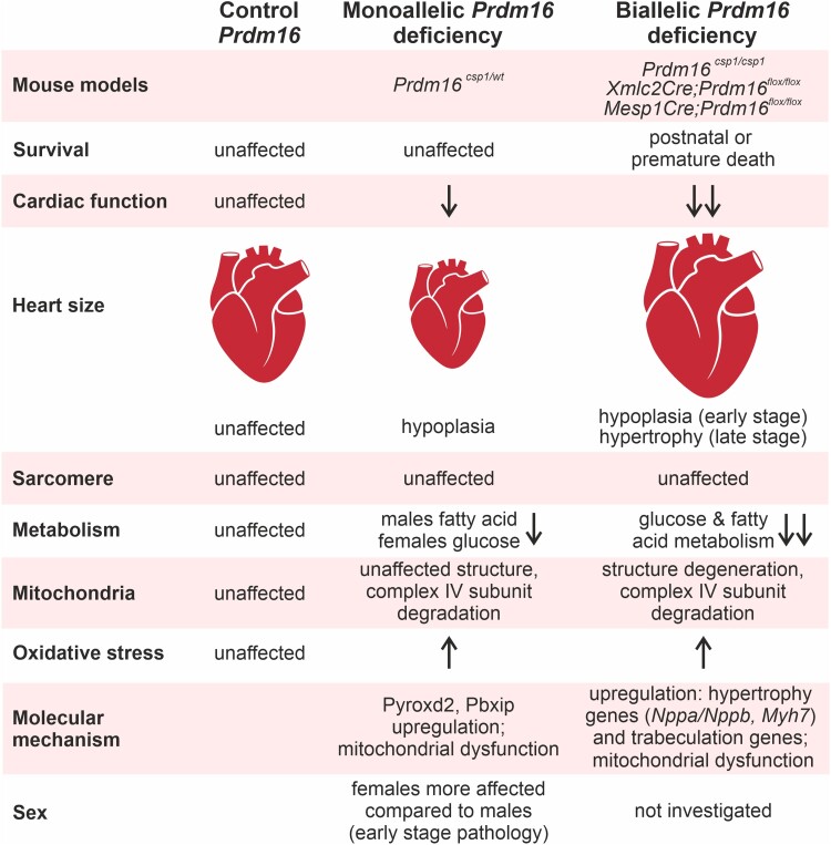
Aims: Mutation of the PRDM16 gene causes human dilated and non-compaction cardiomyopathy. The PRDM16 protein is a transcriptional regulator that affects cardiac development via Tbx5 and Hand1, thus regulating myocardial structure. The biallelic inactivation of Prdm16 induces severe cardiac dysfunction with post-natal lethality and hypertrophy in mice. The early pathological events that occur upon Prdm16 inactivation have not been explored.

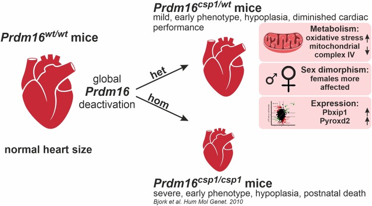
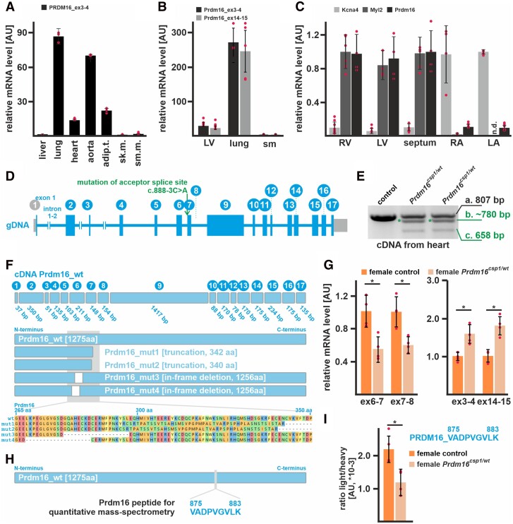
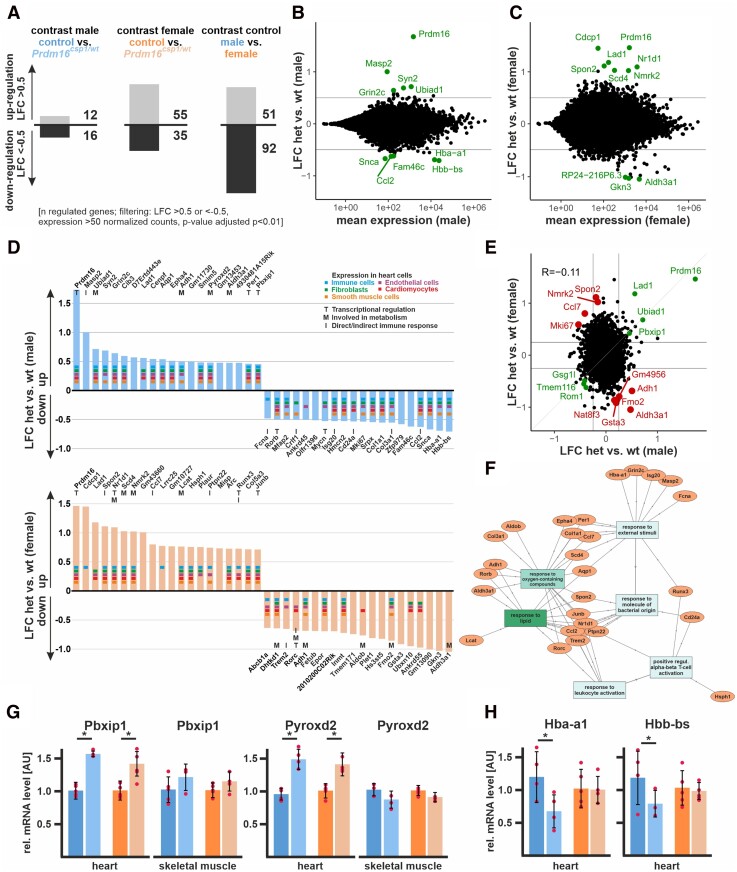
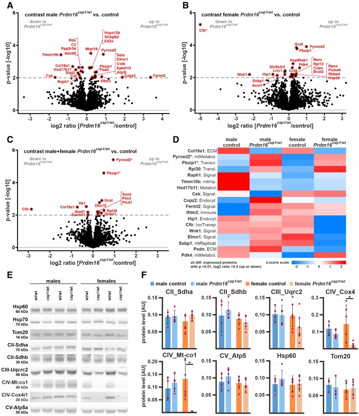
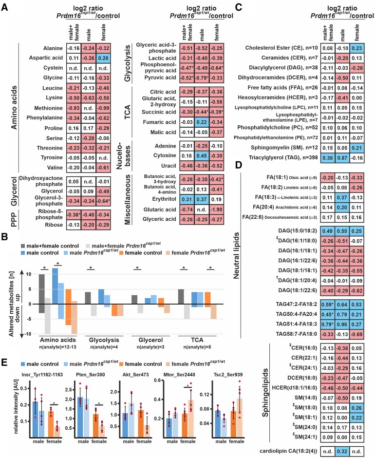
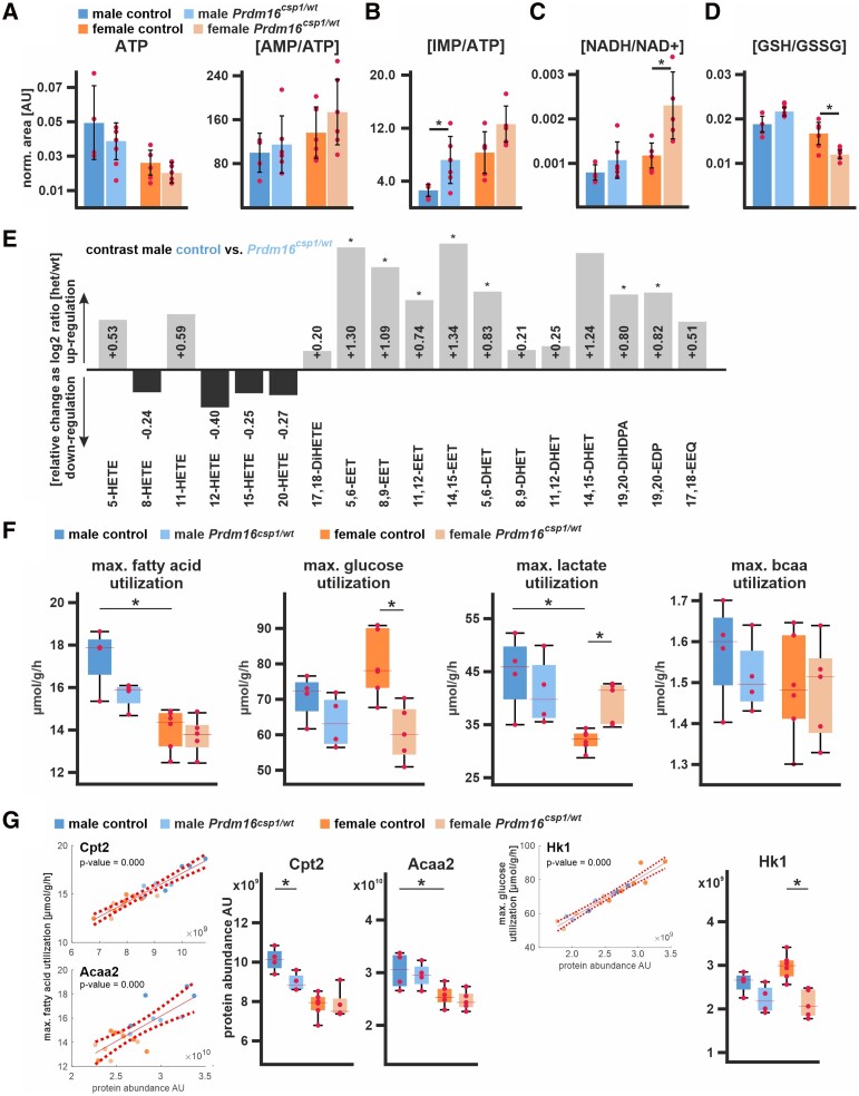
Methods and results: This study performed in-depth pathophysiological and molecular analyses of male and female Prdm16csp1/wt mice that carry systemic, monoallelic Prdm16 gene inactivation. We systematically assessed early molecular changes through transcriptomics, proteomics, and metabolomics. Kinetic modelling of cardiac metabolism was performed in silico with CARDIOKIN. Prdm16csp1/wt mice are viable up to 8 months, develop hypoplastic hearts, and diminished systolic performance that is more pronounced in female mice. Prdm16csp1/wt cardiac tissue of both sexes showed reductions in metabolites associated with amino acid as well as glycerol metabolism, glycolysis, and the tricarboxylic acid cycle. Prdm16csp1/wt cardiac tissue revealed diminished glutathione (GSH) and increased inosine monophosphate (IMP) levels indicating oxidative stress and a dysregulated energetics, respectively. An accumulation of triacylglycerides exclusively in male Prdm16csp1/wt hearts suggests a sex-specific metabolic adaptation. Metabolic modelling using CARDIOKIN identified a reduction in fatty acid utilization in males as well as lower glucose utilization in female Prdm16csp1/wt cardiac tissue. On the level of transcripts and protein expression, Prdm16csp1/wt hearts demonstrate an up-regulation of pyridine nucleotide-disulphide oxidoreductase domain 2 (Pyroxd2) and the transcriptional regulator pre-B-cell leukaemia transcription factor interacting protein 1 (Pbxip1). The strongest concordant transcriptional up-regulation was detected for Prdm16 itself, probably through an autoregulatory mechanism.

Conclusions: Monoallelic, global Prdm16 mutation diminishes cardiac performance in Prdm16csp1/wt mice. Metabolic alterations and transcriptional dysregulation in Prdm16csp1/wt affect cardiac tissue. Female Prdm16csp1/wt mice develop a more pronounced phenotype, indicating sexual dimorphism at this early pathological window. This study suggests that metabolic dysregulation is an early event in the PRDM16 associated cardiac pathology.

Keywords: Cardiomyopathy; Metabolism; Mutation; Prdm16.


Conflict of interest statement

Conflict of interest: All authors declare to have no conflict of interest related to this manuscript. The patent application EP21174633 with the title ‘Computer assisted method for the evaluation of cardiac metabolism’ was filed by Charité—Universitätsmedizin Berlin as the employer of N.B. and Titus Kuehne, with both holding inventorship for this patent application.

Figures

Graphical Abstract
Graphical Abstract
Figure 1
Figure 1
Expression of Prdm16 in the heart of Prdm16csp1/wt mice. (A) Expression analysis reveals high PRDM16 transcript levels in human lung, aorta, adipose tissue (adip.t.), and heart (technical replicate n = 3). Liver, skeletal muscle (sk.m.), and smooth muscle (sm.m.) show low PRDM16 expression. (B) Prdm16 is highly expressed in murine lung tissue, and shows robust levels in the left ventricle (LV) but low expression in skeletal muscle (sm) (n = 3). (C) Prdm16 is robustly expressed in LV, right ventricle (RV), and the septum. The left (LA) and right atrium (RA) does not show considerable Prdm16 expression. Kcna4 and Myl2 transcripts demonstrate atrial and ventricular tissue origin (n = 3). (D) The murine Prdm16 gene comprises 17 exons, and Prdm16csp1/wt mice carry the point mutation c.888-3C>A affecting the acceptor splice site of intron_6–7. (E) PCR genotyping with heart cDNA from heterozygous Prdm16csp1/wt mice generates three products as follows: (i) wild-type fragment (807 bp), (ii) unknown mutant product (∼780 bp, *), and (iii) mutant product without exon 7 (658 bp). (F) The wild-type Prdm16 protein is 1275 amino acids (aa) long. Targeted high-throughput sequencing of the different PCR products identifies four main Prdm16 mutant proteins in Prdm16csp1/wt mice including truncation (mut1, mut2) and in-frame deletion (mut3, mut4) variants (see Supplementary material online, Figures SI and SII in the Data Supplement, for detailed results, see Data Supplement). (G) Quantitative detection of Prdm16 transcript levels with qPCR detectors targeting the exon 7 deletion region shows approximately 40–50% reduced expression. In contrast, PCR detectors targeting exon3–4 and exon14–15 identify increased Prdm16 levels. Analysis was performed on female Prdm16csp1/wt heart tissue (n = 3). (H) Detection of Prdm16 protein was achieved by PRM (parallel-reaction monitoring) using the Prdm16 derived peptide V875-K883. (I) Quantitation of Prdm16 peptide in control and Prdm16csp1/wt lung tissue (n = 4). The ratio of endogenous (light) and standard spike-in (heavy) peptide is shown. Statistical analysis was performed with unpaired t-test (P < 0.05).
Figure 2
Figure 2
Diminished cardiac performance after Prdm16 inactivation. (A) Survival of male and female Prdm16csp1/wt mice is normal until the age of 8 month (n > 13 each group). (B) Eight-month-old Prdm16csp1/wt mice of both sexes have a reduced absolute body weight. Normalization against tibia length demonstrated a diminished relative heart and LV weight in Prdm16csp1/wt mutants (n > 13 each group). (C) M-mode images illustrate the heart phenotype in Prdm16csp1/wt mice as assessed with echocardiography. (D) At the age of 8 months, Prdm16csp1/wt mice of both sexes have diminished cardiac function as illustrated by stroke volume, cardiac output, and ejection fraction (EF) (n > 12 each group). Full echocardiography data are available in Supplementary material online, Tables SIVSVI in the Data Supplement. (E) Plasma brain natriuretic peptide (Bnp) levels were determined with ELISA (n > 5 each group). (F) Histological analysis with H&E staining reveals normal tissue organization in female Prdm16csp1/wt hearts. Scale bars in E and F are 500 µm (full heart) and 50 µm (inset). (G) Histological staining for fibrosis with Picro-Sirius red is negative in female Prdm16csp1/wt heart tissue. (H) The cell area of female heart muscle (cross sections) stained with wheat germ agglutinin (WGA) is significantly reduced (n = 5, each n includes >300 cells). Nuclei were stained with DAPI. Scale bar is 20 µm. Statistical analysis was performed with unpaired t-test (P < 0.05).
Figure 3
Figure 3
Transcriptome analysis of Prdm16csp1/wt heart tissue. (A) Transcriptome analysis with RNAseq of Prdm16csp1/wt LV shows in male 28 and in female 90 differentially regulated genes compared to controls. Analysis of male vs. female control hearts identifies 143 differentially regulated transcripts. Analysed biological replicates are n = 4–6. (B and C) Bioinformatic filtering ranked the normalized log2 cpk according the absolute log2 fold change (LFC) after elimination of regulated targets with an adjusted P-value (padj) < 10−2. Scatterplots show the LFC against mean expression for the contrast Prdm16csp1/wt (het) vs. controls (wt) in male (B) or female (C) heart. The top 10 differentially regulated genes (adj. P < 0.01) are highlighted and labelled. (D) Summary of top 20 up- and down-regulated genes in male (upper panel) and female (lower panel) hearts. Expression of selected genes in cardiac cell types is annotated according to the colour code. Functional association with transcription (T), metabolism (M), and immune response (I) is shown for relevant genes. (E) Scatterplot shows the LFC detected in A and B against each other. Genes with concordant changes (abs LFC > 0.25) are highlighted in green, and genes with discordant changes (top 10; adj. P < 0.01) are highlighted in red. The overall correlation is R = −0.11 (n = 21 276). (F) Gene ontology (GO) network was constructed with GOnet by using the male and female TOP20 up- and down-regulated genes in Prdm16csp1/wt hearts. GO terms response to lipids and response to oxygen-containing compounds show strongest association. (G) Validation of RNAseq expression data for Pbxip1 and Pyroxd2 was performed with qPCR using whole RNA extracts from heart and skeletal muscle. Pbxip1 and Pyroxd2 expression is increased in hearts from Prdm16csp1/wt mice of both sexes. (H) Hba-a1 and Hbb-bs expression is diminished in male Prdm16csp1/wt hearts.
Figure 4
Figure 4
Proteome comparison of Prdm16csp1/wt cardiac tissue. Volcano plots for Prdm16csp1/wt/control pairwise comparisons with log2 ratio (x-axis) and P-value (y-axis) of male hearts (A), female hearts (B), and combined analysis of male + female hearts (C). Colouring indicates significantly altered proteins (threshold dashed grey line, P-value < 0.01), and * highlights highly significantly different proteins with P-value < 0.0001. (D) Heat map of all regulated proteins (P-value < 0.01, abs log2 ratio > 0.5) using z-scored values of group median intensity. Pre-B-cell leukaemia transcription factor interacting protein 1 (Pbxip1) and pyridine nucleotide-disulphide oxidoreductase domain 2 (Pyroxd2) are consistently increased in male and female Prdm16csp1/wt mice with P-value < 0.0001. Analysed biological replicates are n = 4 in each group. (E) Western blot analysis shows protein expression of selected mitochondrial proteins. (F) Quantification of western blot data showing mitochondrial protein levels. Analysed biological replicates are n > 4. Statistical analysis was performed with unpaired t-test, * indicates P < 0.05.
Figure 5
Figure 5
Altered metabolism in Prdm16csp1/wt cardiac tissue. (A) Normalized central carbon metabolite counts are presented as log2 value of the mean of normalized peak area ratio Prdm16csp1/wt/controls of males, females, and combination of both sexes. In female Prdm16csp1/wt LV tissue, broad suppression of several metabolic pathways was observed. In male Prdm16csp1/wt LV tissue, a similar but less pronounced reduction was detected. Statistical analysis of individual metabolites was performed with non-parametric Wilcoxon Rank Sum test, * indicates P < 0.05. (B) Univariate analysis reveals in cardiac tissue of Prdm16csp1/wt mice significant reduction of the amino acid, glycolysis, glycerol, and tricarboxylic acid cycle (TCA) metabolism using combined male and female data. Divergent metabolism of male and female animals was observed for amino acid metabolism, glycolysis, and TCA cycle. (C) Lipid analysis was performed with LC-MS using the lipidizer kit (Sciex). Global lipid analysis and evaluation as log2 value of the intensity ratio Prdm16csp1/wt/controls reveal normal levels for most lipid classes. Strongest regulation was observed for triacylglycerol (TAG) in male Prdm16csp1/wt hearts. The number (n) of validly detected lipids per class is indicated. (D) Selected neutral lipids and sphingolipids critical for the heart, lipids altered in Prdm16csp1/wt mice, and lipids previously associated with heart function (Tham et al.,  Wittenbecher et al.) are presented for Prdm16csp1/wt cardiac tissue of both sexes and in combination. Values for phospholipids are available in Supplementary material online, Figure SIX in the Data Supplement. Analysed biological replicates for all metabolic analysis are n = 4–6. (E) Signalling activity of the mitogen-activated protein kinase (MAPK) and mTOR pathway was assessed with the Milliplex phosphoprotein magnetic bead system and revealed diminished phosphorylation of insulin receptor (Insr_Tyr1162-1163), phosphatase and tensin homologue (Pten_Ser380), and Akt serine/threonine kinase 2 (Akt_Ser473) in female Prdm16csp1/wt mice. Analysed biological replicates for all metabolic analysis are n = 4–6. Statistical analysis of selected lipids was performed with non-parametric Wilcoxon Sum Rank test, * indicates P < 0.05. Colouring indicates reduction (red) or increase (blue) of the log2 of ratio by −0.2 or 0.2, respectively.
Figure 6
Figure 6
Nutrient metabolism in Prdm16csp1/wt cardiac tissue. (A) Assessment of metabolites critical for energy metabolism using LV tissue and LC-MS. Normalized values are shown for adenosine triphosphate (ATP) and the ratio of adenosine monophosphate (AMP) vs. ATP (AMP/ATP). Analysed biological replicates are n = 4–6 for A to D. (B) The ratio of inosine monophosphate (IMP) vs. ATP (IMP/ATP) is increased in Prdm16csp1/wt hearts. (C) The ratio of reduced vs. oxidized nicotinamide adenine dinucleotide (NADH/NAD+) is increased in female Prdm16csp1/wt hearts. (D) Oxidative capacity of cardiac tissues was assessed with the ratio of reduced vs. oxidized glutathione (GSH/GSSG). Female Prdm16csp1/wt hearts show a significantly reduced GSH/GSSG ratio. (E) Eicosanoids were measured in male Prdm16csp1/wt hearts with LC/ESI-MS-MS. Data are presented as log2 Prdm16csp1/wt/controls ratio with down- or up-regulation as black or grey bars, respectively. All epoxyeicosatrienoic (EET) and dihydroxyeicosatrienoic (DHET) acids are increased in Prdm16csp1/wt cardiac tissue. Corresponding absolute measurements are available in Supplementary material online, Table SVIII in the Data Supplement. Analysed biological replicates are n > 6. (F) Modelling of major cardiac metabolic processes occurred with CARDIOKIN1 using protein expression data. Differences in maximal substrate utilization for fatty acids (FA), glucose, lactate, and branched chain amino acids (bcaa). Box plots show median and 25% quartile. Dots depict maximal capacities for individual animals. (G) Individual protein impact for FA and glucose metabolism was correlated for carnitine palmitoyltransferase 2 (Cpt2), acetyl-CoA acyltransferase 2 (Acaa2), and hexokinase 1 (Hk1) using linear regression analysis of the maximal substrate utilization vs. protein abundance (dashed lines indicate confidence interval, 95%). Box plots show median and 25% quartile. Dots depict Cpt2, Acaa2, and Hk1 maximal capacities for individual animals. Statistical analysis of individual metabolites and processes was performed with unpaired t-test, * indicates P < 0.05. Analysed biological replicates are n = 4–6.
Figure 7
Figure 7
Prdm16 is an early regulator of cardiac metabolism. Assessing different mouse models for the PRDM16 associated cardiomyopathy reveals distinct cardiac phenotypes after mono or biallelic Prdm16 inactivation.,, Metabolism is differentially affected at early (Prdm16csp1/wt) and late pathology stages (Xmlc2Cre;Prdm16flox/flox, Mesp1Cre;Prdm16flox/flox).,

Similar articles

Cited by

References

    1. Burke MA, Cook SA, Seidman JG, Seidman CE. Clinical and mechanistic insights into the genetics of cardiomyopathy. J Am Coll Cardiol 2016;68:2871–2886. - PMC - PubMed
    1. Kuhnisch J, Herbst C, Al-Wakeel-Marquard N, Dartsch J, Holtgrewe M, Baban A, Mearini G, Hardt J, Kolokotronis K, Gerull B, Carrier L, Beule D, Schubert S, Messroghli D, Degener F, Berger F, Klaassen S. Targeted panel sequencing in pediatric primary cardiomyopathy supports a critical role of TNNI3. Clin Genet 2019;96:549–559. - PubMed
    1. Ishibashi J, Seale P. Functions of Prdm16 in thermogenic fat cells. Temperature 2015;2:65–72. - PMC - PubMed
    1. Arndt AK, Schafer S, Drenckhahn JD, Sabeh MK, Plovie ER, Caliebe A, Klopocki E, Musso G, Werdich AA, Kalwa H, Heinig M, Padera RF, Wassilew K, Bluhm J, Harnack C, Martitz J, Barton PJ, Greutmann M, Berger F, Hubner N, Siebert R, Kramer HH, Cook SA, MacRae CA, Klaassen S. Fine mapping of the 1p36 deletion syndrome identifies mutation of PRDM16 as a cause of cardiomyopathy. Am J Hum Genet 2013;93:67–77. - PMC - PubMed
    1. Delplancq G, Tarris G, Vitobello A, Nambot S, Sorlin A, Philippe C, Carmignac V, Duffourd Y, Denis C, Eicher JC, Chevarin M, Millat G, Khallouk B, Rousseau T, Falcon-Eicher S, Vasiljevic A, Harizay FT, Thauvin-Robinet C, Faivre L, Kuentz P. Cardiomyopathy due to PRDM16 mutation: first description of a fetal presentation, with possible modifier genes. Am J Med Genet C Semin Med Genet 2020;184:129–135. - PubMed

Publication types