

Lipid storage disease in 4 sibling superb birds-of-paradise (Lophorina superba)
- PMID: 37842940
- PMCID: PMC11032166
- DOI: 10.1177/03009858231203314
Lipid storage disease in 4 sibling superb birds-of-paradise (Lophorina superba)
Abstract
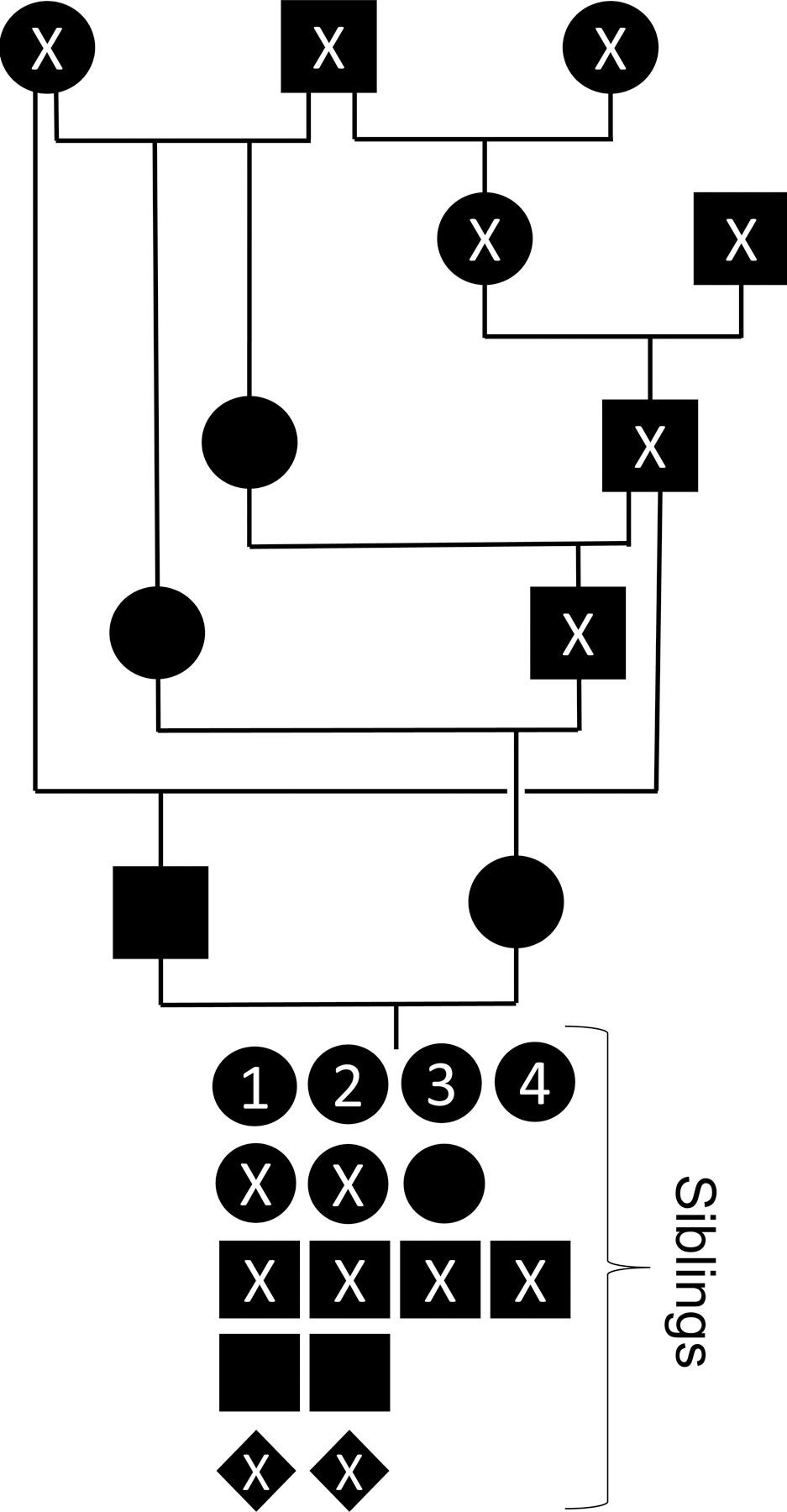
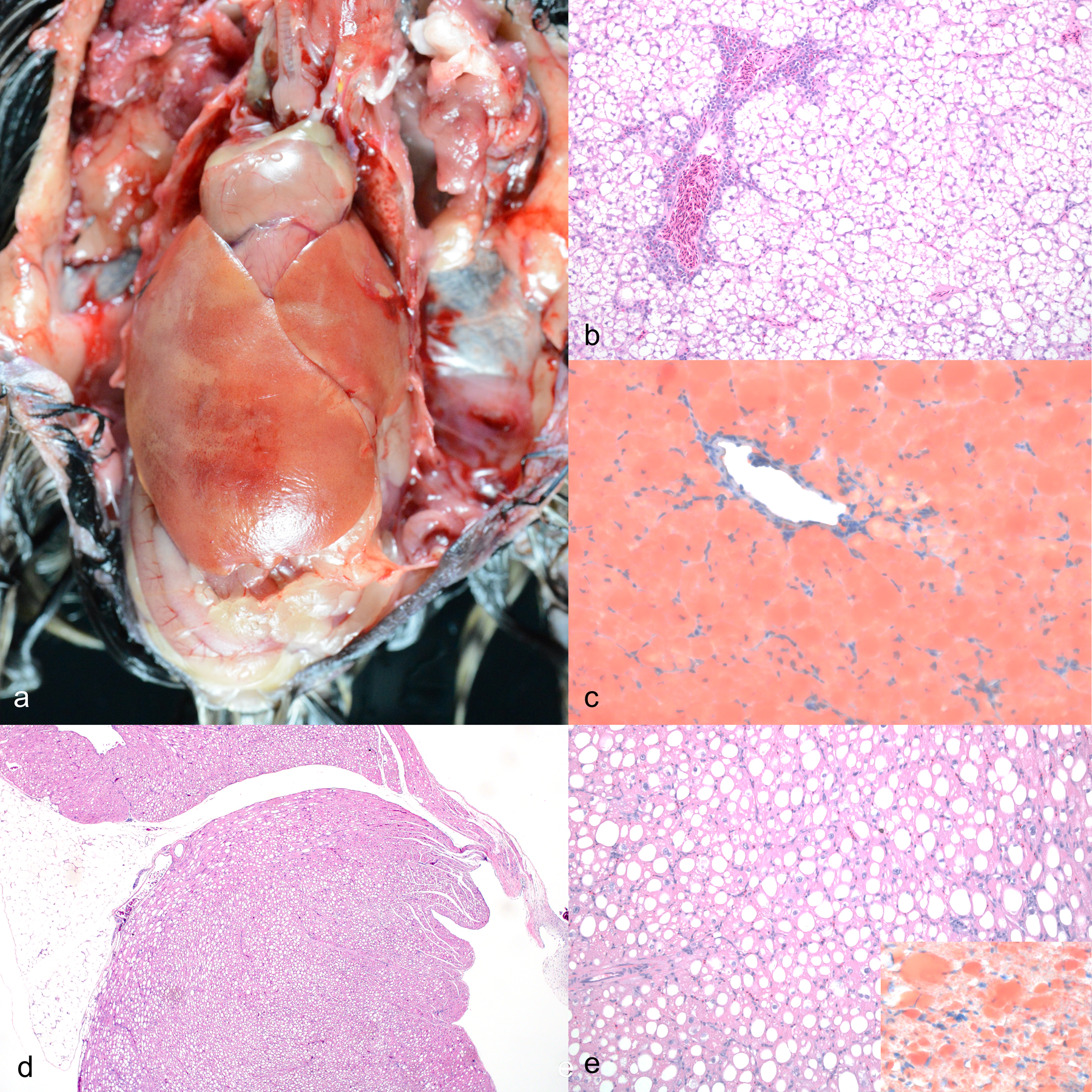
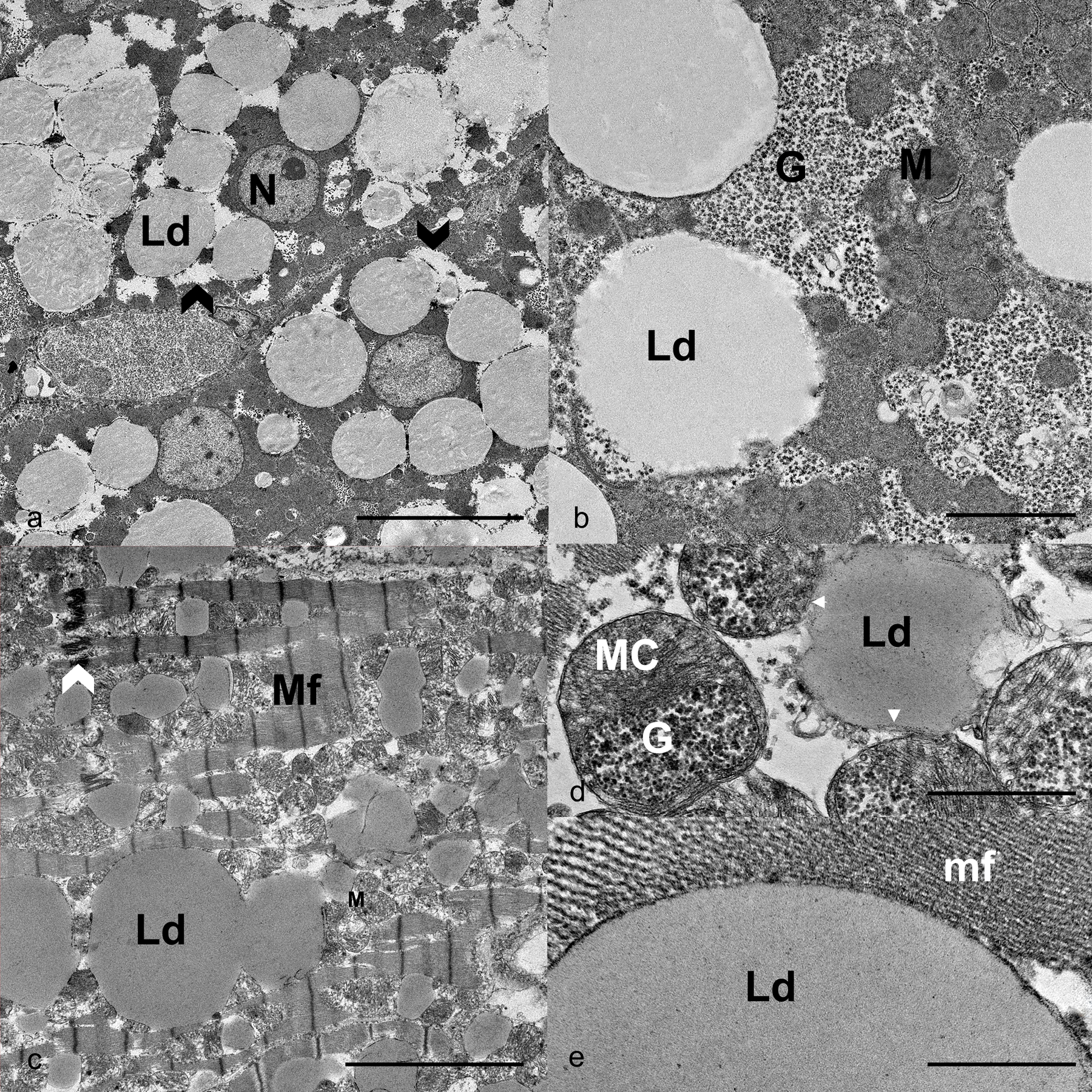
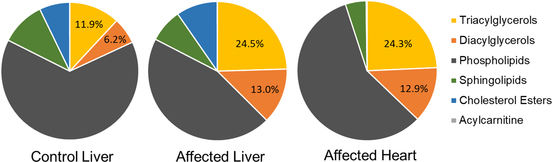
Pedigree analysis, clinical, gross, microscopic, ultrastructural, and lipidomic findings in 4 female superb bird-of-paradise (SBOP, Lophorina superba) siblings led to the diagnosis of a primary inherited glycerolipid storage disease. These birds were the offspring of a related breeding pair (inbreeding coefficient = 0.1797) and are the only known SBOPs to display this constellation of lesions. The birds ranged from 0.75 to 4.3 years of age at the time of death. Two birds were euthanized and 1 died naturally due to the disease, and 1 died of head trauma with no prior clinical signs. Macroscopic findings included hepatomegaly and pallor (4/4), cardiac and renal pallor (2/4), and coelomic effusion (1/4). Microscopic examination found marked tissue distortion due to cytoplasmic lipid vacuoles in hepatocytes (4/4), cardiomyocytes (4/4), renal tubular epithelial cells (4/4), parathyroid gland principal cells (2/2), exocrine pancreatic cells (3/3), and the glandular cells of the ventriculus and proventriculus (3/3). Ultrastructurally, the lipids were deposited in single to coalescing or fused droplets lined by an inconspicuous or discontinuous monolayer membrane. Lipidomic profiling found that the cytoplasmic lipid deposits were primarily composed of triacylglycerols. Future work, including sequencing of the SBOP genome and genotyping, will be required to definitively determine the underlying genetic mechanism of this disease.
Keywords: Lophorina superba; bird-of-paradise; lipid; lipidomics; pathology; storage disease.
Conflict of interest statement
Declaration of Conflicting InterestsThe author(s) declared no potential conflicts of interest with respect to the research, authorship, and/or publication of this article.
Figures
Similar articles
-
Distinctive courtship phenotype of the Vogelkop Superb Bird-of-Paradise Lophorina niedda Mayr, 1930 confirms new species status.PeerJ. 2018 Apr 16;6:e4621. doi: 10.7717/peerj.4621. eCollection 2018. PeerJ. 2018. PMID: 29682415 Free PMC article.
-
Breeding history and husbandry of the Superb Bird-of-paradise (Lophorina superba).Zoo Biol. 2021 Sep;40(5):485-490. doi: 10.1002/zoo.21627. Epub 2021 Jun 25. Zoo Biol. 2021. PMID: 34170023
-
Artificial incubation and hand-rearing of Superb Bird-of-paradise (Lophorina superba).Zoo Biol. 2022 Nov;41(6):588-594. doi: 10.1002/zoo.21685. Epub 2022 Jan 31. Zoo Biol. 2022. PMID: 35098574
-
Why neotypification of emLophorina/em emsuperba/em (Pennant, 1781) (Aves: Paradisaeidae) is justified-and necessary.Zootaxa. 2021 Apr 6;4951(2):zootaxa.4951.2.5. doi: 10.11646/zootaxa.4951.2.5. Zootaxa. 2021. PMID: 33903404 Review.
-
The anatomy, physiology, and diseases of the avian proventriculus and ventriculus.Vet Clin North Am Exot Anim Pract. 2003 Jan;6(1):85-111. doi: 10.1016/s1094-9194(02)00027-0. Vet Clin North Am Exot Anim Pract. 2003. PMID: 12616835 Review.
References
-
- Armién AG, Wolf TM, Mor SK, et al. Molecular and biological characterization of a cervidpoxvirus isolated from moose with necrotizing dermatitis. Vet Pathol 2020;57(2):296–310. - PubMed
-
- Beaufrère H, Ammersbach M, Reavill DR, et al. Prevalence of and risk factors associated with atherosclerosis in psittacine birds. J Am Vet Med Assoc 2013;242(12):1696–1704. - PubMed
-
- Beaufrère H, Gardhouse S, Ammersbach M. Lipoprotein characterization in Quaker parrots (Myiopsitta monachus) using gel‐permeation high‐performance liquid chromatography. Vet Clin Pathol 2020;49(3):417–427. - PubMed
-
- Beaufrère H, Reavill D, Heatley J, Susta L. Lipid-related lesions in Quaker parrots (Myiopsitta monachus). Vet Pathol 2019;56(2):282–288. - PubMed
