. 2023 Dec 1;211(11):1725-1735.
doi: 10.4049/jimmunol.2300307.

The Influence of Human IgG Subclass and Allotype on Complement Activation

Affiliations

The Influence of Human IgG Subclass and Allotype on Complement Activation

Timon Damelang et al. J Immunol. .

Abstract

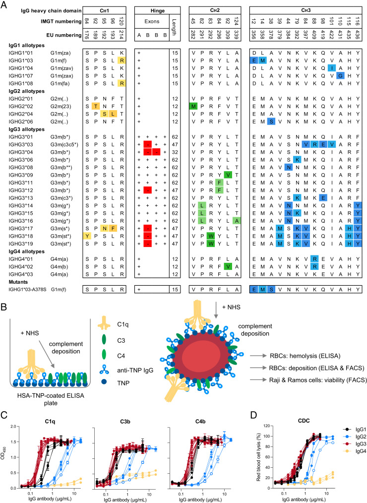
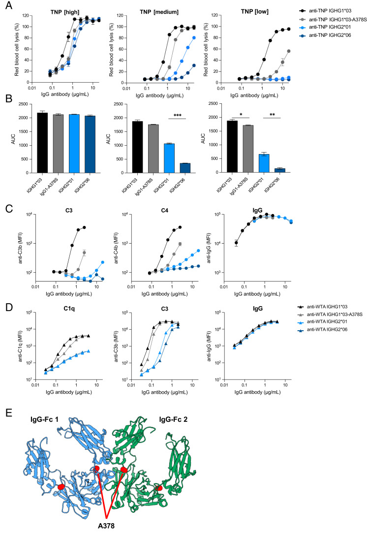
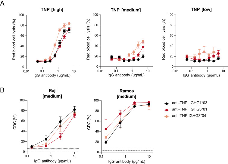
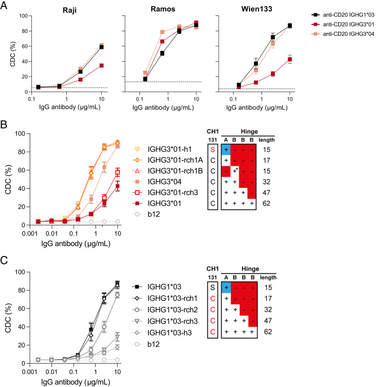
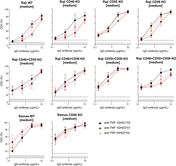
Complement activation via the classical pathway is initiated when oligomeric Igs on target surfaces are recognized by C1 of the complement cascade. The strength of this interaction and activation of the complement system are influenced by structural variation of the Ab, including Ab isotype, subclass, and glycosylation profile. Polymorphic variants of IgG have also been described to influence Fc-dependent effector functions. Therefore, we assessed complement binding, deposition, and complement-dependent cytotoxicity (CDC) of 27 known IgG allotypes with anti-trinitrophenyl specificity. Differences between allotypes within subclasses were minor for IgG1, IgG3, and IgG4 allotypes, and more substantial for IgG2. Allelic variant IGHG2*06, containing a unique serine at position 378 in the CH3 domain, showed less efficient complement activation and CDC compared with other IgG2 polymorphisms. We also observed variable cell lysis between IgG1 and IgG3, with IgG3 being superior in lysis of human RBCs and Ramos cells, and IgG1 being superior in lysis of Raji and Wien133 cells, demonstrating that a long-standing conundrum in the literature depends on cellular context. Furthermore, we compared IgG1 and IgG3 under different circumstances, showing that Ag density and Ab hinge length, but not complement regulators, define the context dependency of Ab-mediated CDC activity. Our results point toward a variation in the capacity of IgG subclasses to activate complement due to single amino acid changes and hinge length differences of allotypes to activate complement, which might give new insights on susceptibility to infectious, alloimmune, or autoimmune diseases and aid the design of Ab-based therapeutics.


Conflict of interest statement

A.F.L., M.M.M. and J.S. are former employees of Genmab and own stock and/or warrants. T.D. and S.W.d.T. were awarded Genmab-sponsored research funding. The other authors have no financial conflicts of interest.

Figures

FIGURE 1.
FIGURE 1.
Complement activation by IgG subclasses and allotypes. (A) Overview of the amino acid variation (one-letter code) between different polymorphic variants (allotypes) for each individual IgG subclass. The presence or absence of a hinge exon in IgG3 allotypes is indicated with a + or – symbol, respectively (adapted from Ref. 41). (B) Schematic illustration of the complement deposition ELISA. Trinitrophenyl (TNP)ylated human serum albumin is coated on an ELISA plate, followed by incubation with anti-TNP IgG Abs and subsequently with pooled normal human serum (NHS). Schematic illustration of hemolytic assay and complement-dependent cytotoxicity (CDC) assay with RBCs, Raji cells, and Ramos cells as target cells. RBCs, Raji cells, and Ramos cells were opsonized with anti-TNP Abs and then incubated with NHS. Complement activation was either determined by flow cytometry (deposition of C4b and C3b), or hemoglobulin release from RBCs was measured via ELISA. (C) Complement activation by IgG1 (black), IgG2 (blue), IgG3 (red), and IgG4 allotypes (yellow) as measured by deposition of C1q, C4, and C3 in ELISA (1 mM TNPylated HSA) or (D) CDC activity in a hemolysis assay (2 mM TNPylated RBCs). IGHG2*06 is indicated with empty circles. All Abs were tested in a 2-fold serial dilution starting from 20 µg/ml for IgG2 and IgG4 allotypes and starting from 5 µg/ml for IgG1 and IgG3 allotypes.
FIGURE 2.
FIGURE 2.
Serine 378 decreases complement activation of IgG1 and IgG2. (A) Complement-dependent cytotoxicity (CDC) activity of two IgG2 allotypes (IGHG2*01 in light blue and IGHG2*06 in dark blue), IGHG1*03 (black), and an IGHG1*03-378S mutant (gray) specific to trinitrophenyl (TNP) on RBCs at three different Ag densities (1 mM [high], 0.5 mM [medium], and 0.25 mM [low] TNBS concentration). Anti-TNP Abs were tested in duplicates in a 2-fold serial dilution starting at 20 µg/ml. (B) Area under the curve (AUC) of their CDC activity. (C) Complement deposition (C3b and C4b) and Ab binding (IgG) on RBCs (0.5 mM TNBS) was determined by flow cytometry for the same panel of anti-TNP allotypes. (D) Complement deposition (C1q and C3b) and IgG binding on S. aureus with the same allotypes, but with anti–wall teichoic acid (WTA) specificity was determined by flow cytometry. (E) Structural representation of two IgG1 Fc domains (blue and green) derived from the crystal structure matrix of IgG1 (PDB:1hzh). The alanine at position 378 in both Fc domains is depicted with red spheres and is positioned close to the interface between the Fc domains. *p < 0.05, **p < 0.01, ***p < 0.001.
FIGURE 3.
FIGURE 3.
Complement activation mediated by IgG1 and IgG3 is cell type–dependent. (A) Complement-dependent cytotoxicity (CDC) activity of IGHG1*03 (black), IGHG3*01 (long hinge, red), and IGHG3*04 (short hinge, pink) specific to trinitrophenyl (TNP) on RBCs at various Ag densities of 0.25 mM (low), 0.5 mM (medium), and 1 mM (high) TNBS in the presence of 20% serum. (B) CDC activity of same anti-TNP Abs was determined on Raji and Ramos B cells at an Ag density of 0.5 mM (medium) TNBS in the presence of 20% serum. All allotypes were tested in duplicates in a 4-fold serial dilution starting at 10 µg/ml. Negative controls are shown as dotted line for TNP unlabeled cells and dashed line represents TNPylated cells in the absence of Abs. Data represent mean ± SEM of n = 3.
FIGURE 4.
FIGURE 4.
Influence of the hinge length on CD20-targeted complement-dependent cytotoxicity. The capacity of CD20-specific IgG1*03 and IgG3*01 variants to induce complement-dependent cytotoxicity (CDC) was assessed in vitro. (A) CDC activity of anti-CD20 IGHG1*03 (black), IGHG3*01 (long hinge, red), and IGHG3*04 (short hinge, pink) of Raji, Ramos, and Wien133 cells. All allotypes were tested in duplicates or triplicates in a 4-fold serial dilution starting at 10 µg/ml in presence of 20% serum. The dashed gray line represents cells without any IgG Abs. (B) Matched set of natural (IGHG3*01 and IGHG3*04) and IGHG3*01 hinge mutants (containing deletions or substitutions of selected hinge exons) representing a range of hinge lengths (from yellow to dark red). Data represent mean ± SEM of n = 3. Red amino acid residues represent mutations to enable IgG1 hinge-like (top panel) or IgG3 hinge-like (bottom panel) HC-LC pairing. The blue + represents the IgG1 hinge exon; * indicates that C219S has been included to retain IgG3-like HC-LC pairing. (C) Matched set of natural (IGHG1*03) and IGHG1*03 hinge mutants (containing substitutions or insertions of selected hinge exons) representing a range of hinge lengths (from black to light gray). See (B) for details. Anti-HIV-1 gp120 clone b12 is used as negative control. Data represent mean ± SEM of n = 3.
FIGURE 5.
FIGURE 5.
Complement activation of IgG1 and IgG3 is independent of complement regulators. Complement-dependent cytotoxicity (CDC) activity of IGHG1*03 (black), IGHG3*01 (long hinge, red), and IGHG3*04 (short hinge, pink) specific to trinitrophenyl (TNP) on Raji and Ramos B cells including CD46, CD55, and CD59 knockout cell lines at an Ag density of 0.5 mM (medium) TNBS. Negative controls are shown as dotted line for TNP unlabeled cells, and a dashed line represents TNPylated cells in the absence of Abs. Data represent mean ± SEM of n = 3.

References

    1. Merle, N. S., Church S. E., Fremeaux-Bacchi V., Roumenina L. T.. 2015. Complement system part I—molecular mechanisms of activation and regulation. Front. Immunol. 6: 262. - PMC - PubMed
    1. Ricklin, D., Reis E. S., Mastellos D. C., Gros P., Lambris J. D.. 2016. Complement component C3—the “Swiss army knife” of innate immunity and host defense. Immunol. Rev. 274: 33–58. - PMC - PubMed
    1. Schmidt, C. Q., Lambris J. D., Ricklin D.. 2016. Protection of host cells by complement regulators. Immunol. Rev. 274: 152–171. - PMC - PubMed
    1. Strasser, J., de Jong R. N., Beurskens F. J., Wang G., Heck A. J. R., Schuurman J., Parren P. W. H. I., Hinterdorfer P., Preiner J.. 2019. Unraveling the macromolecular pathways of IgG oligomerization and complement activation on antigenic surfaces. Nano Lett. 19: 4787–4796. - PubMed
    1. Diebolder, C. A., Beurskens F. J., De Jong R. N., Koning R. I., Strumane K., Lindorfer M. A., Voorhorst M., Ugurlar D., Rosati S., Heck A. J. R.. 2014. Complement is activated by IgG hexamers assembled at the cell surface. Science 343: 1260–1263. - PMC - PubMed

Publication types

Substances

Grants and funding