. 2023 Dec 1;80(12):1353-1363.
doi: 10.1001/jamaneurol.2023.3618.

Etiology of White Matter Hyperintensities in Autosomal Dominant and Sporadic Alzheimer Disease

Collaborators, Affiliations

Etiology of White Matter Hyperintensities in Autosomal Dominant and Sporadic Alzheimer Disease

Zahra Shirzadi et al. JAMA Neurol. .

Abstract

Importance: Increased white matter hyperintensity (WMH) volume is a common magnetic resonance imaging (MRI) finding in both autosomal dominant Alzheimer disease (ADAD) and late-onset Alzheimer disease (LOAD), but it remains unclear whether increased WMH along the AD continuum is reflective of AD-intrinsic processes or secondary to elevated systemic vascular risk factors.

Objective: To estimate the associations of neurodegeneration and parenchymal and vessel amyloidosis with WMH accumulation and investigate whether systemic vascular risk is associated with WMH beyond these AD-intrinsic processes.

Design, setting, and participants: This cohort study used data from 3 longitudinal cohort studies conducted in tertiary and community-based medical centers-the Dominantly Inherited Alzheimer Network (DIAN; February 2010 to March 2020), the Alzheimer's Disease Neuroimaging Initiative (ADNI; July 2007 to September 2021), and the Harvard Aging Brain Study (HABS; September 2010 to December 2019).

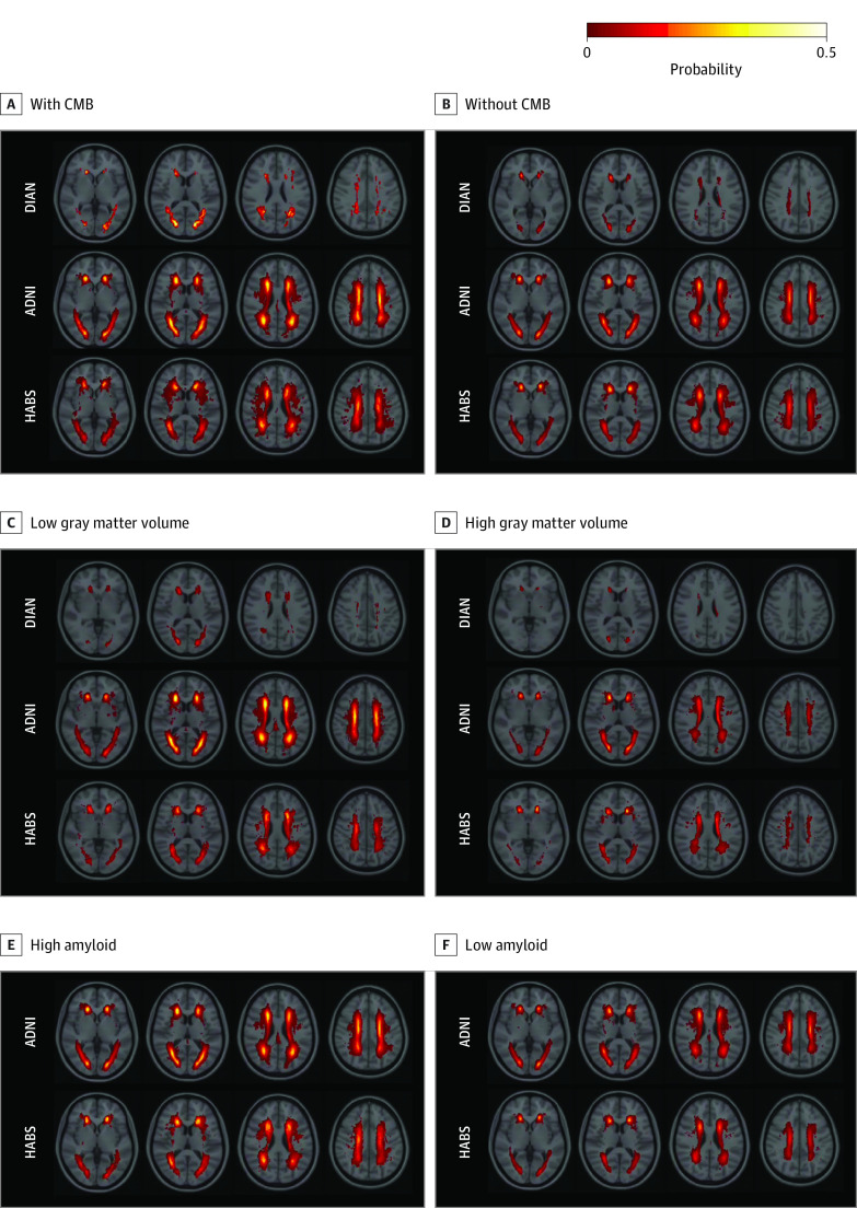
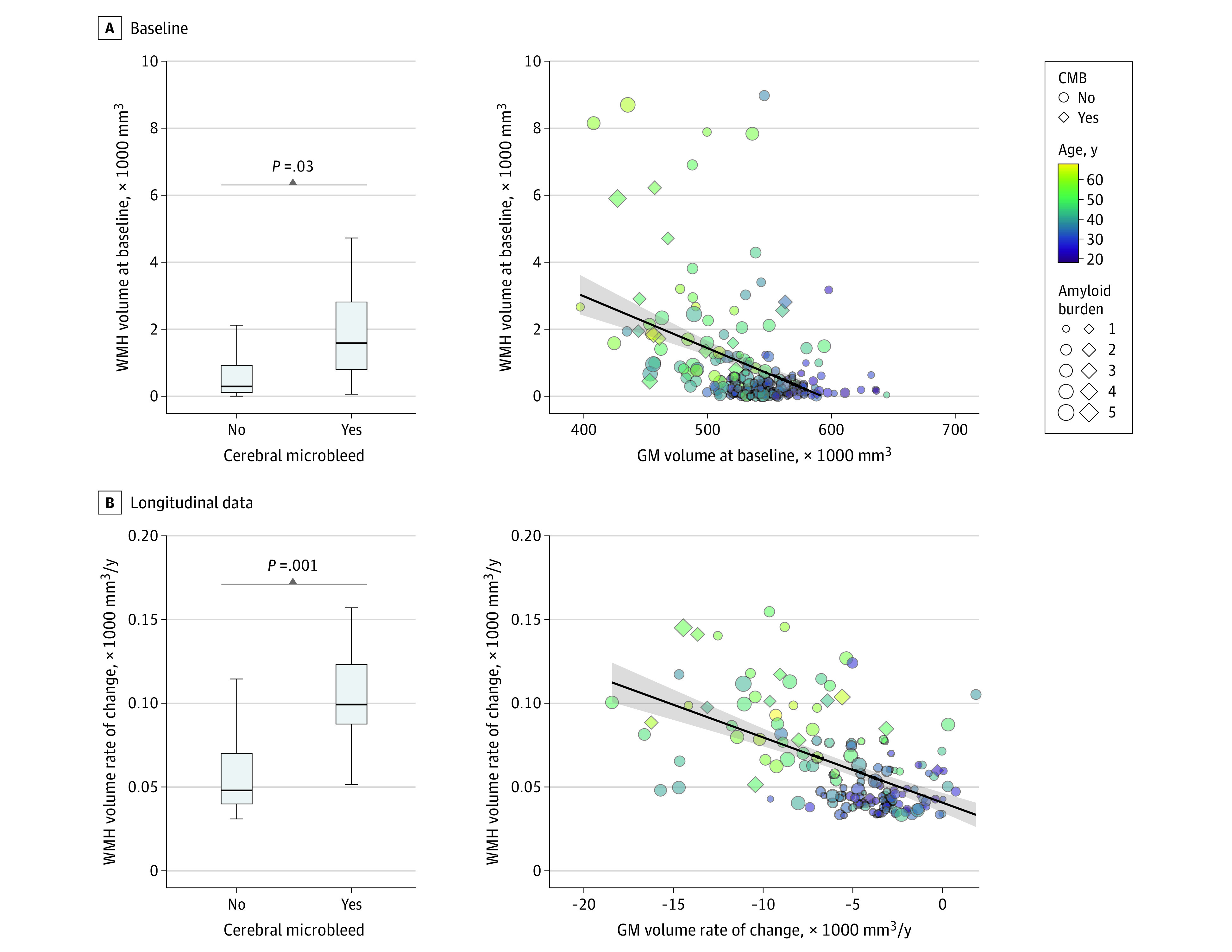
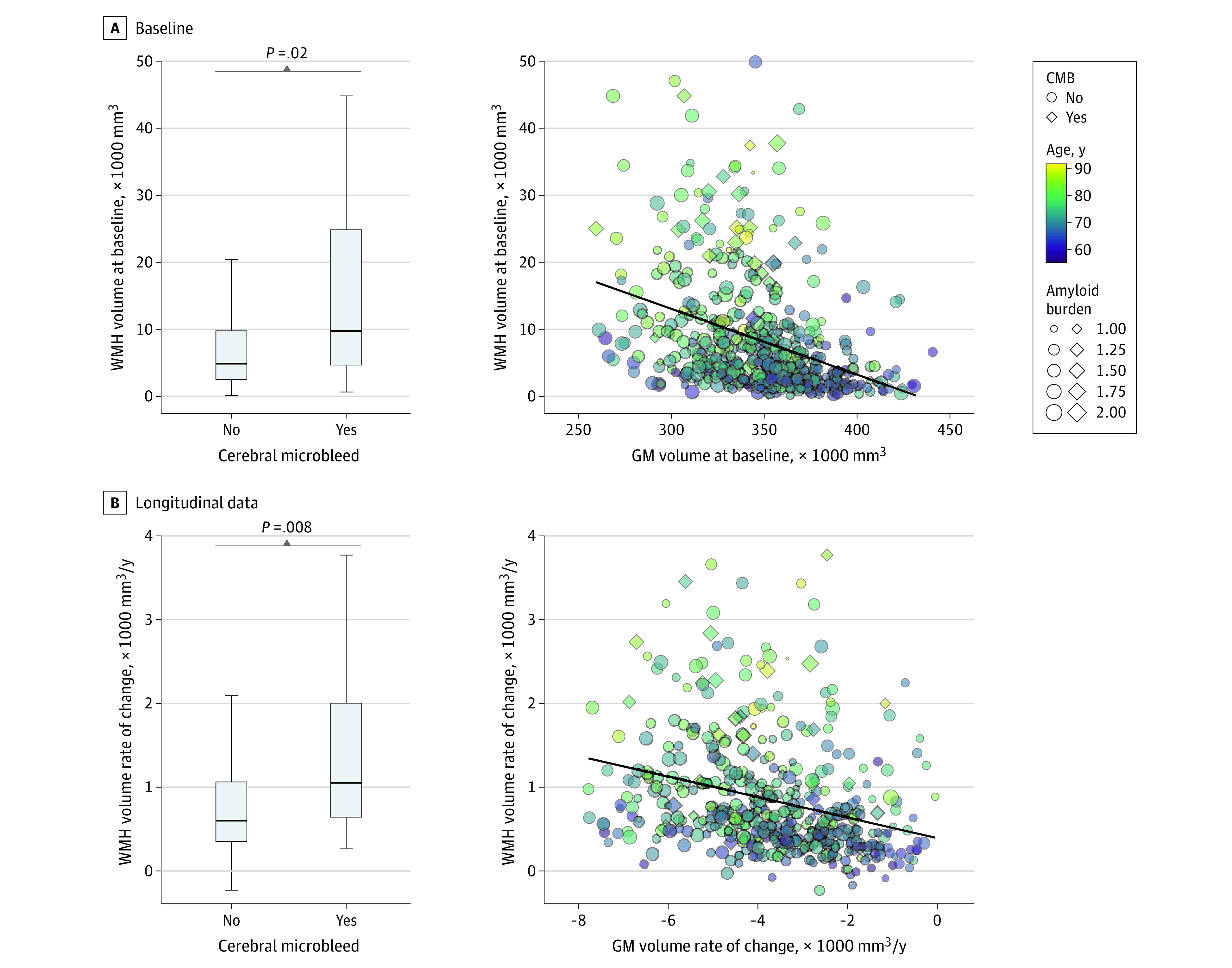
Main outcome and measures: The main outcomes were the independent associations of neurodegeneration (decreases in gray matter volume), parenchymal amyloidosis (assessed by amyloid positron emission tomography), and vessel amyloidosis (evidenced by cerebral microbleeds [CMBs]) with cross-sectional and longitudinal WMH.

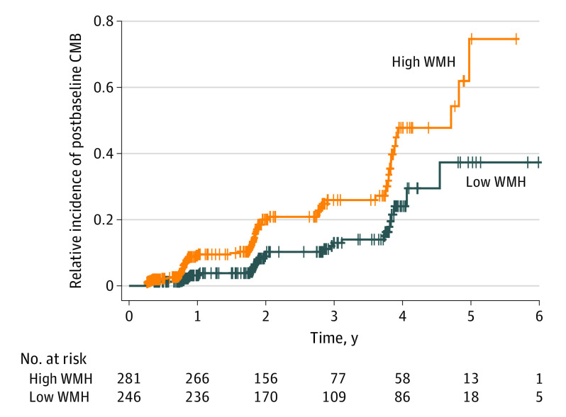
Results: Data from 3960 MRI sessions among 1141 participants were included: 252 pathogenic variant carriers from DIAN (mean [SD] age, 38.4 [11.2] years; 137 [54%] female), 571 older adults from ADNI (mean [SD] age, 72.8 [7.3] years; 274 [48%] female), and 318 older adults from HABS (mean [SD] age, 72.4 [7.6] years; 194 [61%] female). Longitudinal increases in WMH volume were greater in individuals with CMBs compared with those without (DIAN: t = 3.2 [P = .001]; ADNI: t = 2.7 [P = .008]), associated with longitudinal decreases in gray matter volume (DIAN: t = -3.1 [P = .002]; ADNI: t = -5.6 [P < .001]; HABS: t = -2.2 [P = .03]), greater in older individuals (DIAN: t = 6.8 [P < .001]; ADNI: t = 9.1 [P < .001]; HABS: t = 5.4 [P < .001]), and not associated with systemic vascular risk (DIAN: t = 0.7 [P = .40]; ADNI: t = 0.6 [P = .50]; HABS: t = 1.8 [P = .06]) in individuals with ADAD and LOAD after accounting for age, gray matter volume, CMB presence, and amyloid burden. In older adults without CMBs at baseline, greater WMH volume was associated with CMB development during longitudinal follow-up (Cox proportional hazards regression model hazard ratio, 2.63; 95% CI, 1.72-4.03; P < .001).

Conclusions and relevance: The findings suggest that increased WMH volume in AD is associated with neurodegeneration and parenchymal and vessel amyloidosis but not with elevated systemic vascular risk. Additionally, increased WMH volume may represent an early sign of vessel amyloidosis preceding the emergence of CMBs.


Conflict of interest statement

Conflict of Interest Disclosures: Dr Shirzadi reported receiving grants from the Alzheimer Society of Canada and Health Equity Scholars Program and receiving a BrightFocus Foundation fellowship during the conduct of the study. Dr Joseph-Mathurin reported receiving grants from the Alzheimer’s Association and the National Institute on Aging (NIA), National Institutes of Health (NIH) during the conduct of the study. Dr Kantarci reported consulting for Biogen and receiving nonfinancial support from Eli Lilly and Company outside the submitted work. Dr Hassenstab reported receiving personal fees from Eisai, Parabon NanoLabs Inc, Roche, ALZpath, and Prothena outside the submitted work. Dr Morris reported receiving grants from the NIH during the conduct of the study. Dr Karch reported receiving grants from the NIH during the conduct of the study. Dr Levey reported receiving grants from the NIA during the conduct of the study. Dr Schofield reported receiving grants from the NIH and the Anonymous Foundation, paid through Washington University in St Louis, and grants from the Roth Family Foundation during the conduct of the study. Dr Salloway reported receiving grants from Biogen, Eli Lilly and Company, Genentech, Roche, Esai, and Novartis and personal fees from Biogen, Eli Lilly and Company, Genentech, Roche, Eisai, Novartis, Acumen, Novo Nordisk, and Prothena for consulting during the conduct of the study. Dr McDade reported receiving grants from the NIA during the conduct of the study; grants from Eli Lilly and Company, Hoffmann-La Roche, and Eisai outside the submitted work; and personal fees from Alector and Eli Lilly and Company for serving on the data safety and monitoring board and from Alzamend for serving as a scientific advisor outside the submitted work. Dr J. Levin reported receiving personal fees from MODAG GmbH as compensation for work as chief marketing officer; personal fees from Biogen and Teva for lecturing; and personal fees from Eisai for advising, all outside the submitted work. Dr Allegri reported receiving grants from the NIH (subcontract, Washington University in St Louis) and from the Fleni Foundation during the conduct of the study. Dr Fox reported receiving grants from the UK National Institute for Health and Care Research (NIHR) University College London (UCL) Hospitals Biomedical Research Centre and from Rosetrees Trust during the conduct of the study; personal fees from Roche/Genentech, Biogen, Eli Lilly and Company, and Siemens paid to UCL; and nonfinancial support from Ionis for provision of research materials outside the submitted work. Dr Goate reported receiving personal fees from Genentech SAB, Muna Therapeutics SAB, and Biogen for consulting outside the submitted work. Dr Day reported receiving grants from the NIA during the conduct of the study and from the Alzheimer’s Association and the Chan Zuckerberg Initiative; receiving personal fees from Parabon NanoLabs Inc, DynaMed (EBSCO), PeerView Media, Inc, and Continuing Education, Inc; having stock holdings in ANI Pharmaceuticals; and receiving nonfinancial support from Avid Radiopharmaceuticals (in-kind contribution of flortaucipir precursor for research) outside the submitted work. Dr Berman reported receiving grants from the NIA during the conduct of the study. Dr Benzinger reported receiving nonfinancial support from Avid Radiopharmaceuticals for radiotracers and precursors during the conduct of the study and receiving personal fees from Eli Lilly and Company, Eisai, Bristol Myers Squibb, and Biogen for consulting and grants from Siemens outside the submitted work. Dr Sohrabi reported receiving personal fees from Alector Therapeutics for serving as director of a private company (SMarT Minds Western Australia, a site for 1 of their clinical trials) and grants paid to the Health Future Institute from Track Dutch-CAA Consortium outside the submitted work. Dr Bateman reported receiving a Zenith Grant from the Alzheimer’s Association; grants from the American Health Assistance Foundation, Glenn Foundation, Ruth K. Broad Biomedical Research Foundation, Anonymous Foundation, Merck research collaboration, Alzheimer’s Association, Association for Frontotemporal Degeneration FTD Biomarkers Initiative, BrightFocus Foundation, Cure Alzheimer’s Fund, Foundation for Barnes-Jewish Hospital, GHR Foundation, MetLife Foundation, Rainwater Foundation, Tau Consortium, Tau SILK Consortium (AbbVie, Biogen, Eli Lilly and Company, Novartis), and Centene; grants from Tracy Family Stable Isotope Labeling Quantitation (SILQ) Center donors Richard Frimel, David and Amy Payne, John and Linda Tracy, Pat and Jane Tracy, Tom and Catherine Tracy, and Robert Willman; and grants from the NfL Consortium (AbbVie, Biogen, Roche, UCL, and BMS). Dr Johnson reported receiving grants from the NIH during the conduct of the study. Dr Sperling reported receiving grants from the NIA during the conduct of the study and consulting for AC Immune, Acumen, Alector, Alnylam, Cytox, Genentech, Janssen, JOMDD, NervGen, Neuraly, Neurocentria, Oligomerix, Prothena, Renew, Shionogi, Vigil Neuroscience, Ionis, and Vaxxinity outside the submitted work. Dr Chhatwal reported receiving grants from the NIH and the BrightFocus Foundation during the conduct of the study and personal fees from ExpertConnect and Humana outside the submitted work. No other disclosures were reported.

Figures

Figure 1.
Figure 1.. Probability Maps of White Matter Hyperintensities for Individuals With or Without Cerebral Microbleeds (CMBs), With Low or High Gray Matter Volume, and With Low or High Amyloid Burden
Individual white matter hyperintensity maps were coregistered to the MNI space to generate these probability maps. ADNI indicates Alzheimer’s Disease Neuroimaging Initiative; DIAN, Dominantly Inherited Alzheimer Network; and HABS, Harvard Aging Brain Study.
Figure 2.
Figure 2.. Association of Cerebral Microbleeds (CMBs) and Gray Matter (GM) Volume With White Matter Hyperintensity (WMH) Volume in Carriers of Autosomal Dominant Alzheimer Disease Sequence Variations
In the box and whisker plots, the horizontal bar inside the box indicates the median, the box starts at the first quartile (bottom) and ends at the third (top), and the limit lines indicate minimum (lower line) and maximum (upper line) values. Amyloid burden indicates the composite amyloid positron emission tomography standard uptake value ratio.
Figure 3.
Figure 3.. Association of Cerebral Microbleeds (CMBs) and Gray Matter (GM) Volume With White Matter Hyperintensity (WMH) Volume in Older Adults From the Alzheimer’s Disease Neuroimaging Initiative
In the box and whisker plots, the horizontal bar inside the box indicates the median, the box starts at the first quartile (bottom) and ends at the third (top), and the limit lines indicate minimum (lower line) and maximum (upper line) values. Amyloid burden indicates the composite amyloid positron emission tomography standard uptake value ratio.
Figure 4.
Figure 4.. Emergence of Cerebral Microbleeds (CMBs) for Alzheimer's DIsease Neuroimaging Initiative Participants Without CMBs at Baseline
Cox proportional hazards regression model hazard ratio, 2.63; 95% CI, 1.72-4.03; P < .001. WMH indicates white matter hyperintensity.

References

    1. Smith EE. What turns the white matter white? metabolomic clues to the origin of age-related cerebral white matter hyperintensities. Circulation. 2022;145(14):1053-1055. doi:10.1161/CIRCULATIONAHA.122.059281 - DOI - PubMed
    1. McAleese KE, Miah M, Graham S, et al. . Frontal white matter lesions in Alzheimer’s disease are associated with both small vessel disease and AD-associated cortical pathology. Acta Neuropathol. 2021;142(6):937-950. doi:10.1007/s00401-021-02376-2 - DOI - PMC - PubMed
    1. McAleese KE, Walker L, Graham S, et al. . Parietal white matter lesions in Alzheimer’s disease are associated with cortical neurodegenerative pathology, but not with small vessel disease. Acta Neuropathol. 2017;134(3):459-473. doi:10.1007/s00401-017-1738-2 - DOI - PMC - PubMed
    1. Ungvari Z, Toth P, Tarantini S, et al. . Hypertension-induced cognitive impairment: from pathophysiology to public health. Nat Rev Nephrol. 2021;17(10):639-654. doi:10.1038/s41581-021-00430-6 - DOI - PMC - PubMed
    1. Schoemaker D, Zanon Zotin MC, Chen K, et al. . White matter hyperintensities are a prominent feature of autosomal dominant Alzheimer’s disease that emerge prior to dementia. Alzheimers Res Ther. 2022;14(1):89. doi:10.1186/s13195-022-01030-7 - DOI - PMC - PubMed

Publication types

Substances