Review
. 2023 Oct 16:39:197-221.
doi: 10.1146/annurev-cellbio-011723-021442.

Mechanisms of Regeneration and Fibrosis in the Endometrium

Affiliations
Review

Mechanisms of Regeneration and Fibrosis in the Endometrium

Claire J Ang et al. Annu Rev Cell Dev Biol. .

Abstract

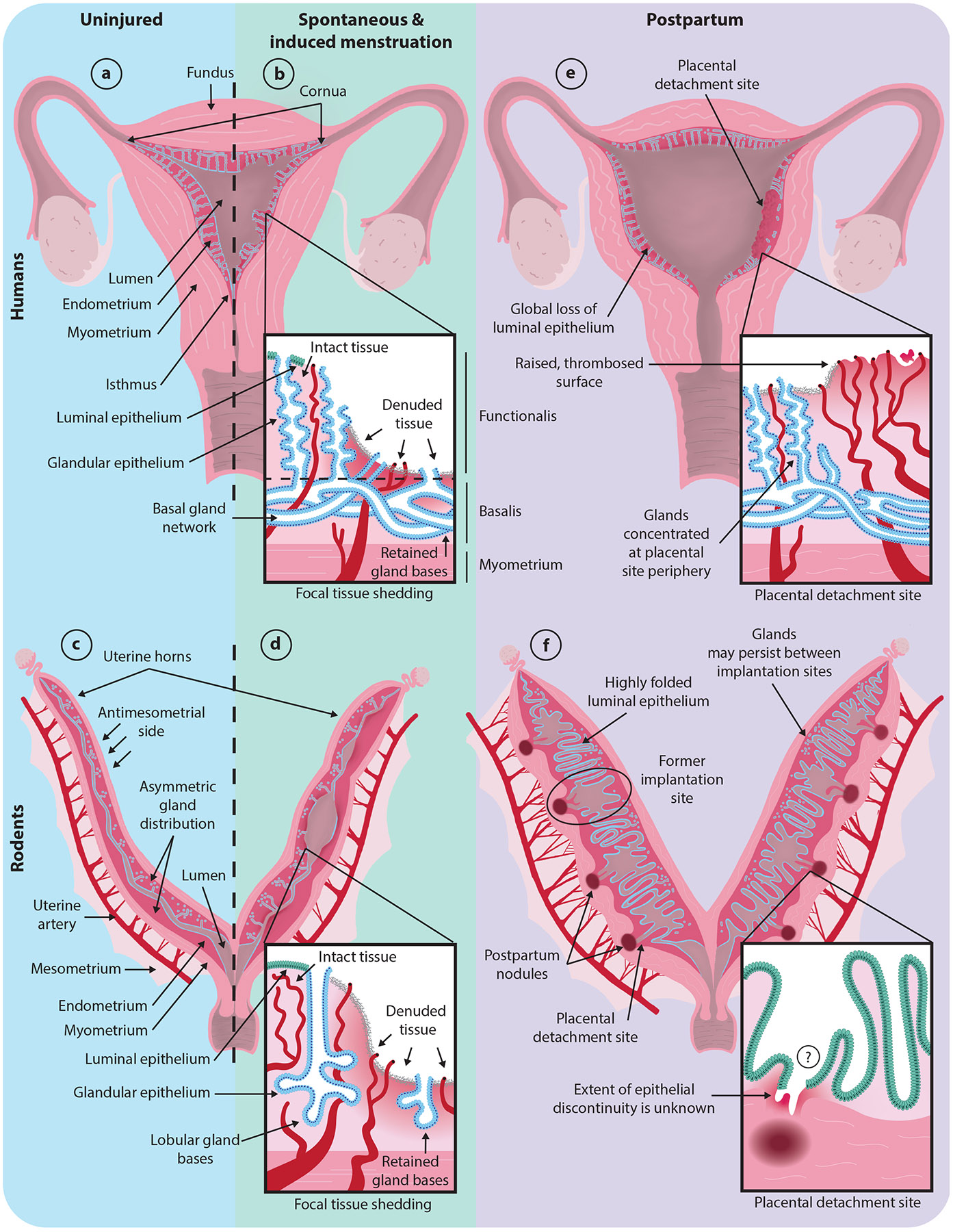
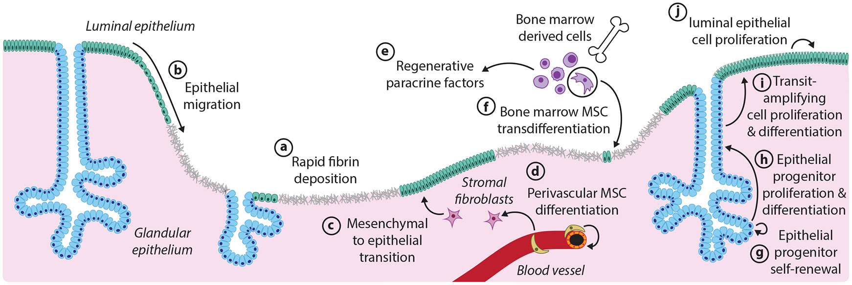
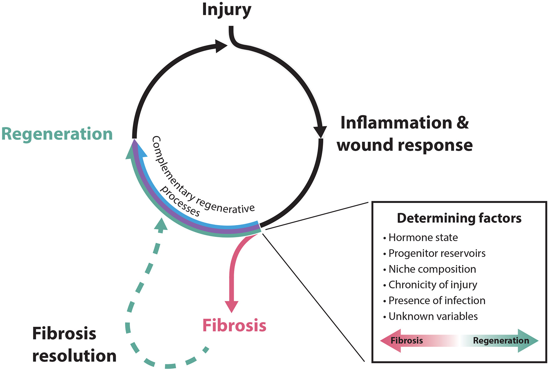
The uterine lining (endometrium) regenerates repeatedly over the life span as part of its normal physiology. Substantial portions of the endometrium are shed during childbirth (parturition) and, in some species, menstruation, but the tissue is rapidly rebuilt without scarring, rendering it a powerful model of regeneration in mammals. Nonetheless, following some assaults, including medical procedures and infections, the endometrium fails to regenerate and instead forms scars that may interfere with normal endometrial function and contribute to infertility. Thus, the endometrium provides an exceptional platform to answer a central question of regenerative medicine: Why do some systems regenerate while others scar? Here, we review our current understanding of diverse endometrial disruption events in humans, nonhuman primates, and rodents, and the associated mechanisms of regenerative success and failure. Elucidating the determinants of these disparate repair processes promises insights into fundamental mechanisms of mammalian regeneration with substantial implications for reproductive health.

Keywords: Asherman syndrome; intrauterine adhesion; menstruation; parturition; pregnancy; stem cell.


Figures

Figure 1
Figure 1
Working models of physiological disruption of the endometrium (dark magenta). Luminal epithelium is shown in green; glandular epithelium is shown in blue; and blood vessels are shown in dark red. The insets illustrate zoomed-in models of the indicated damaged regions. (a) Intact human uterus. (b) Human uterus undergoing menstruation. (c) Intact mouse uterus. (d) Mouse uterus undergoing experimentally induced tissue shedding as a model of menstruation. (e) Human uterus within one day of parturition. (f) Mouse uterus within one day of parturition. If or how postpartum gland bases connect to the lumen remains unclear. Note that the three-dimensional gland structure in the human and mouse postpartum uterus remains unknown. Mouse, but not human, uteri are approximately to scale across conditions.
Figure 2
Figure 2
Proposed mechanisms of endometrial regeneration following physiological tissue damage. The mechanisms presented here derive from both human and mouse studies, although the endometrial architecture depicted is from the mouse. The relative timing and extent of the contributions of each mechanism remain to be elucidated. (a) Injured sites are rapidly covered with the fibrinous extracellular matrix. (b) Epithelial migration from nearby intact luminal epithelium (green) or glandular epithelium (blue) covers the denuded areas. (c) In the stromal compartment, fibroblasts (magenta) undergo a mesenchymal to epithelial transition to contribute to new luminal epithelium. (d) Perivascular MSCs differentiate to restore stromal fibroblasts. (e) Paracrine signaling factors from bone marrow-derived cells (purple), and other sources promote regenerative outcomes. (f) Bone marrow–derived MSCs (circled) transdifferentiate to make minor contributions to the endometrial epithelium. (g,h) Long-lived epithelial progenitors residing in the gland bases self-renew and give rise to transit-amplifying cells residing higher up in the gland. (i) Transit-amplifying cells proliferate and differentiate to form short-lived, luminal epithelial cells. (j) The local proliferation of luminal epithelial cells also sustains the surface epithelium. Abbreviation: MSC, mesenchymal stem cell.
Figure 3
Figure 3
Wound healing outcomes in the endometrium. Both physiological and nonphysiological endometrial injuries induce an acute inflammatory/wound response, which can be resolved via complementary regenerative processes (outlined in Figure 2) to restore tissue architecture prior to subsequent injury. If coincident with underlying predisposing conditions, which may involve a number of known and unknown determining factors, this inflammation may, instead, progress to fibrosis. Without intervention, fibroses can be terminal or, at some frequency, undergo spontaneous reversion through poorly understood processes to restore functional endometrial tissues.

Similar articles

Cited by

References

    1. AAGL (Am. Assoc. Gynecol. Laparosc.). 2017. AAGL practice report: practice guidelines on intrauterine adhesions developed in collaboration with the European Society of Gynaecological Endoscopy (ESGE). J. Minim. Invasive Gynecol 24:695–705 - PubMed
    1. Abbas Y, Brunel LG, Hollinshead MS, Fernando RC, Gardner L, et al. 2020. Generation of a three-dimensional collagen scaffold-based model of the human endometrium. Interface Focus 10:20190079. - PMC - PubMed
    1. Acosta Go V-A, Ibrahim Y. 2022. Schistosomiasis induced Asherman’s syndrome in a patient undergoing assisted reproductive technology (ART): a case report and literature review. Fertil. Steril 118:e199
    1. Aghajanova L, Horcajadas JA, Esteban FJ, Giudice LC. 2010. Bone marrow-derived human mesenchymal stem cell: potential progenitor of the endometrial stromal fibroblast. Biol. Reprod 82:1076–87 - PMC - PubMed
    1. Ahn J, Yoon M-J, Hong S-H, Cha H, Lee D, et al. 2021. Three-dimensional microengineered vascularised endometrium-on-a-chip. Hum. Reprod 36:2720–31 - PMC - PubMed

Publication types

LinkOut - more resources