. 2023 Oct 24;120(43):e2303794120.
doi: 10.1073/pnas.2303794120. Epub 2023 Oct 16.

Signal transduction at GPCRs: Allosteric activation of the ERK MAPK by β-arrestin

Affiliations

Signal transduction at GPCRs: Allosteric activation of the ERK MAPK by β-arrestin

Alem W Kahsai et al. Proc Natl Acad Sci U S A. .

Abstract

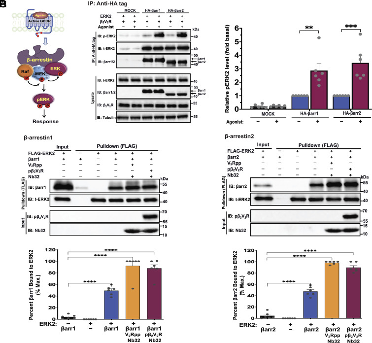
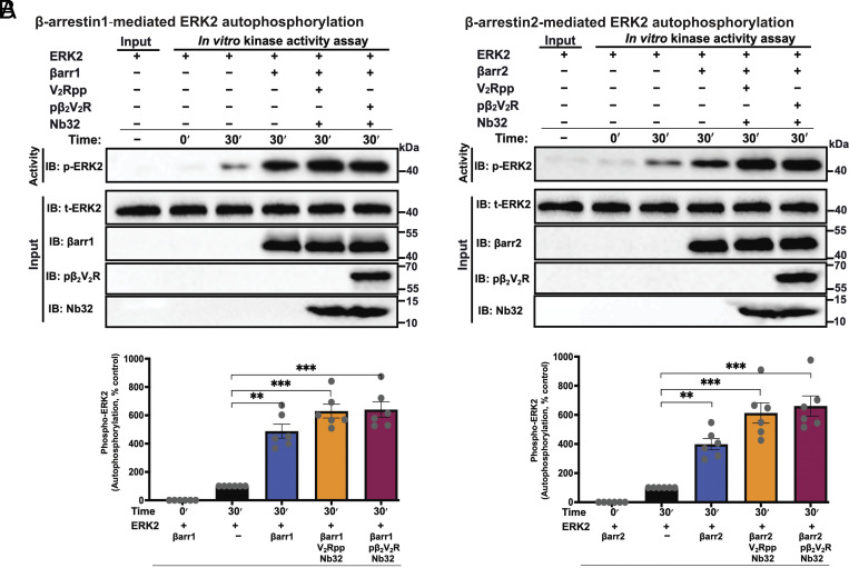
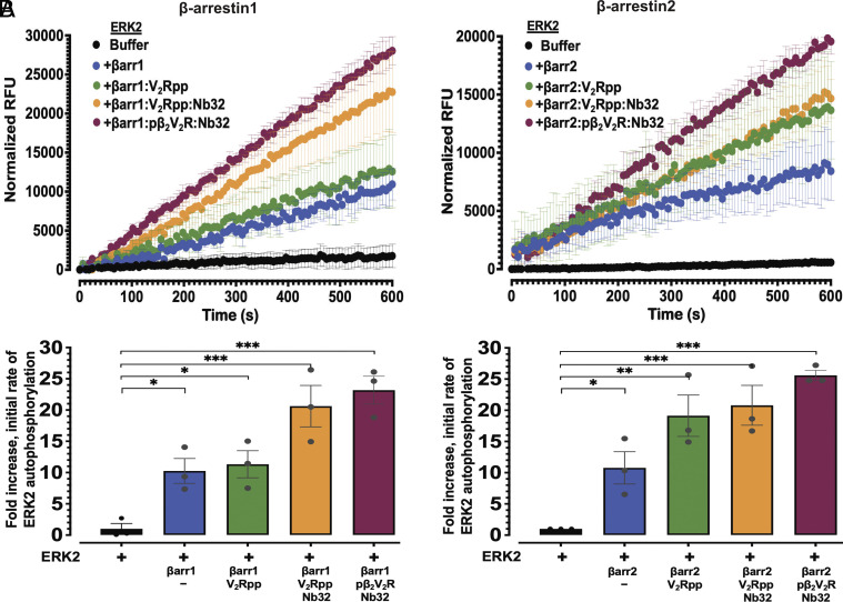
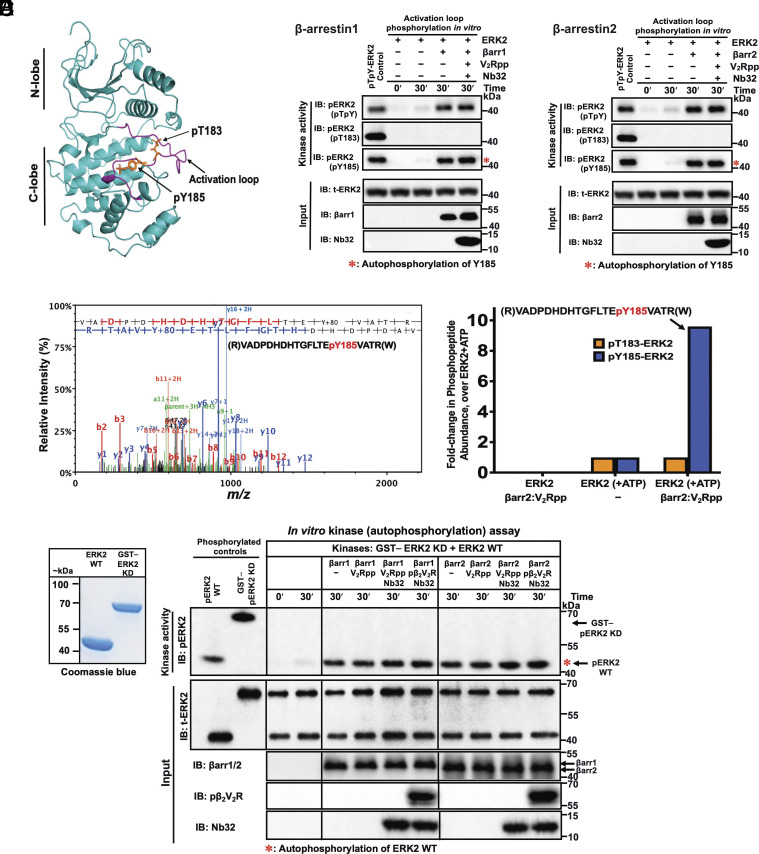
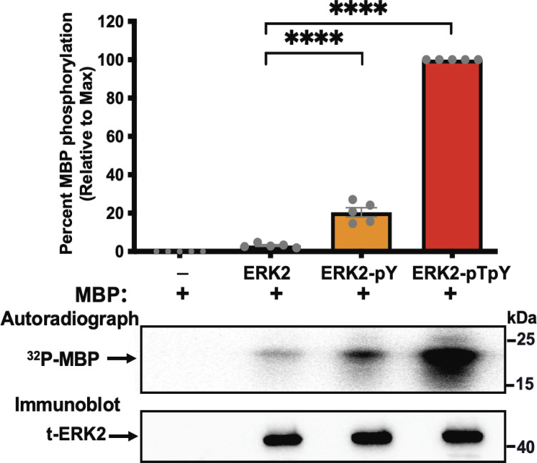
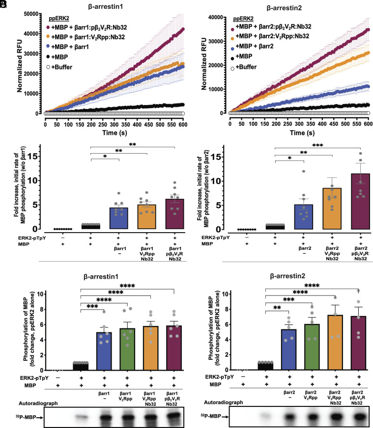
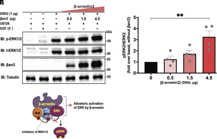
β-arrestins are multivalent adaptor proteins that bind active phosphorylated G protein-coupled receptors (GPCRs) to inhibit G protein signaling, mediate receptor internalization, and initiate alternative signaling events. β-arrestins link agonist-stimulated GPCRs to downstream signaling partners, such as the c-Raf-MEK1-ERK1/2 cascade leading to ERK1/2 activation. β-arrestins have been thought to transduce signals solely via passive scaffolding by facilitating the assembly of multiprotein signaling complexes. Recently, however, β-arrestin 1 and 2 were shown to activate two downstream signaling effectors, c-Src and c-Raf, allosterically. Over the last two decades, ERK1/2 have been the most intensely studied signaling proteins scaffolded by β-arrestins. Here, we demonstrate that β-arrestins play an active role in allosterically modulating ERK kinase activity in vitro and within intact cells. Specifically, we show that β-arrestins and their GPCR-mediated active states allosterically enhance ERK2 autophosphorylation and phosphorylation of a downstream ERK2 substrate, and we elucidate the mechanism by which β-arrestins do so. Furthermore, we find that allosteric stimulation of dually phosphorylated ERK2 by active-state β-arrestin 2 is more robust than by active-state β-arrestin 1, highlighting differential capacities of β-arrestin isoforms to regulate effector signaling pathways downstream of GPCRs. In summary, our study provides strong evidence for a new paradigm in which β-arrestins function as active "catalytic" scaffolds to allosterically unlock the enzymatic activity of signaling components downstream of GPCR activation.

Keywords: ERK MAPK; catalytic scaffolds; scaffold proteins; signal transduction; β-arrestin.


Conflict of interest statement

R.J.L. is a founder of Trevena, Inc. and Septerna, Inc., companies that discover and develop novel GPCR-targeted therapeutics. R.J.L. is also on the board of Lexicon Pharmaceuticals. S.A. is a shareholder of Septerna. All other authors declare no competing financial interests.

Figures

Fig. 1.
Fig. 1.
The scaffold proteins β-arrestin1 and 2 interact with ERK2. (A) Cartoon showing a model of a βarr scaffold linking agonist-activated phosphorylated receptor with components of the RAF–MEK–ERK MAPK pathway, which ultimately leads to ERK activation. (B) βarr1 and 2 form complexes with ERK2 as assessed by coimmunoprecipitation. CRISPR/Cas9 βarr1/2-knockout HEK293 cells were transfected with empty vector or HA-tagged βarr1 or 2 plus β2V2R and ERK2 expression plasmids. After 48 h, cells were serum-starved and subsequently stimulated with the β2AR agonist isoproterenol (10 μM), lysed, and immunoprecipitated with anti-HA antibody affinity gel. Precipitated protein complexes were detected by western blotting using anti-phospho-ERK1/2 (anti-ERK2 pThr183/pTyr185 for rat species), total ERK2, or anti‐HA-HRP (βarr1/2) antibodies. Total ERK2, βarr1/2, β2V2R, and tubulin immunoblots are shown as input controls. Bar graphs (Right) illustrate quantifications of phospho-ERK2 levels. (C and D) ERK2 binds βarr1/2 and their receptor-mediated active states. Pulldown of βarr1 (C, Top) or βarr2 (D, Top) with FLAG-tagged ERK2 on anti-FLAG beads. Purified 3xFLAG-tagged ERK2 was incubated with βarr1 or 2 alone or each in their active state form bound to V2Rpp or pβ2V2R:BI-167107 together with active βarr-stabilizing nanobody (Nb32). The pull-down products were analyzed by western blotting with antibodies specific for total ERK2 or βarr1/2 as shown by representative blots. βarr1/2, ERK2, pβ2V2R, and Nb32 input control immunoblots are shown. Graphs (Bottom) show quantification levels of βarr1/2. Data are means ± SEM of at least five independent experiments. **P ≤ 0.01; ***P ≤ 0.001; and ****P ≤ 0.0001 (ANOVA).
Fig. 2.
Fig. 2.
β-arrestin1/2 and their active states allosterically stimulate ERK2 autophosphorylation. (A and B, Top) shows representative western blots demonstrating that βarr1/2 and their active states (bound to V2Rpp or pβ2V2R) stimulate the autophosphorylation activity of ERK2. Reactions were performed in an endpoint format (30 min) at 30 °C using ERK2 (30 nM) with or without 1 μM βarr1 (A) or βarr2 (B) or each bound to V2Rpp or pβ2V2R together with Nb32. Samples were subsequently analyzed by western blotting with antibody specific for phospho-ERK1/2 (anti-ERK2-pT183/pY185). Total ERK2, βarr1/2, pβ2V2R, and Nb32 immunoblots represent individual input controls. Lower panels in A and B show bar graphs representing the extent of ERK2 autophosphorylation under the different conditions expressed as the average fold enhancement (means ± SEM) relative to a control reaction done with buffer (i.e., ERK2 alone treated as 100%). Data shown are means ± SEM (N = 6); significances by one-way ANOVA, followed by Dunnett's multiple comparison test. **P ≤ 0.01, ***P ≤ 0.001, and ****P ≤ 0.0001.
Fig. 3.
Fig. 3.
β-arrestin1/2 allosterically enhance the rate of ERK2 autophosphorylation. (A and B) show ERK2 autophosphorylation kinetics assessed using an in vitro real-time fluorescence-based kinase assay, in the absence or presence of basal state βarr1 (A) or βarr2 (B) or each in their active state forms bound to either V2Rpp or pβ2V2R together with Nb32. Kinase reactions were carried out with ERK2 (15 nM) and βarr1/2 (300 nM). Graphs (Top) represent the time course transitions of ERK2 autophosphorylation in relative fluorescence units (RFUs). Each experiment included control wells lacking ERK2 (with βarr 1 or 2, and ATP), and fluorescence signal data points from control reactions were subtracted to obtain corrected RFUs. (A and B, Lower) show quantification of ERK autophosphorylation presented as fold-enhancement of initial rates from each profile relative to vehicle control (ERK2 alone treated as onefold). Mean values are plotted, with error bars representing SEM (N = 3). Significances by one-way ANOVA, followed by Dunnett's multiple comparison test. *P ≤ 0.05; **P ≤ 0.01; and ***P ≤ 0.001.
Fig. 4.
Fig. 4.
Binding of β-arrestins to ERK2 enhances autophosphorylation of a tyrosine residue in its activation loop and occurs intramolecularly (in cis). (A) Ribbon diagram of ERK2 (PDB code: 2ERK as template; illustration using PyMOL v.2.5 Schrödinger, LLC) showing kinase activity regulatory sites (pT183 and pY185 for rat ERK2 sequence) within the activation loop (in purple) of active ERK2 (18, 40). Phosphorylation on Thr183 locks catalytic residues in a productive conformation, while on Tyr185 enables substrate binding and specificity by influencing the conformation of the MAPK. (B) and (C) show representative immunoblots from an in vitro kinase assay examining activation loop autophosphorylation of ERK2 (30 nM) performed in the absence or presence of βarr1/2 (1 μM) or their active states bound to V2Rpp together with Nb32 at 30 °C for 30 min. Immunoblot analysis was performed using three activation loop site-specific phospho-ERK1/2 antibodies (against dual sites: anti-ERK2-pThr183/pTyr185; anti-ERK2-pThr183; or anti-ERK2-pTyr185). Total ERK2, βarr1/2, and Nb32 immunoblots are shown as input controls. (D) LC-MS/MS analysis validates that βarr-modulation produces a monophosphorylated ERK2 at Tyr185. MS analysis of the autophosphorylation reaction containing ERK2 with or without βarr2 bound to V2Rpp was tryptic digested and analyzed by LC-MS/MS. (Left) Tandem MS spectrum of the identified precursor phosphopeptide ion shows N- (“b” ions) and C- (“y” ions) terminus fragments indicating the phosphorylation of rat ERK2 Tyr residue at position 185 (presented above the spectrum). (Right) bar graphs show fold change in the relative abundance of pThr183 and pTyr185 containing phosphopeptides derived from the kinase reaction containing ERK2 alone (control treated as 1) or ERK2 treated with active βarr2–V2Rpp (E) βarr-stimulated Tyr185 ERK2 activation loop autophosphorylation occurs in cis. (Left) shows Coomassie stained gel of wt-ERK2 and GST-tagged kinase-dead (KD) ERK2. (Right) shows representative western blots from in vitro kinase assay performed by incubating KD-ERK2 (GST–ERK2-K52A; 45 nM) and wt-ERK2 (30 nM) together in the presence or absence of βarr1/2 (1 μM) or indicated active states and allowed to react for 30 min at 30 °C. Immunoblotting was performed using phospho-specific antibodies of ERK2. Inputs used for kinase reaction were analyzed by immunoblotting for total ERK2, βarr1/2, pβ2V2R, and Nb32. Dually phosphorylated wt-ERK2 (lane 1, ppERK2) and KD-ERK2 (lane 2, GST–ppERK2-K52A) were prepared by coincubating with active MEK1 are shown as positive controls. Representative blots of three independent experiments are shown.
Fig. 5.
Fig. 5.
Monophosphorylation of ERK2 on Tyr185 substantially increases kinase activity. Bovine myelin basic protein (MBP) was incubated with ERK2 that was either unphosphorylated (ERK2), mono-phosphorylated ERK2 on Tyr185 (ERK2-pY), or dually phosphorylated on Thr183 and Tyr185 (ERK2-pTpY). Recombinant proteins were incubated at 37 °C for 1 h with [γ-32P] ATP as a phosphate source. Samples were separated on SDS-PAGE and 32P incorporation was assessed by autoradiography using a PhosphorImager. Total ERK2 immunoblot is shown as input control for each ERK2 species. Relative quantification of 32P incorporation is shown in the upper panel. Data are means ± SEM (N = 5). Statistical test: one-way ANOVA with Tukey's multiple comparison test, ****P ≤ 0.0001.
Fig. 6.
Fig. 6.
β-arrestins enhance the activity of dually phosphorylated ERK2-pTpY to phosphorylate MBP. (A and B, Top panels) Time courses of ERK2-pTpY-catalyzed phosphorylation of MBP as assessed using real-time fluorescence-based kinase assay in the absence or presence of basal state βarr1 (A) or βarr2 (B) or each in their active state forms (bound to V2Rpp or pβ2V2R together with Nb32). Relative fluorescence unit (RFU) intensities were background corrected to account for any contribution from control reactions: ERK2-pTpY alone and ERK2-pTpY with βarr1/2. Corrected RFU values are plotted as a function of time. Initial rates were determined from curve fitting of the linear phase of the reactions using linear regression. (A and B, Lower panels) quantification of phosphorylation of MBP presented as fold-enhancement of initial rates from each profile (as shown in the Top panel) relative to vehicle control (absence of βarr treated as onefold). Error bars indicate ± SEM of the mean from at least seven independent experiments. (C and D) Endpoint format kinase activity of ERK2-pTpY- against MBP was performed using [γ-32P] ATP as phosphate source in the absence or presence of βarr1 (C) or βarr2 (D) or their active state forms. Samples were separated on SDS-PAGE and 32P incorporation was assessed by autoradiography using a PhosphorImager. (C and D, Top panels) represent bar graphs of quantifications of phosphorylation levels of MBP under the indicated conditions. Lower panels in C and D show representative autoradiograms. Data are means ± SEM of at least five independent experiments. Asterisks indicated the significant difference based on one-way ANOVA analysis with Dunnett’s multiple comparison test (*P ≤ 0.05, **P ≤ 0.01, ***P ≤ 0.001, and ****P ≤ 0.0001).
Fig. 7.
Fig. 7.
β-arrestin2 induces ERK2 activation in intact cells. (A) CRISPR/Cas9 βarr1/2 knockout HEK293 cells were transiently transfected with plasmids containing ERK2 and varying amounts of βarr2. After 48 h, cells were serum-starved and pretreated with vehicle (Dimethyl sulfoxide, DMSO) or 1 μM U0126 for 30 min. Lysates were immunoblotted for phospho-ERK2, total-ERK2, βarr2 (A1CT), and tubulin. (B) Quantification of MEK1/2- independent βarr-dependent ERK2 activation. Densitometry analysis of data from A is shown. Phospho-ERK signals were normalized to that of total-ERK2 and expressed as relative ratios to determine the fold response of βarr-mediated ERK2 activation compared to control. Data are means ± SEM of four independent experiments. Statistical test: one-way ANOVA with Bonferroni multiple comparison test, **P ≤ 0.01. (C) Cartoon showing a model of MEK-independent βarr mediated ERK Activation. The inhibition of MEK1/2 (chemically using U0126 herein) represents a tool to rule out the role of MEK1/2 in the allosteric activation of ERK1/2 by βarr1/2.

References

    1. Lefkowitz R. J., A brief history of G-protein coupled receptors (Nobel Lecture). Angew. Chem. Int. Ed Engl. 52, 6366–6378 (2013). - PubMed
    1. Pierce K. L., Premont R. T., Lefkowitz R. J., Seven-transmembrane receptors. Nat. Rev. Mol. Cell Biol 3, 639–650 (2002). - PubMed
    1. Lohse M. J., Benovic J. L., Codina J., Caron M. G., Lefkowitz R. J., Beta-arrestin: A protein that regulates beta-adrenergic receptor function. Science 248, 1547–1550 (1990). - PubMed
    1. Peterson Y. K., Luttrell L. M., The diverse roles of arrestin scaffolds in G protein-coupled receptor signaling. Pharmacol. Rev. 69, 256–297 (2017). - PMC - PubMed
    1. Kahsai A. W., Pani B., Lefkowitz R. J., GPCR signaling: Conformational activation of arrestins. Cell Res. 28, 783–784 (2018). - PMC - PubMed

Publication types

MeSH terms