. 2023 Dec 6;43(49):8472-8486.
doi: 10.1523/JNEUROSCI.2238-22.2023.

Different Faces of Medial Beta-Band Activity Reflect Distinct Visuomotor Feedback Signals

Affiliations

Different Faces of Medial Beta-Band Activity Reflect Distinct Visuomotor Feedback Signals

Antoine Schwey et al. J Neurosci. .

Abstract

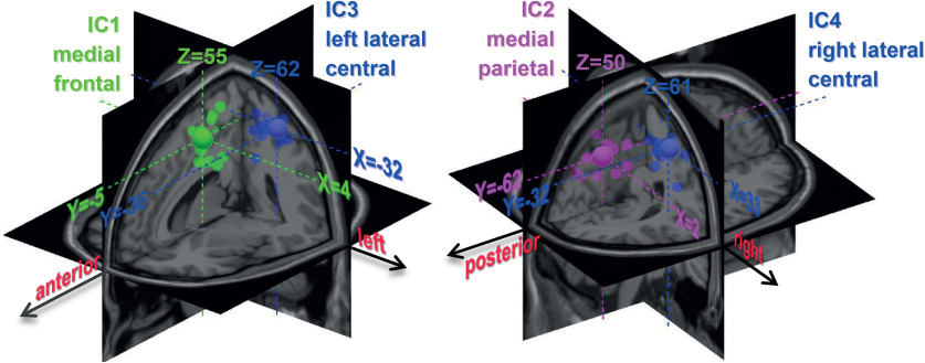
Beta-band (13-35 Hz) modulations following reward, task outcome feedback, and error have been described in cognitive and/or motor adaptation tasks. Observations from different studies are, however, difficult to conciliate. Among the studies that used cognitive response selection tasks, several reported an increase in beta-band activity following reward, whereas others observed increased beta power after negative feedback. Moreover, in motor adaptation tasks, an attenuation of the postmovement beta rebound follows a movement execution error induced by visual or mechanical perturbations. Given that kinematic error typically leads to negative task-outcome feedback (e.g., target missed), one may wonder how contradictory modulations, beta power decrease with movement error versus beta power increase with negative feedback, may coexist. We designed a motor adaptation task in which female and male participants experience varied feedbacks-binary success/failure feedback, kinematic error, and sensory-prediction error-and demonstrate that beta-band modulations in opposite directions coexist at different spatial locations, time windows, and frequency ranges. First, high beta power in the medial frontal cortex showed opposite modulations well separated in time when compared in success and failure trials; that is, power was higher in success trials just after the binary success feedback, whereas it was lower in the postmovement period compared with failure trials. Second, although medial frontal high-beta activity was sensitive to task outcome, low-beta power in the medial parietal cortex was strongly attenuated following movement execution error but was not affected by either the outcome of the task or sensory-prediction error. These findings suggest that medial beta activity in different spatio-temporal-spectral configurations play a multifaceted role in encoding qualitatively distinct feedback signals.SIGNIFICANCE STATEMENT Beta-band activity reflects neural processes well beyond sensorimotor functions, including cognition and motivation. By disentangling alternative spatio-temporal-spectral patterns of possible beta-oscillatory activity, we reconcile a seemingly discrepant literature. First, high-beta power in the medial frontal cortex showed opposite modulations separated in time in success and failure trials; power was higher in success trials just after success feedback and lower in the postmovement period compared with failure trials. Second, although medial frontal high-beta activity was sensitive to task outcome, low-beta power in the medial parietal cortex was strongly attenuated following movement execution error but was not affected by the task outcome or the sensory-prediction error. We propose that medial beta activity reflects distinct feedback signals depending on its anatomic location, time window, and frequency range.

Keywords: EEG; beta-band oscillations; feedback processing; motor learning; reaching; sensory-prediction error.


Figures

Figure 1.
Figure 1.
Task, experimental protocol, and behavioral data. A, Participants were instructed to shoot, without stopping, at one of three possible visual targets. In rotation-trial series, the cursor representing the index fingertip was displayed rotated by 30° CW or 30° CCW, relative to its real position. The rotation-trial series always consisted of four trials separated by a variable number of NR trials (blue-gray) in which the cursor displayed the real position of the hand. In the first rotation trials (Catch, red), the visual rotation was unexpectedly (re)introduced. In the three following trials (RS1–3, green), participants knew the rotation would be applied and used the reaiming strategy to counter it. Participants were also told that after the fourth rotation trials the visual rotation would be removed (AR, blue). Each Mixed block consisted of 18 rotation-trial series. B, In the experimental session, participants performed four Mixed blocks, in which the direction of the visual rotation was kept constant at 30° CW or 30° CCW. The rotation directions alternated over the four Mixed blocks, with the order counterbalanced across participants. The mixed blocks were preceded and separated by Baseline blocks of 64 and 32 no-rotation trials, respectively. C, Left, Initial movement-direction error data averaged across participants (n = 23), calculated as the perpendicular hand-path deviation measured at tangential velocity peak (PD-vel). Error bars indicate SE. Colors used for the individual trials represent the different trial categories in A. Positive values correspond to errors in the CW direction. Right, The values of the PD-vel observed at the group level (n = 23) for the different categories of trials of the Mixed blocks (Catch, RS, and AR) and the successful and failed trials of the Baseline blocks (B-hit and B-miss). The sign of the errors observed for the opposite visual rotation (CW and CCW) has been flipped so that data could be collapsed (see above, Materials and Methods). Box plot represents statistical results. Middle mark indicates the median. Edges of the box represent the 25th and 75th percentiles. Whiskers extend to the extreme data. Each participant's data are plotted individually. Kinematic errors observed for the Catch and AR trials significantly differed from those observed in all other trial categories (***p < 0.0001, Bonferroni corrected), with deviations in the direction of the visual rotation for Catch trials and deviations in the opposite direction for AR trials.
Figure 2.
Figure 2.
Individual IC topographies. Topographies of the ICs identified for each participant for the IC1, IC2, and left (IC3), and right (IC4) lateral central cortex.
Figure 3.
Figure 3.
Dipoles fitted for individual IC. Estimated dipole locations within a three-shell BEM of the MNI standard brain for the medial frontal (green), medial parietal (purple), and left and right lateral central (blue) ICs. Small spheres represent dipoles for each participant. Large spheres represent centroids of the individual dipoles for the different types of ICs.
Figure 4.
Figure 4.
Individual frequency band selection and linear mixed-model design. Top, Model equation; parameters are described in the text. Bottom left, Frequency band individually selected for the theta, alpha, low- and high-beta bands. White lines in the middle of the box plots indicate the medians. Edges of the box represent the 25th and 75th percentiles. Whiskers extend to the extreme data. Each participant's data are plotted individually. For illustration purposes, the spectrograms of the data of one participant (S02) are overlaid. The spectrogram computed for the activation time courses of the different ICs concatenated into a single time series, used to select the center of the different frequency bands, is shown (thick black line), together with the spectrogram computed for the activation time-courses of each IC separately (IC1, IC2, IC3, and IC4, green, purple, and blue thin lines, respectively). The extent of each frequency band is indicated by a shaded area around the participant's (S02) specific theta, alpha, and low- and high-beta bands. The thick-colored dashed lines indicate the center of the frequency bands. Upper and lower limits of the frequency bands are indicated by thick colored dotted lines; band widths were fixed to 3, 5, 7 and 9 Hz, respectively, for theta, alpha, low beta, and high beta. Bottom right, Individual topographies for each type of ICs.
Figure 5.
Figure 5.
Pairwise classification and power profiles for the trial categories B-Hit versus B-Miss. A, Accuracy of the prediction of the trial categories B-Hit versus B-Miss based on the actual data (dashed blue line) and based on the shuffled data (dashed green line) for each model, computed at each 100 ms step, taking as input data from a 300-ms-wide time window. Red dots indicate the models for which the accuracy was significantly higher (p < 0.01, two-tailed t test, Bonferroni corrected) when using the actual data relative to using the same but shuffled data. The p values associated with the overall models are overlaid (dashed black line). B, Regression coefficients for each of the 16 predictors of the models, 4 ICs (IC1–IC4) * 4 frequency bands (theta, alpha, low beta, high beta). Significant coefficients (p < 0.01, two-tailed t test) are highlighted in red boxes. Time = 0 corresponds to the time of the outcome feedback. C, Illustrative hand paths of single trials of the two categories by one participant (S02). D, Group averaged topographies together with power profiles for the IC1 (top) and the IC2 (bottom) aligned to the task outcome feedback. Power profiles obtained by averaging the individually selected ICs and frequency bands separately for the trial categories, B-Hit (green) and B-Miss (red). The period during which power differed significantly (p < 0.05, two-tailed t test; FDR corrected) between trial categories is indicated in gray. Small black dots indicate sampling point of uncorrected significant difference.
Figure 6.
Figure 6.
Pairwise classification and power profiles for the trial categories Catch versus NR-miss. A, Accuracy of the prediction of the trial categories, Catch versus NR-miss, based on the actual data (dashed blue line) and the shuffled data (dashed green line) for each model, computed at each 100 ms step, with input data taken from a 300-ms-wide time-window. Red dots indicate the models for which the accuracy was significantly higher (p < 0.01, two-tailed t test, Bonferroni corrected) when using the actual data relative to using the same but shuffled data. The p values associated with the overall models are overlaid (dashed black line). Also overlaid are the prediction accuracy (light blue and green dashed lines) and the fit (gray black line) achieved by the linear models based on the reduced data (see above, Materials and Methods). B, Regression coefficients for each of the 16 predictors of the models, 4 ICs (IC1–IC4) * 4 frequency bands (theta, alpha, low beta, high beta). Significant coefficients (p < 0.01, two-tailed t test) are highlighted in red boxes. Coefficients significant according to a threshold (α = 0.06) adjusted for the size of the dataset are highlighted in yellow dotted boxes. Time = 0 corresponds to the time of the outcome feedback. C, Illustrative hand paths of single trials of the two categories by one participant (S02). D, Group averaged topographies together with power profiles for the IC1 (top row) and the IC2 (bottom row) aligned to the task outcome feedback. Power profiles obtained by averaging the individually selected ICs and frequency bands separately for the trial categories, Catch (red) and NR-miss (blue-gray). The period during which power differed significantly (p < 0.05, two-tailed t test; FDR corrected) between trial categories is indicated in gray. Small black dots indicate sampling point of uncorrected significant difference.
Figure 7.
Figure 7.
Pairwise classification and power profiles for the trial categories RS-hit versus NR-hit. A, Accuracy of the prediction of the trial categories RS-hit versus NR-hit based on the actual data (dashed blue line) and based on the shuffled data (dashed green line) for each model, computed at each 100 ms step, taking as input data from a 300-ms-wide time-window. Red dots indicate the models for which the accuracy was significantly higher (p < 0.01, two-tailed t test, Bonferroni corrected) when using the actual data relative to using the same but shuffled data. The p values associated with the overall models are overlaid (dashed black line). B, Regression coefficients for each of the 16 predictors of the models, 4 ICs (IC1–IC4) * 4 frequency bands (theta, alpha, low beta, high beta). Significant coefficients (p < 0.06, two-tailed t test; threshold adjusted for the size of the dataset) are highlighted in yellow dashed boxes. Time = 0 corresponds to the time of the outcome feedback. C, Illustrative hand paths of single trials of the two categories by one participant (S02). D, Group averaged topographies together with power profiles for the IC1 (top row) and IC2 (bottom row) aligned to the task outcome feedback. Power profiles obtained by averaging the individually selected ICs and frequency bands separately for the trial categories, RS-Hit (green) and NR-Hit (blue-gray). The period during which power differed significantly (p < 0.05, two-tailed t test; FDR corrected) between trial categories is indicated in gray. Small black dots indicate sampling point of uncorrected significant difference.

Similar articles

Cited by

References

    1. Aguera PE, Jerbi K, Caclin A, Bertrand O (2011) ELAN: a software package for analysis and visualization of MEG, EEG, and LFP signals. Comput Intell Neurosci 2011:158970. 10.1155/2011/158970 - DOI - PMC - PubMed
    1. Alayrangues J, Torrecillos F, Jahani A, Malfait N (2019) Error-related modulations of the sensorimotor post-movement and foreperiod beta-band activities arise from distinct neural substrates and do not reflect efferent signal processing. Neuroimage 184:10–24. 10.1016/j.neuroimage.2018.09.013 - DOI - PubMed
    1. Atasoy S, Donnelly I, Pearson J (2016) Human brain networks function in connectome-specific harmonic waves. Nat Comms 7:1253. - PMC - PubMed
    1. Bunzeck N, Dayan P, Dolan RJ, Duzel E (2010) A common mechanism for adaptive scaling of reward and novelty. Hum Brain Mapp 31:1380–1394. 10.1002/hbm.20939 - DOI - PMC - PubMed
    1. Cavanagh JF, Frank MJ (2014) Frontal theta as a mechanism for cognitive control. Trends Cogn Sci 18:414–421. 10.1016/j.tics.2014.04.012 - DOI - PMC - PubMed

Publication types

LinkOut - more resources