. 2023 Jan-Dec:19:17448069231210423.
doi: 10.1177/17448069231210423.

Mitochondrial transplantation attenuates traumatic neuropathic pain, neuroinflammation, and apoptosis in rats with nerve root ligation

Affiliations

Mitochondrial transplantation attenuates traumatic neuropathic pain, neuroinflammation, and apoptosis in rats with nerve root ligation

Chi-Chen Huang et al. Mol Pain. 2023 Jan-Dec.

Abstract

Traumatic neuropathic pain (TNP) is caused by traumatic damage to the somatosensory system and induces the presentation of allodynia and hyperalgesia. Mitochondrial dysfunction, neuroinflammation, and apoptosis are hallmarks in the pathogenesis of TNP. Recently, mitochondria-based therapy has emerged as a potential therapeutic intervention for diseases related to mitochondrial dysfunction. However, the therapeutic effectiveness of mitochondrial transplantation (MT) on TNP has rarely been investigated. Here, we validated the efficacy of MT in treating TNP. Both in vivo and in vitro TNP models by conducting an L5 spinal nerve ligation in rats and exposing the primary dorsal root ganglion (DRG) neurons to capsaicin, respectively, were applied in this study. The MT was operated by administrating 100 µg of soleus-derived allogeneic mitochondria into the ipsilateral L5 DRG in vivo and the culture medium in vitro. Results showed that the viable transplanted mitochondria migrated into the rats' spinal cord and sciatic nerve. MT alleviated the nerve ligation-induced mechanical and thermal pain hypersensitivity. The nerve ligation-induced glial activation and the expression of pro-inflammatory cytokines and apoptotic markers in the spinal cord were also repressed by MT. Consistently, exogenous mitochondria reversed the capsaicin-induced reduction of mitochondrial membrane potential and expression of pro-inflammatory cytokines and apoptotic markers in the primary DRG neurons in vitro. Our findings suggest that MT mitigates the spinal nerve ligation-induced apoptosis and neuroinflammation, potentially playing a role in providing neuroprotection against TNP.

Keywords: Apoptosis; mitochondrial dysfunction; mitochondrial transplantation; neuroinflammation; traumatic neuropathic pain.


Conflict of interest statement

Declaration of conflicting interestsThe author(s) declared no potential conflicts of interest with respect to the research, authorship, and/or publication of this article.

Figures
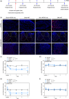
Figure 1.
Figure 1.
The spatiotemporal distribution of transplanted mitochondria and the effects of MT on SNL-induced mechanical and thermal pain hypersensitivity in rats. (a) The experimental timeline to study the effects of MT on TNP in vivo. (b) Representative micrographs of DAPI (blue) and MTDR (red) signals in the ipsilateral DRG and spinal cord of sham rats and rats receiving SNL with or without MT on PSD7. Scale: 100 µm in the upper and lower rows, 50 µm in the middle row. (c) Quantitative results of the mechanical von Frey test applied on the ipsilateral side. (d) Quantitative results of the mechanical von Frey test applied on the contralateral side. The paw withdrawal threshold (PWT) means the largest force when the rat withdrew its leg. (e) Quantitative results of the thermal Hargreaves test applied on the ipsilateral side. (f) Quantitative results of the thermal Hargreaves test applied on the contralateral side. The paw withdrawal latency (PWL) means the period from the initiation of heating the rat’s paw on the hot plate to the time of the paw withdrawal from the hot plate. N = 5 rats/group. Data were expressed as mean ± standard deviation. **p < .01, ***p < .001, ****p < .0001, repeated measures two-way ANOVA. The details of the statistic results of post-hoc comparisons were described in the main text.
Figure 2.
Figure 2.
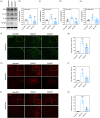
The effects of MT on expression of pro-inflammatory cytokines and degree of glial activation in the ipsilateral spinal cord of rats. (a) Representative micrographs of immunoblots of TNF, IL-1β, IL-6, and NF-κB. (b)(e) Quantitative results of the relative expression of TNF, IL-1β, IL-6, and NF-κB. (f) Representative images of immunofluorescence of GFAP in the dorsal horn of spinal cord of rats. (g) Quantitative results of the area fraction of GFAP+ area in the dorsal horn of spinal cord of rats. (h) Representative images of immunofluorescence of Iba1 in the dorsal horn of spinal cord of rats. (i) Quantitative results of the area fraction of Iba1+ area in the dorsal horn of spinal cord of rats. (j) Representative images of immunofluorescence of Iba1 in the ventral horn of spinal cord of rats. (k) Quantitative results of the area fraction of Iba1+ area in the ventral horn of spinal cord of rats. Data were expressed as mean ± standard deviation. *p < .05, **p < .01, ***p < .001, versus Sham/MT, Tukey’s multiple comparisons. #p < .05, ##p < .01, ###p < .001, versus SNL/MT, Tukey’s multiple comparisons. N = 5 rats/group (a)–(e) and 3 rats/group (f)–(h).
Figure 3.
Figure 3.
The effects of MT on the expression of apoptosis mediators in the ipsilateral spinal cord of rats. (a) Representative micrographs of immunoblots of cleaved caspase 3, Bcl-1, and BAX. (b)–(d) Quantitative results of the relative expression of caspase 3, Bcl-1, and BAX. (e) Quantitative results of the expression ratio of Bcl-2 to BAX. Data were expressed as mean ± standard deviation. ***p < .001, versus Sham/MT, Tukey’s multiple comparisons. #p < .05, ##p < .01, ####p < .0001, versus SNL/MT, Tukey’s multiple comparisons. N = 5 rats/group.
Figure 4.
Figure 4.
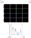
The effect of treatments with capsaicin and exogenous mitochondria on mitochondrial membrane potential in the primary DRG neurons. (a) Representative images of JC-1 staining and DAPI on primary DRG neurons treated with capsaicin and exogenous mitochondria. JC-1 demonstrates a potential-dependent accumulation within the mitochondria, leading to the formation of J aggregates emitting red fluorescence; upon depolarization, it remains as monomer showing green fluorescence. As a result, mitochondrial depolarization is indicated by a decrease in the red/green fluorescence intensity ratio. Scale: 200 µm. (b) Quantitative results of the red-to-green fluorescence intensity ratio of JC-1 staining. Data were expressed as mean ± standard deviation. *p < .05, ****p < .0001, versus capsaicin/mitochondria, Tukey’s multiple comparisons. ##p < .01, versus capsaicin+/mitochondria, Tukey’s multiple comparisons. N = 5 biological replications.
Figure 5.
Figure 5.
The effect of treatments with capsaicin and exogenous mitochondria on the production of pro-inflammatory cytokines in the primary DRG neurons. (a) Quantitative results of levels of TNF in the conditioned media. (b) Quantitative results of levels of IL-1β in the conditioned media. (c) Quantitative results of levels of IL-6 in the conditioned media. Data were expressed as mean ± standard deviation. ***p < .001, ****p < .0001, versus capsaicin/mitochondria, Tukey’s multiple comparisons. ###p < .001, ####p < .0001, versus capsaicin+/mitochondria, Tukey’s multiple comparisons. N = 5 biological replications.
Figure 6.
Figure 6.
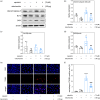
The effect of treatments with capsaicin and exogenous mitochondria on apoptosis in the primary DRG neurons. (a) Representative micrographs of immunoblots of cleaved caspase 3, Bcl-1, and BAX. (b)–(d) Quantitative results of the relative expression of cleaved caspase 3, Bcl-1, and BAX. (e) Representative images of the TUNEL and DAPI stainings. Scale: 200 µm. (f) Quantitative results of TUNEL staining. Data were expressed as mean ± standard deviation. *p < .05, ****p < .0001, versus capsaicin/mitochondria, Tukey’s multiple comparisons. ###p < .001, ####p < .0001, versus capsaicin+/mitochondria, Tukey’s multiple comparisons. N = 5 biological replications.

References

    1. Ciaramitaro P, Mondelli M, Logullo F, Grimaldi S, Battiston B, Sard A, Scarinzi C, Migliaretti G, Faccani G, Cocito D, Italian Network for Traumatic N. Traumatic peripheral nerve injuries: epidemiological findings, neuropathic pain and quality of life in 158 patients. J Peripher Nerv Syst 2010; 15: 120–127. - PubMed
    1. Dworkin RH, O'Connor AB, Kent J, Mackey SC, Raja SN, Stacey BR, Levy RM, Backonja M, Baron R, Harke H, Loeser JD, Treede RD, Turk DC, Wells CD. Interventional management of neuropathic pain: NeuPSIG recommendations. Pain 2013; 154: 2249–2261. - PMC - PubMed
    1. Kalliolias GD, Ivashkiv LB. TNF biology, pathogenic mechanisms and emerging therapeutic strategies. Nat Rev Rheumatol 2016; 12: 49–62. - PMC - PubMed
    1. Lim TK, Rone MB, Lee S, Antel JP, Zhang J. Mitochondrial and bioenergetic dysfunction in trauma-induced painful peripheral neuropathy. Mol Pain 2015; 11: 58. DOI: 10.1186/s12990-015-0057-7. - DOI - PMC - PubMed
    1. van Horssen J, van Schaik P, Witte M. Inflammation and mitochondrial dysfunction: A vicious circle in neurodegenerative disorders? Neurosci Lett 2019; 710: 132931. - PubMed

LinkOut - more resources