. 2023 Nov;8(11):2033-2049.
doi: 10.1038/s41564-023-01493-w. Epub 2023 Oct 16.

Bifidobacteria metabolize lactulose to optimize gut metabolites and prevent systemic infection in patients with liver disease

Affiliations

Bifidobacteria metabolize lactulose to optimize gut metabolites and prevent systemic infection in patients with liver disease

Matthew A Odenwald et al. Nat Microbiol. 2023 Nov.

Abstract

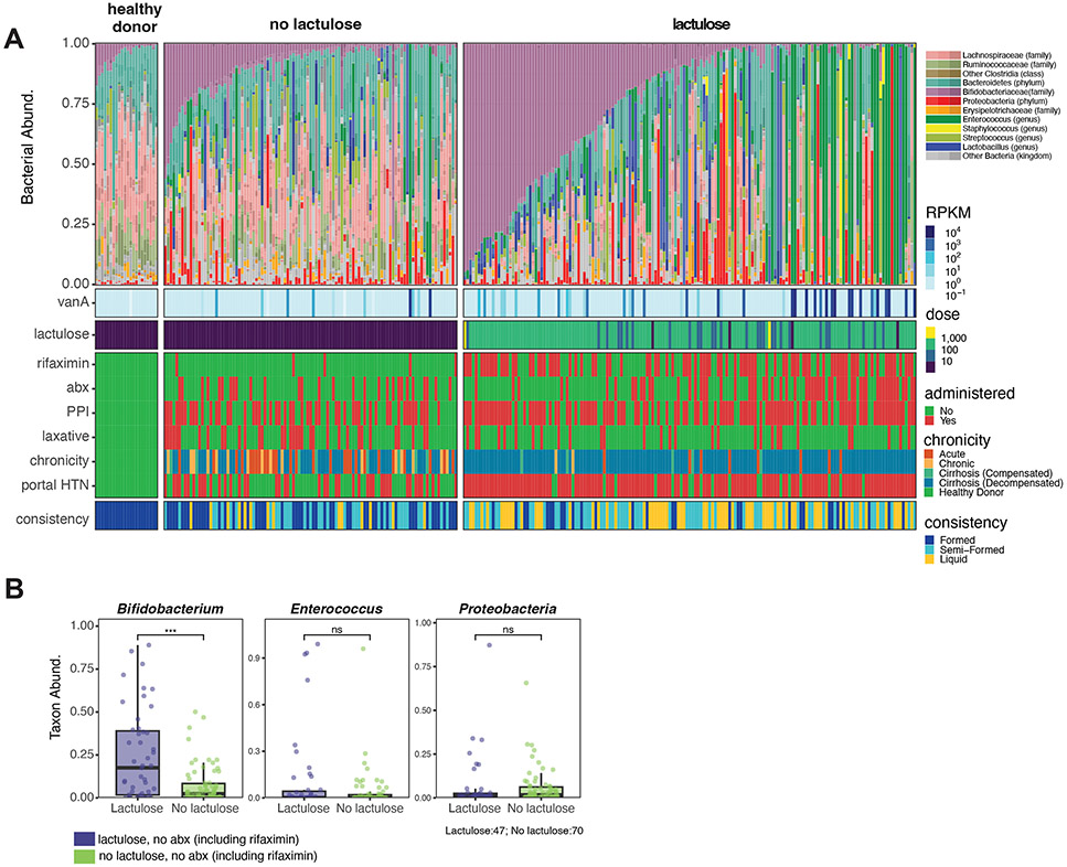
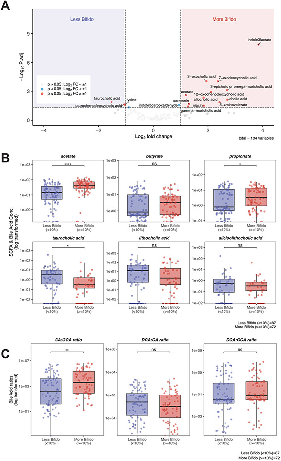
Progression of chronic liver disease is precipitated by hepatocyte loss, inflammation and fibrosis. This process results in the loss of critical hepatic functions, increasing morbidity and the risk of infection. Medical interventions that treat complications of hepatic failure, including antibiotic administration for systemic infections and lactulose treatment for hepatic encephalopathy, can impact gut microbiome composition and metabolite production. Here, using shotgun metagenomic sequencing and targeted metabolomic analyses on 847 faecal samples from 262 patients with acute or chronic liver disease, we demonstrate that patients hospitalized for liver disease have reduced microbiome diversity and a paucity of bioactive metabolites, including short-chain fatty acids and bile acid derivatives, that impact immune defences and epithelial barrier integrity. We find that patients treated with the orally administered but non-absorbable disaccharide lactulose have increased densities of intestinal bifidobacteria and reduced incidence of systemic infections and mortality. Bifidobacteria metabolize lactulose, produce high concentrations of acetate and acidify the gut lumen in humans and mice, which, in combination, can reduce the growth of antibiotic-resistant bacteria such as vancomycin-resistant Enterococcus faecium in vitro. Our studies suggest that lactulose and bifidobacteria serve as a synbiotic to reduce rates of infection in patients with severe liver disease.


Figures

Figure 1:
Figure 1:. Fecal samples from hospitalized patients with liver disease display a wide range of microbiome and metabolomic profiles.
A total of 847 fecal samples from hospitalized patients with liver disease and 22 healthy donor fecal samples were analyzed by shotgun metagenomics and targeted metabolomics. (A) Relative taxa abundance by shotgun metagenomics is shown for each sample. Metagenomic alpha-diversity was quantified using the inverse Simpson metric, and samples are arranged from left to right in order of increasing metagenomic alpha-diversity. Samples from patients with liver disease were categorized as either low, medium, or high alpha-diversity based on tertiles of inverse Simpson levels. Quantitative targeted metabolite concentrations for each corresponding sample are shown below the metagenomic data. (B) Relative abundance of indicated taxa and (C) select metabolite concentrations were plotted for each tertile of alpha-diversity. Units for SCFA are mM, and units for BA derivatives are in μg/mL. For panels B and C, each point represents a single value where n = 283 (low diversity), 282 (medium diversity), 282 (high diversity), and 22 (healthy donor). Median and interquartile range are indicated by the horizontal line and box, respectively. The lower vertical line depicts Q1 – 1.5*IQR and the upper vertical line depicts Q3 + 1.5*IQR. Statistical comparisons between individual groups were analyzed using a two-tailed Wilcoxon rank sum test. Individual groups were compared to the healthy donor group. using the Benjamini-Hochberg procedure and represented as follows: *, p < 0.05; **, p < 0.01; ***, p < 0.001; ****, p < 0.0001 *.
Figure 2:
Figure 2:. Lactulose use is associated with increased Bifidobacteria species abundance and reduced VRE abundance in the absence of systemic antibiotic use.
(A) Relative abundance by shotgun taxonomy of one sample per subject is shown (n = 22 healthy donors, 262 patients with liver disease). The sample with highest Bifidobacteria abundance for each subject was analyzed. Samples were arranged first by whether they were obtained without lactulose (left) or within 7 days after lactulose administration (right) and then by decreasing Bifidobacterium relative abundance. Among the Bifidobacteriaceae family, a small number of non-Bifidobacteria members are part of the Actinobacteria phylum and are also shown in shades of purple. The antibiotic resistance (vanA) gene was queried and colored based on expression level normalized to gene length (RPKM). Cumulative oral lactulose dose (grams) for 7 days prior to sample collection is shown. Rifaximin, broad-spectrum antibiotics, proton pump inhibitors (PPI), and alternative laxatives exposure prior to sample collection is shown. Red = treatment was administered within 7 days prior to the sample being collected; green = treatment not given in this period. The chronicity of liver disease and whether a patient had clinically significant portal hypertension (green = no, red = yes) at time of consent is shown. Stool consistency was categorized as either liquid (yellow), semi-formed (turquoise), or formed (royal blue) for each sample. (B) Relative abundances of Bifidobacteria, Enterococcus, and Proteobacteria were quantified for samples from patients that were not exposed to antibiotics (including rifaximin) in the 7 preceding days. Samples were stratified based on lactulose exposure. One sample per patient was analyzed, and each dot represents a single value (n=47 (lactulose exposure), n=70 (no lactulose exposure). Median and interquartile range are indicated by the horizontal line and box, respectively. The lower vertical line depicts Q1 – 1.5*IQR and the upper vertical line depicts Q3 + 1.5*IQR. Statistical comparisons between groups were analyzed using a two-tailed Wilcoxon rank sum test. P-values are adjusted for multiple comparisons using the Benjamini-Hochberg procedure and represented as: *, p < 0.05; **, p < 0.01; ***, p < 0.001; ****, p < 0.0001.
Figure 3:
Figure 3:. Lactulose-mediated Bifidobacteria expansion is associated with significant changes in bioactive fecal metabolites.
(A) Volcano plot (log2fold change vs. log10p-value) of qualitative metabolites comparing samples with low (< 10%) vs. high (≥10%) Bifidobacteria abundance after lactulose exposure. P-values were calculated using a two-tailed Wilcoxon rank sum test and are corrected for multiple comparisons using the Benjamini-Hochberg procedure. Values with log2 fold-change > 1 (corresponding to a 2-fold change with a p-value < 0.05) were considered significant. (B) select SCFA and BA were quantified. Units for SCFA are mM, and units for BA derivatives are in μg/mL. (C) BA conversion from conjugated-primary BA to primary BA and then to secondary BAs was tested for each sample. Each point represents a molar ratio for an individual sample. For all comparisons (A-C), there is one sample per patient that was chosen based on the highest relative abundance of Bifidobacteria, and sample size was n=87 (lactulose exposure <10% Bifidobacteria) and n=72 (lactulose exposure with ≥10% Bifidobacteria). For panels B and C, each point represents a single sample. Median and interquartile range are indicated by the horizontal line and box, respectively. The lower vertical line depicts Q1 – 1.5*IQR and the upper vertical line depicts Q3 + 1.5*IQR. Statistical comparisons between individual groups were analyzed using a two-tailed Wilcoxon rank sum test. P-values are adjusted for multiple comparisons using the Benjamini-Hochberg procedure and represented as follows: *, p < 0.05; **, p < 0.01; ***, p < 0.001; ****, p < 0.0001. For both panels B and C, Samples were grouped by whether they had expanded Bifidobacteria in the presence of lactulose. Median and interquartile range are indicated by the line and box, respectively. The lower vertical line depicts Q1 – 1.5*IQR and the upper vertical line depicts Q3 + 1.5*IQR. CA: cholic acid; GCA: glycocholic acid; DCA: deoxycholic acid.
Figure 4:
Figure 4:. Lactulose-mediated Bifidobacteria expansion is associated with exclusion of antibiotic-resistant Enterococcus species.
(A) Relative abundance of potentially pathogenic taxa Enterococcus and Proteobacteria were plotted based on lactulose exposure (no lactulose, left; lactulose, right) and relative abundance of Bifidobacteria (< 10%, blue; ≥10%, red). Each point represents a single sample with the following sample sizes: total n = 262; no lactulose, < 10%, n=79; no lactulose, ≥10%, n=24; lactulose, < 10%, n=87; lactulose, ≥10%, n=72. Median and interquartile range are indicated by the horizontal line and box, respectively. The lower vertical line depicts Q1 – 1.5*IQR and the upper vertical line depicts Q3 + 1.5*IQR. Statistical comparisons between individual groups were analyzed using a two-tailed Wilcoxon rank sum test. P-values are adjusted for multiple comparisons using the Benjamini-Hochberg procedure and represented as follows: *, p < 0.05; **, p < 0.01; ***, p < 0.001; ****, p < 0.0001. (B) A human-derived B. longum species was grown in BHIS media (blue) or BHIS supplemented with 50mM lactulose (red) or 50mM sucrose (green), and growth curves (OD600 over time) are shown. The growth curve shows mean +/− standard deviation for an experiment done in triplicate. This graph is representative of three independent experiments, also done in triplicate, all with consistent results. (C) Schematic of experimental design for B. longum (purple bacilli) and VRE (green cocci) co-culture. B. longum and VRE were grown to steady state and diluted to low density (OD600 = 0.05) prior to inoculation in either BHIS or BHIS supplemented with 50mM lactulose. Cultures were inoculated with both bacteria simultaneously (top) or B. longum was given either a 24-hour or 48-hour lead time prior to VRE inoculation (bottom). (D) After 24-hours of co-culture, serial dilutions of three replicates were plated for VRE c.f.u. counts. Each replicate is plotted as an individual point, and the median is represented by the horizontal line. *, p < 0.05 two-tailed student’s t-test. The plot is from one experiment that is representative of three independent experiments done in triplicate all with consistent results.
Figure 5:
Figure 5:. Bifidobacteria expansion and associated metabolite production are associated with decreased incidence of systemic infection and prolonged survival.
(A) A Linear discriminant analysis effect size (LEfSe) showing the significant (Wilcoxon rank-sum, two-tailed, p ≤ 0.05) effect sizes of taxa between groups. (B) Acetate (mM) and taurocholic acid (μg/mL) were quantified, and (C) conversion from conjugated-primary BA to primary BA and then to secondary BAs was tested for each sample. Each point represents a molar ratio for an individual sample. (A-C) Sample size: n = 101 (no SBP), n = 21 (SBP)). (D) LEfSe showing the significant (Wilcoxon rank- sum, two-tailed, p ≤ 0.05) effect sizes of taxa between groups. (E) Acetate (mM) and taurocholic acid (μg/mL) were quantified, and (F) conversion from conjugated-primary BA to primary BA and then to secondary BAs was tested for each sample. Each point represents a molar ratio for an individual sample. (D-F) Sample size: n = 227 (no bacteremia), n = 19 (bacteremia)). (B,C,E, and F) Each point represents a single sample. Median and interquartile range are indicated by the horizontal line and box, respectively. The lower vertical line depicts Q1 – 1.5*IQR and the upper vertical line depicts Q3 + 1.5*IQR. Statistical comparisons between individual groups were analyzed using a two-tailed Wilcoxon rank sum test. P-values are adjusted for multiple comparisons using the Benjamini-Hochberg procedure. Survival curves were stratified based on (G) initial stool sample alpha-diversity or (H) lactulose administration and Bifidobacteria expansion of the initial stool sample. The number at risk for each condition at each 10-day interval is shown below the survival curve. Survival analysis was performed using Kaplan-Meier curves, and the p-value was obtained from a log-rank test. CA: cholic acid, TCA: taurocholic acid, DCA: deoxycholic acid.

Comment in

References

    1. Moon AM, Singal AG & Tapper EB Contemporary Epidemiology of Chronic Liver Disease and Cirrhosis. Clin Gastroenterol H 18, 2650–2666 (2019). - PMC - PubMed
    1. Younossi ZM et al. Epidemiology of chronic liver diseases in the USA in the past three decades. Gut 69, 564 (2020). - PubMed
    1. Termeie O. et al. Alarming Trends: Mortality from Alcoholic Cirrhosis in the United States. Am J Medicine (2022) doi:10.1016/j.amjmed.2022.05.015. - DOI - PubMed
    1. Collaborators, G. 2017 C. et al. The global, regional, and national burden of cirrhosis by cause in 195 countries and territories, 1990–2017: a systematic analysis for the Global Burden of Disease Study 2017. Lancet Gastroenterology Hepatology 5, 245–266 (2020). - PMC - PubMed
    1. Franchis R. de et al. BAVENO VII - RENEWING CONSENSUS IN PORTAL HYPERTENSION Report of the Baveno VII Consensus Workshop: personalized care in portal hypertension. J Hepatol 76, 959–974 (2021). - PubMed

LinkOut - more resources