. 2023 Oct 16;11(1):226.
doi: 10.1186/s40168-023-01673-0.

Antimicrobial peptides modulate lung injury by altering the intestinal microbiota

Affiliations

Antimicrobial peptides modulate lung injury by altering the intestinal microbiota

Ahmed Abdelgawad et al. Microbiome. .

Abstract

Background: Mammalian mucosal barriers secrete antimicrobial peptides (AMPs) as critical, host-derived regulators of the microbiota. However, mechanisms that support microbiota homeostasis in response to inflammatory stimuli, such as supraphysiologic oxygen, remain unclear.

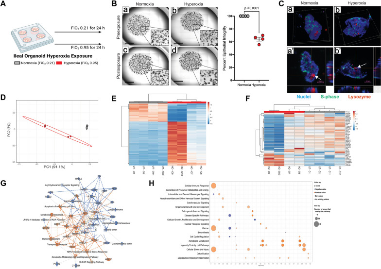
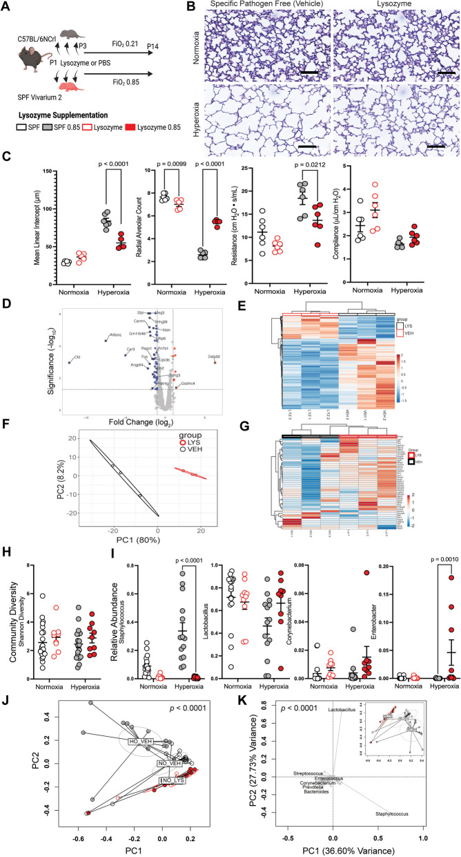
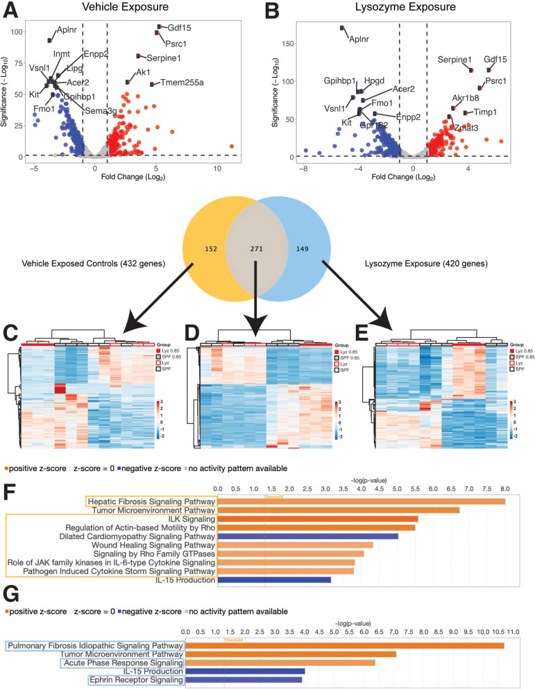
Results: We show that supraphysiologic oxygen exposure to neonatal mice, or direct exposure of intestinal organoids to supraphysiologic oxygen, suppresses the intestinal expression of AMPs and alters intestinal microbiota composition. Oral supplementation of the prototypical AMP lysozyme to hyperoxia-exposed neonatal mice reduced hyperoxia-induced alterations in their microbiota and was associated with decreased lung injury.

Conclusions: Our results identify a gut-lung axis driven by intestinal AMP expression and mediated by the intestinal microbiota that is linked to lung injury in newborns. Together, these data support that intestinal AMPs modulate lung injury and repair. Video Abstract.

Keywords: Bronchopulmonary dysplasia; Chronic lung disease; Gut-lung axis; Lysozyme; Microbiome; Neonatal lung injury; Neonate; Post-prematurity lung disease.


Conflict of interest statement

CL is the founder and CEO of Alveolus Bio and Resbiotic, Inc., NA and KW are advisors, and TN is now an employee.

Figures

Fig. 1
Fig. 1
Oxygen exposure reduces intestinal antimicrobial peptide expression. A Neonatal C57BL/6 J mice were exposed to normoxia or hyperoxia from the 3rd-14.th day of life (n = 4 litters with 5–7 neonatal mice/litter per exposure group). FiO2, fraction of inspired oxygen. SPF, specific-pathogen-free. B Representative photomicrographs of the distal lung sections of 14-day-old mice. C Hyperoxia exposure is associated with alterations in lung morphology and function. Data are shown as mean ± SEM, with significance testing by a two-tailed t-test. D Volcano plot of ileal gene expression array showing gene expression altered by hyperoxia exposure. E Heatmap showing genes regulated by hyperoxia exposure. F Principal components analysis showing differential clustering of normoxia and hyperoxia exposed ileal genes. PC, principal component. G Ileal antimicrobial peptide expression is decreased in hyperoxia-exposure mice. H Community diversity of the adherent and luminal ileal bacterial microbiome is not significantly altered by hyperoxia exposure. I The relative abundance of an operational taxonomic unit (OTU 002) that aligns to the genus Staphylococcus increases after hyperoxia exposure, as do OTUs aligning to Corynebacterium (OTU 124) and Romboutsia (OTU 013). Data are shown as mean ± SEM, with significance testing by a two-tailed t-test. J Principal coordinates analysis of Bray–Curtis dissimilarity shows global alterations in community composition in hyperoxia-exposed mice. Significance testing by permutational ANOVA (PERMANOVA), with equivocal dispersion confirmed by permutational multivariate analysis of dispersion (PERMDISP). PC, principal component. K Loading plot of principal components analysis of Hellinger transformed Euclidian distances showing the contribution of specific genera to the global community composition. Schematic in (A) was generated using BioRender. See also Figures S1, S2 and S5
Fig. 2
Fig. 2
Antimicrobial peptide expression is reduced in hyperoxia-exposed intestinal organoids. A Small intestinal spheroid organoids derived from neonatal C57BL/6 J mice were exposed to either hyperoxia or normoxia for 24 h (n = 6 wells/treatment group). B Representative images of organoids before and after exposure, with insets at 40 × magnification. The percentage of organoids with healthy-appearing epithelium declined in hyperoxia-exposed organoids. Data are shown as mean ± SEM, with significance testing by a two-tailed t-test. The scale bar represents 1000 mm. C Representative immunohistochemistry after exposure to normoxia or hyperoxia. Nuclei in blue, actively proliferating cells in green, and lysozyme-positive cells in red. Arrows identify lysozyme-positive Paneth cells. The scale bar represents 25 mm. D Principal components analysis showing differential clustering of normoxia and hyperoxia exposed ileal genes. PC, principal component. E Heatmap showing genes regulated by hyperoxia exposure. F Heatmap of antimicrobial peptide expression is decreased in hyperoxia-exposure organoids. G Ingenuity pathway analysis showing regulated pathways in hyperoxia or normoxia. H Bubble plot showing up and down-regulated pathways from hyperoxia exposure. The schematic in (A) was generated using BioRender
Fig. 3
Fig. 3
Intestinal lysozyme supplementation reduces hyperoxia-induced lung injury. A Neonatal C57BL/6NCrl mice randomized to either every other day exposure to lysozyme by gastric gavage or their littermate controls were then exposed to normoxia or hyperoxia from the 3rd-14.th day of life (n = 4 litters with 5–7 neonatal mice/litter per exposure group). FiO2, fraction of inspired oxygen. PBS, phosphate-buffered saline (vehicle). SPF, specific-pathogen-free. B Representative photomicrographs of the distal lung sections of 14-day-old mice. C Lysozyme exposure ameliorates hyperoxia-induced disruptions in lung morphology and function. Data are shown as mean ± SEM, with significance testing by two-way ANOVA. D Volcano plot of ileal RNAseq showing gene expression altered by lysozyme exposure. E Heatmap showing genes regulated by lysozyme exposure. F Principal components analysis showing differential clustering of ileal genes in lysozyme-exposed mice. PC, principal component. G Ileal antimicrobial peptide expression is altered in lysozyme-exposed mice. H The community diversity of the adherent and luminal ileal bacterial microbiome is not significantly altered by lysozyme exposure. I The hyperoxia-induced increase in the relative abundance of operational taxonomic unit 014 (Staphylococcus) is ameliorated by lysozyme exposure. Multiple other genera are increased in lysozyme and hyperoxia-exposed mice. Data are shown as mean ± SEM, with significance testing by two-way ANOVA. J Principal coordinates analysis of Bray–Curtis dissimilarity shows global alterations in community composition in lysozyme-exposed mice. Significance testing by permutational ANOVA (PERMANOVA), with equivocal dispersion confirmed by permutational multivariate analysis of dispersion (PERMDISP). PC, principal component. K Loading plot of a principal components analysis of a Hellinger transformed Euclidian distance showing global community composition significantly altered in lysozyme-exposed mice. The schematic in (A) was generated using BioRender. See also Figure S7
Fig. 4
Fig. 4
Lysozyme exposure alters the lung transcriptome. A Volcano plot showing hyperoxia alters gene expression in vehicle-exposed controls. B Volcano plot showing lysozyme exposure alters gene expression in the lung. C Heatmap showing differentially expressed genes in vehicle-exposed controls. D Heatmap showing similarly expressed genes between all groups. E Heatmap showing differentially expressed genes in lysozyme-exposed mice. F Major pathways altered in mice only exposed to normoxia or hyperoxia. G Major pathways altered in lysozyme-exposed mice

Update of

References

    1. Nikolić MZ, Sun D, Rawlins EL. Human lung development: recent progress and new challenges. Development. 2018;145:dev163485. - PMC - PubMed
    1. Hauspurg AK, Allred EN, Vanderveen DK, Chen M, Bednarek FJ, Cole C, et al. Blood Gases and Retinopathy of Prematurity: The ELGAN Study. Neonatology. 2011;99:104–111. doi: 10.1159/000308454. - DOI - PMC - PubMed
    1. Thébaud B, Goss KN, Laughon M, Whitsett JA, Abman SH, Steinhorn RH, et al. Bronchopulmonary dysplasia. Nat Rev Dis Primers. 2019;5:78. doi: 10.1038/s41572-019-0127-7. - DOI - PMC - PubMed
    1. Mookherjee N, Anderson MA, Haagsman HP, Davidson DJ. Antimicrobial host defence peptides: functions and clinical potential. Nat Rev Drug Discov. 2020;19:311–332. doi: 10.1038/s41573-019-0058-8. - DOI - PubMed
    1. Lecaille F, Lalmanach G, Andrault P-M. Antimicrobial proteins and peptides in human lung diseases: a friend and foe partnership with host proteases. Biochimie. 2016;122:151–168. doi: 10.1016/j.biochi.2015.08.014. - DOI - PubMed

Publication types