. 2023 Oct 16;54(1):91.
doi: 10.1186/s13567-023-01228-z.

Immunomodulatory effects of inactivated Ligilactobacillus salivarius CECT 9609 on respiratory epithelial cells

Affiliations

Immunomodulatory effects of inactivated Ligilactobacillus salivarius CECT 9609 on respiratory epithelial cells

María Bravo et al. Vet Res. .

Abstract

The microbiota in humans and animals play crucial roles in defense against pathogens and offer a promising natural source for immunomodulatory products. However, the development of physiologically relevant model systems and protocols for testing such products remains challenging. In this study, we present an experimental condition where various natural products derived from the registered lactic acid bacteria Ligilactobacillus salivarius CECT 9609, known for their immunomodulatory activity, were tested. These products included live and inactivated bacteria, as well as fermentation products at different concentrations and culture times. Using our established model system, we observed no morphological changes in the airway epithelium upon exposure to Pasteurella multocida, a common respiratory pathogen. However, early molecular changes associated with the innate immune response were detected through transcript analysis. By employing diverse methodologies ranging from microscopy to next-generation sequencing (NGS), we characterized the interaction of these natural products with the airway epithelium and their potential beneficial effects in the presence of P. multocida infection. In particular, our discovery highlights that among all Ligilactobacillus salivarius CECT 9609 products tested, only inactivated cells preserve the conformation and morphology of respiratory epithelial cells, while also reversing or altering the natural immune responses triggered by Pasteurella multocida. These findings lay the groundwork for further exploration into the protective role of these bacteria and their derivatives.

Keywords: Airway epithelial cells; Pasteurella multocida; immunomodulator; lactic acid bacteria; multiciliated cells; secretory cells.


Conflict of interest statement

The authors declare that they have no competing interests.

Figures
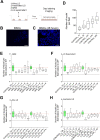
Figure 1
Figure 1
LS or inactivated LS do not affect airway basal stem cell (BSCs) proliferation. A Schematic representation of the basal stem cells (BSCs) treatment and processing protocol. B, C Images of DAPI labelled cells in control treatment for 1 h (B) and 48 h (C). D Quantification of cell numbers in control conditions at different time points using our semi-automatic system (see materials and methods for more detail). E–H Cell number quantification at two different times and relative to control conditions in MRS medium (E), LS supernatant (F), alive LS (G) and inactivated LS (H). Scale bar in B and C represents 100 μm.
Figure 2
Figure 2
A-LS cells affect epithelial barrier function and multiciliated cells. A Schematic representation of the airway epithelial monolayers treatment and processing protocol with alive (A-LS) and inactivated (i-LS) bacteria. B, C Scanning electron microscopy images of airway epithelial cells treated with A-LS (B) or i-LS (C). D Transepithelial resistance (TEER) measurements were used to test the epithelial barrier function in airway control epithelial cells or treated with A-LS or i-LS. E–H Images of live cells in control conditions (E and G), treated with A-LS (F) or treated with i-LS (H). I, J Maximal projection of confocal images for acetylated tubulin (in green), phalloidin (in red) and nucleus (in blue) in controls (I) and A-LS treated cells (J). K Maximal projection of confocal images for acetylated tubulin (in red), phalloidin (in green) and nucleus (in blue) in i-LS treated cells. Scale bar in B and C represents 5 μm. Scale bar in E, F, G and H represents 50 μm. Scale bar in I, J, and K represents 20 μm.
Figure 3
Figure 3
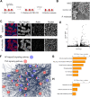
Pasteurella multocida infection characterization in airway epithelial cells. A Schematic representation of the Pasteurella multocida infection protocol. B Scanning electron microscopy image of mouse tracheal epithelial cells infected with P. multocida (C, D) Maximal projection of confocal images for acetylated tubulin (in red), phalloidin (in green) and nucleus (in blue) in controls (C) and P. multocida infected (D) MTECs. E Volcano plot of control vs P. multocida infected MTECs. F Image obtained from STRING showing upregulated transcripts related to the NF-Kappa B and TNF signalling pathways in P. multocida infected MTECs. G, H Gene ontology analyses of upregulated (G) and downregulated (H) transcripts in P. multocida infected MTECs. Scale bar in B represents 5 μm. Scale bar in C and D represents 20 μm.
Figure 4
Figure 4
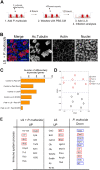
Inactivated BAL 5 treatment reverts the transcriptional program induced by Pasteurella multocida infection in the airway epithelia. A Schematic representation of the airway epithelial monolayers infection, treatment and processing protocol. B Maximal projection of confocal images for acetylated tubulin (in red), phalloidin (in green) and nucleus (in blue) in airway epithelia infected with P. multocida and treated with i-LS. C Number of differentially expressed transcripts in different experimental conditions and comparisons. (D) tSNE result of the analyses of the 15 RNA-seq samples from different conditions. E Table including transcripts related with immune system in different experimental conditions. Red box indicates upregulated transcripts in cells treated with i-LS and infected with P. multocida, which were downregulated in P. multocida infected cells. Blue box indicates downregulated transcripts in P. multocida infected cells, which were not differentially expressed in cells treated with i-LS treated and infected with P. multocida. Scale bar in B represents 20 μm.

References

    1. Ganesan S, Comstock AT, Sajjan US. Barrier function of airway tract epithelium. Tissue Barriers. 2013;1:e24997. doi: 10.4161/tisb.24997. - DOI - PMC - PubMed
    1. Parker D, Prince A. Innate immunity in the respiratory epithelium. Am J Respir Cell Mol Biol. 2011;45:189–201. doi: 10.1165/rcmb.2011-0011RT. - DOI - PMC - PubMed
    1. Frey A, Lunding LP, Ehlers JC, Weckmann M, Zissler UM, Wegmann M. More than just a barrier: the immune functions of the airway epithelium in asthma pathogenesis. Front Immunol. 2020;11:761. doi: 10.3389/fimmu.2020.00761. - DOI - PMC - PubMed
    1. Invernizzi R, Lloyd CM, Molyneaux PL. Respiratory microbiome and epithelial interactions shape immunity in the lungs. Immunology. 2020;160:171–182. doi: 10.1111/imm.13195. - DOI - PMC - PubMed
    1. Bertalanffy FD, Leblond CP. Structure of respiratory tissue. Lancet. 1955;266:1365–1368. doi: 10.1016/S0140-6736(55)93164-0. - DOI - PubMed

Substances

LinkOut - more resources