

The clinical application of critical flicker fusion frequency in demyelinating optic neuritis
- PMID: 37846319
- PMCID: PMC10577832
- DOI: 10.1016/j.aopr.2021.100011
The clinical application of critical flicker fusion frequency in demyelinating optic neuritis
Abstract
Purpose: To investigate the application of critical flicker fusion frequency (CFF) in demyelinating optic neuritis (DON).
Methods: A cross-sectional study. A total of 127 eyes in 69 DON patients and 63 eyes in 33 healthy control (HC) groups were included between January 2021 to September 2021 from Department of Ophthalmology, PLA General Hospital. Patients underwent best-corrected visual acuity (BCVA), visual field, optical coherence tomography (OCT), flash visual evoked potential (F-VEP), and CFF examinations. The affected eyes were divided into aquaporins 4 (AQP4-), myelin oligodendrocyte glycoprotein (MOG-), and double negative DON according to serum antibody; mild, moderate, severe degree visual impairment according to BCVA ≥ 0.5, 0.1-0.5, < 0.1; and 4 groups: < 1, 1 ∼< 3, 3 ∼ < 6 and > 6 months according to time interval from onset to CFF examination. One-way ANOVA was used to perform above subgroup analysis. The correlations between CFF and F-VEP peak time, peak value, BCVA and mean visual filed defect (MD) were analyzed in order via Pearson correlation analysis.
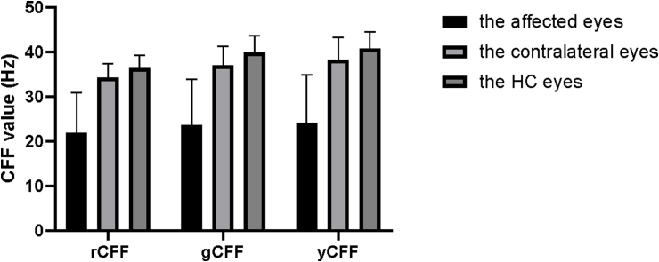
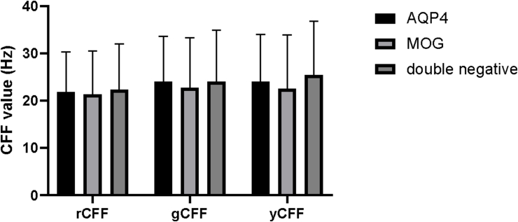
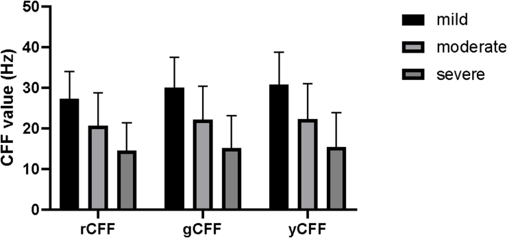
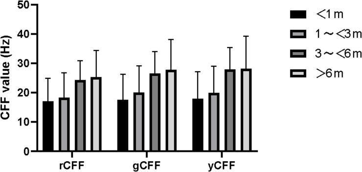
Results: he trichromatic values of red, green, and yellow in DON affected eyes were 21.83 ± 9.03, 23.66 ± 10.21, 24.09 ± 10.77 Hz, respectively, which was significantly reduced compared with the HC group (t = -14.82, -14.22, -14.00; P < 0.001). The subgroup analysis showed no significant difference between different antibody subtypes (P = 0.914 <0.848 <0.604), whereas, a significant decrease of CFF trichromatic value was found in severe visual acuity impairment group (P < 0.001). There was a significant difference in CFF- trichromatic values at different time points (P < 0.001), to be specific, CFF fluctuated under 20Hz within 3 months after onset and tended to be stable around 24-28Hz. Correlation analysis showed that the peak time of F-VEP (r = -0.486, -0.515, -0.526; P < 0.001), BVCA (r = -0.640, -0.659, -0.642; P < 0.001), were negatively correlated with CFF trichromatic values, MD and CFF were positively correlated (r = 0.486, 0.453, 0.476; P = 0.003, 0.006, 0.004).
Conclusions: A significant decrease of CFF value was found in DON-affected eyes, and it has a good correlation with BCVA, MD and latency of F-VEP, and can better reflect the impairment of visual function.
Keywords: Critical flicker fusion frequency; Demyelinating optic neuritis.
© 2021 The Authors.
Conflict of interest statement
The authors declare that they have no known competing financial interests or personal relationships that could have appeared to influence the work reported in this paper.
Figures
Similar articles
-
The theory of critical flicker fusion frequency and its application in cataracts.Adv Ophthalmol Pract Res. 2022 Oct 22;3(1):29-32. doi: 10.1016/j.aopr.2022.10.002. eCollection 2023 Feb-Mar. Adv Ophthalmol Pract Res. 2022. PMID: 37846427 Free PMC article. Review.
-
Critical flicker fusion frequency in demyelinating and ischemic optic neuropathies.Int Ophthalmol. 2018 Jun;38(3):1069-1077. doi: 10.1007/s10792-017-0561-z. Epub 2017 May 19. Int Ophthalmol. 2018. PMID: 28527029
-
Trichromatic critical flicker frequency as potential visual test in cataract and macula disease patients.Graefes Arch Clin Exp Ophthalmol. 2024 Jul;262(7):2171-2179. doi: 10.1007/s00417-024-06398-w. Epub 2024 Feb 8. Graefes Arch Clin Exp Ophthalmol. 2024. PMID: 38329529
-
[Clinical characteristics of myelin oligodendrocyte glycoprotein antibody-positive optic neuritis].Zhonghua Yan Ke Za Zhi. 2019 Mar 11;55(3):174-179. doi: 10.3760/cma.j.issn.0412-4081.2019.03.005. Zhonghua Yan Ke Za Zhi. 2019. PMID: 30841683 Chinese.
-
[Practical significance of critical fusion frequency (CFF). Chronological resolution of the visual system in differential diagnosis].Ophthalmologe. 2010 Aug;107(8):715-9. doi: 10.1007/s00347-010-2214-8. Ophthalmologe. 2010. PMID: 20533042 Review. German.
Cited by
-
The theory of critical flicker fusion frequency and its application in cataracts.Adv Ophthalmol Pract Res. 2022 Oct 22;3(1):29-32. doi: 10.1016/j.aopr.2022.10.002. eCollection 2023 Feb-Mar. Adv Ophthalmol Pract Res. 2022. PMID: 37846427 Free PMC article. Review.
-
An Arduino-Powered Device for the Study of White Perception beyond the Visual Chromatic Critical Flicker Fusion Frequency.J Imaging. 2024 Jul 10;10(7):163. doi: 10.3390/jimaging10070163. J Imaging. 2024. PMID: 39057734 Free PMC article.
References
-
- Wakakura M., Ishikawa S., Oono S., et al. [Incidence of acute idiopathic optic neuritis and its therapy in Japan. Optic neuritis treatment trial multicenter cooperative research group (ONMRG)] Nippon Ganka Gakkai Zasshi. 1995;99(1):93–97. - PubMed
LinkOut - more resources
Full Text Sources
