

Hepatic angiomyolipoma, misdiagnosed as hepatocellular carcinoma
- PMID: 37846416
- PMCID: PMC10576989
- DOI: 10.1093/jscr/rjad556
Hepatic angiomyolipoma, misdiagnosed as hepatocellular carcinoma
Abstract
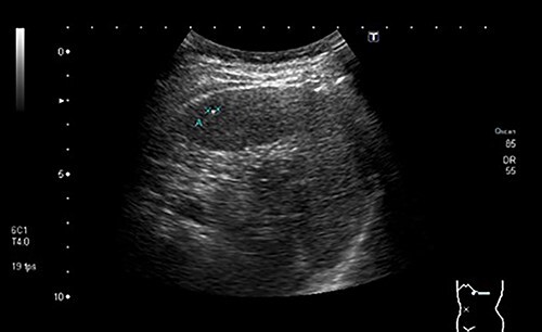
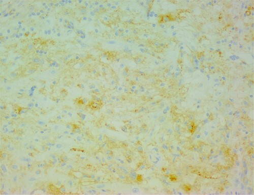
Perivascular epithelioid cell neoplasm (PEComa) is a rare type of tumor, and hepatic PEComa is even rarer. Its preoperative diagnosis is difficult, given the absence of specific clinical manifestations, often constituting an accidental finding, and the lack of a gold standard for identification using imaging studies. Instead, the diagnosis of hepatic PEComa is based on morphological and immunohistochemical features. We describe a case of an asymptomatic hepatic PEComa, angiomyolipoma type, which appeared in a middle-aged woman with chronic liver disease, during her follow-up and screening. Given the patient's context, human immunodeficiency virus-positive with chronic hepatitis C, and the similarities between the two tumors, the hepatic lesion was interpreted as hepatocellular carcinoma. The patient underwent surgical excision of the tumor, and the positive immunohistochemical staining for human melanoma black 45 and Melan A made the definitive diagnosis. In the absence of aggressiveness tumor markers, surveillance was decided. We also provide a literature review of these tumors.
Published by Oxford University Press and JSCR Publishing Ltd. © The Author(s) 2023.
Conflict of interest statement
None declared.
Figures


References
-
- Ding GH, Liu Y, Wu MC, Yang GS, Yang JM, Cong WM. Diagnosis and treatment of hepatic angiomyolipoma. J Surg Oncol 2011;103:807–12. - PubMed
-
- Klompenhouwer AJ, Dwarkasing RS, Doukas M, Pellegrino S, Vilgrain V, Paradis V. et al. Hepatic angiomyolipoma: an international multicenter analysis on diagnosis, management and outcome. Hpb [Internet] 2020;22:622–9. - PubMed
Publication types
LinkOut - more resources
Full Text Sources
