. 2023 Nov;54(11):2864-2874.
doi: 10.1161/STROKEAHA.123.043040. Epub 2023 Oct 17.

Perinatal Azithromycin Provides Limited Neuroprotection in an Ovine Model of Neonatal Hypoxic-Ischemic Encephalopathy

Affiliations

Perinatal Azithromycin Provides Limited Neuroprotection in an Ovine Model of Neonatal Hypoxic-Ischemic Encephalopathy

Jana Krystofova Mike et al. Stroke. 2023 Nov.

Abstract

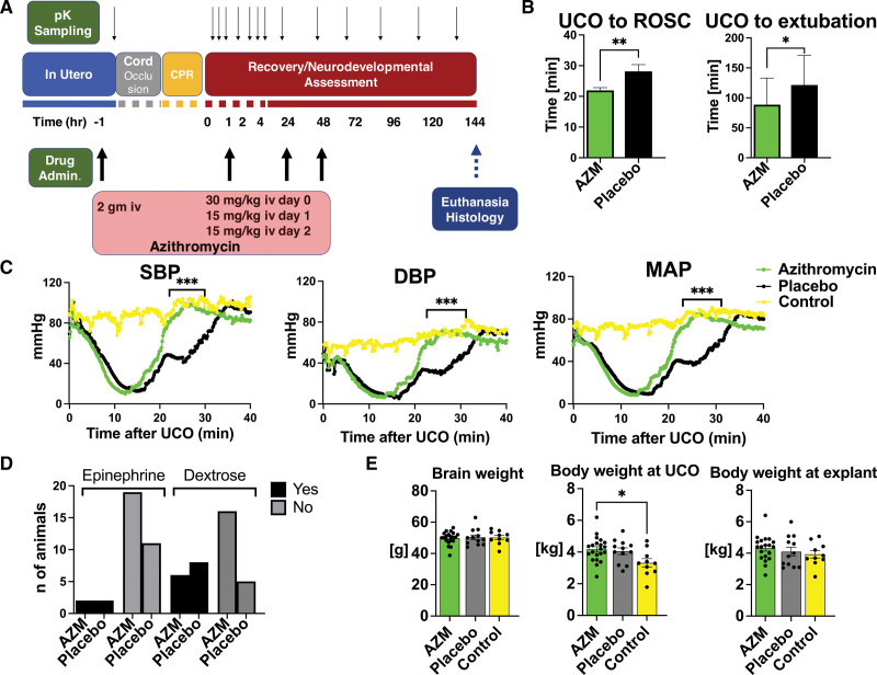
Background: Hypoxic-ischemic brain injury/encephalopathy affects about 1.15 million neonates per year, 96% of whom are born in low- and middle-income countries. Therapeutic hypothermia is not effective in this setting, possibly because injury occurs significantly before birth. Here, we studied the pharmacokinetics, safety, and efficacy of perinatal azithromycin administration in near-term lambs following global ischemic injury to support earlier treatment approaches.

Methods: Ewes and their lambs of both sexes (n=34, 141-143 days) were randomly assigned to receive azithromycin or placebo before delivery as well as postnatally. Lambs were subjected to severe global hypoxia-ischemia utilizing an acute umbilical cord occlusion model. Outcomes were assessed over a 6-day period.

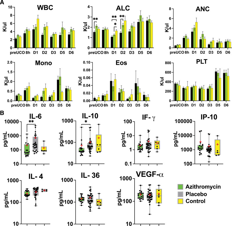
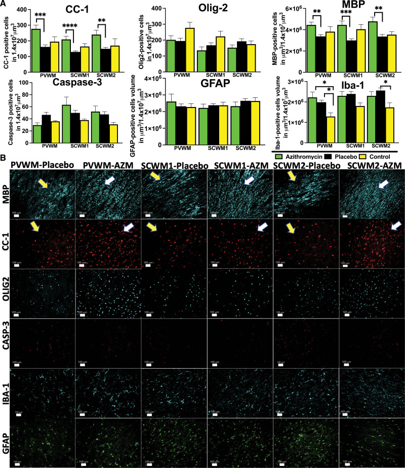
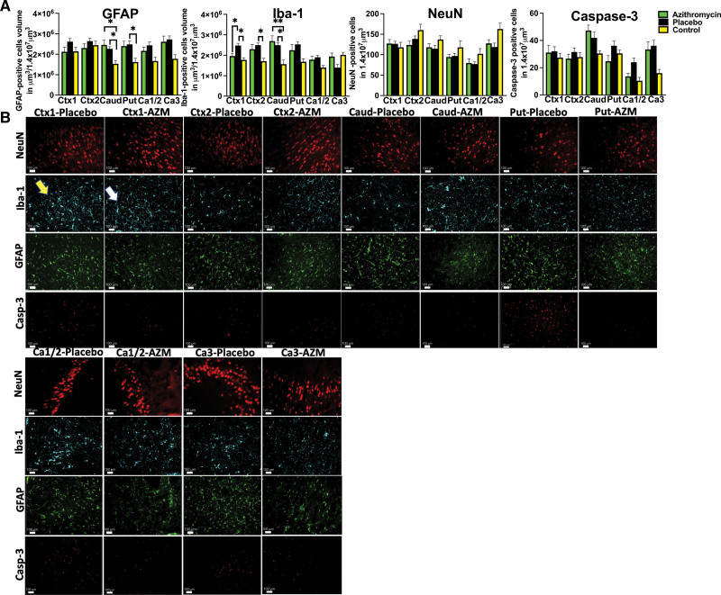
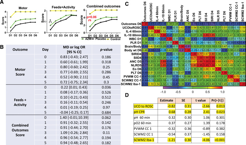
Results: While maternal azithromycin exhibited relatively low placental transfer, azithromycin-treated lambs recovered spontaneous circulation faster following the initiation of cardiopulmonary resuscitation and were extubated sooner. Additionally, peri- and postnatal azithromycin administration was well tolerated, demonstrating a 77-hour plasma elimination half-life, as well as significant accumulation in the brain and other tissues. Azithromycin administration resulted in a systemic immunomodulatory effect, demonstrated by reductions in proinflammatory IL-6 (interleukin-6) levels. Treated lambs exhibited a trend toward improved neurodevelopmental outcomes while histological analysis revealed that azithromycin supported white matter preservation and attenuated inflammation in the cingulate and parasagittal cortex.

Conclusions: Perinatal azithromycin administration enhances neonatal resuscitation, attenuates neuroinflammation, and supports limited improvement of select histological outcomes in an ovine model of hypoxic-ischemic brain injury/encephalopathy.

Keywords: asphyxia; azithromycin; brain hypoxia-ischemia; neonates; ovine model.


Conflict of interest statement

Disclosures None.

Figures

Figure 1.
Figure 1.
Umbilical cord occlusion (UCO) model and resuscitation outcomes. A, Timeline of azithromycin (AZM) administration. B, Return of spontaneous circulation (ROSC) was faster in AZM-treated animals, and the time to extubation was shorter compared with the placebo group. Groups were compared using unpaired t test for ROSC assessment and Mann-Whitney U test for extubation time. AZM: n=21, placebo: n=13. C, UCO (time 0 min) induced asystole with corresponding loss in cardiac output and hypotension. ROSC restored blood pressure to levels similar to age-matched controls. Hemodynamic data were analyzed using grouped analysis of the individual group’s means for a specific time point. AZM: n=18; placebo: n=13; control: n=6 (controls data were used with permission from Mike et al). D, The incidence of second dose of epinephrine and dextrose administration was similar between the groups. The proportion of variables was assessed using Fisher exact test. AZM: n=21; placebo: n=13. E, There were no differences in selected anthropometric parameters between AZM and placebo. Brain and body weight differences were assessed using ANOVA. AZM: n=21; placebo: n=12; control: n=10. Data shown in the graphs B and E as mean±SEM, graph C are shown as mean, graph D is a contingency table. AZM-treated group, green. Placebo, black. Controls, yellow. CPR indicates cardiopulmonary resuscitation; DBP, diastolic blood pressure; MAP, mean arterial pressure; pK, pharmacokinetic; and SBP, systolic blood pressure. *P<0.05, **P<0.01, ***P<0.001.
Figure 2.
Figure 2.
Peripheral markers of inflammation. A, Absolute lymphocyte count (ALC) differed at baseline and on days 1 and 2 in azithromycin (AZM) vs placebo group. Cellular subgroups were evaluated by mixed-effect analysis with Smidak correction for multiple comparisons. The summary column graphs show means±SEM. AZM: n=8 to 21; placebo: n=7 to 12; control: n=4 to 21 (control data were used with permission from Mike et al). B, At 6 days after the umbilical cord occlusion (UCO), we noticed changes in IL-6 (interleukin-6) and IL-10 (interleukin-10). No changes were observed in IL-4 (interleukin-4), IF-g (interferon-g), IP-10 (interferon-g inducible protein), IL-36 (interleukin-36), or VEGF-α (vascular endothelial growth factor-α). We used Kruskal-Wallis test. AZM: n=15 to 19; placebo: n=27 to 32; control: n=5 to 6. The box graphs show the mean as red asterisk “*” and the median with interquartile range. AZM-treated group, green. Placebo, black. Controls, yellow. ANC indicates absolute neutrophil count; Eos, eosinophil; Mono, monocyte; PLT, platelet; and WBC, white blood cell. *P<0.05, **P<0.01.
Figure 3.
Figure 3.
Quantitative analysis of white matter markers and markers of inflammation. A, The number of antiadenomatous polyposis coli clone CC-1 (CC-1), and the volume of MBP (myelin basic protein)-stained myelin fibers reflect the changes triggered by umbilical cord occlusion in periventricular white matter (PVWM), subcortical white matter of cingulate gyrus (SCWM1), and subcortical white matter of the first parasagittal gyrus (SCWM2) in azithromycin (AZM)-treated (green) vs placebo-treated (black) vs control (yellow) animals. The cellular death was quantified by the number of cleaved Casp-3 (caspase-3)-positive cells. The neuroinflammation was quantified by the total volume of GFAP (glial fibrillary acidic protein)-positive glial and microglial Iba-1-positive cells. Analyzed were AZM: n=7 to 20, placebo: n=6 to 11, and control: n=5 to 9 animals (control data were used with permission from Mike et al) using either ANOVA or Kruskal-Wallis test. Data are presented as mean±SEM. Brackets show significance as follows: *P<0.05, **P<0.01,***P<0.001, ****P<0.0001. B, The representative photomicrographs of histological changes with preservation of the myelin fibers (MBP) and mature oligodendrocytes (CC-1) in AZM-treated group (MBP), myelin breaks, and loss of CC-1 in the placebo group. Cellular death (Casp-3) and gliosis (GFAP) were unchanged among the groups, and more microglial (Iba-1 [ionized calcium-binding adaptor molecule-1]) accumulation was observed in PVWM in both groups and placebo group in SCWM2. Yellow arrows—placebo histopathology, white arrows—AZM-treated histopathology.
Figure 4.
Figure 4.
Histological changes in gray matter. A, We compared quantitative changes in inflammatory markers of gliosis (GFAP [glial fibrillary acidic protein]) and microglial accumulation (Iba-1 [ionized calcium-binding adaptor molecule-1]); neuronal counts (neuronal nuclei [NeuN]), cellular death markers (Casp-3 [caspase-3]) in cingulate gyrus (Ctx1), first parasagittal gyrus (Ctx2), caudate (Caud), putamen (Put), and Ca1/2 and Ca3 of the hippocampus. Analyzed were azithromycin (AZM): n=7 to 20, placebo: n=6 to 11, controls: n=5 to 8 (control data were used with permission from Mike et al) using either ANOVA or Kruskal-Wallis test. Data are presented as mean±SEM. Brackets show significance as follows: *P<0.05, **P<0.01. AZM-treated animals (green) vs placebo (black) vs control (yellow). B, The observed quantitative changes are represented in photomicrographs by accumulation of microglial cells in Ctx-1 (Iba-1 marker), thickened glial cells in Caud in both groups, and Put in the placebo group (GFAP). Placebo animal histologies (yellow arrow) are compared with the AZM-treated animals (white arrow).
Figure 5.
Figure 5.
Neurological outcomes. A, We assessed combined outcomes score, as a composite score consisting of motor function and feeds+activity. The summary graphs picture the relationship to uninjured controls. Azithromycin (AZM)-treated group, green. Placebo, black. Controls, yellow (control data were used with permission from Mike et al). B, Odds ratios of outcomes measures are reported on a logarithmic scale. The statistical approach is described in the Supplemental Material. Data are presented as mean±SEM. Compared were AZM: n=21, placebo: n=13. P<0.05 was considered significant. C, The correlation matrix pictures Spearman correlation coefficients of selected study parameters with combined neurological outcomes scores on day 6 after the umbilical cord occlusion (UCO) that reached statistical significance (P<0.05). The earliest parameters that correlate with neurological outcomes are time from UCO to return of spontaneous circulation (ROSC), total white blood cell (WBC) counts, and platelets to lymphocytes ratio (PLR) cell ratio on day 1. At a later time point, brain-to-body ratio, the body weight at explant, systemic inflammation response index (SIRI), systemic immune-inflammation index (SII) on day 4, absolute neutrophil count (ANC), neutrophil to lymphocytes ratio (NLR) on day 5, eosinophils (Eos), and platelet (PLT) on day 6, and histological markers of mature oligodendrocytes correlate with neurological outcomes on day 6. D, Lasso analysis of data revealed UCO to ROSC, pH at CPR, and microgliosis in SCWM2 to predict neurological outcomes on day 6. Yellow: P<0.05, n=34. Pre-UCO prior to the umbilical cord occlusion. CC-1 indicates antiadenomatous polyposis coli clone CC-1; CPR, cardiopulmonary resuscitation; Iba-1, ionized calcium-binding adaptor molecule-1; IL-4, interleukin-4; IL-10, interleukin-10; PVWM, periventricular white matter; and SCWM2, subcortical white matter of the first parasagittal gyrus.

References

    1. Lawn J, Shibuya K, Stein C. No cry at birth: global estimates of intrapartum stillbirths and intrapartum-related neonatal deaths. Bull World Health Organ. 2005;83:409–417. - PMC - PubMed
    1. Gebregziabher GT, Hadgu FB, Abebe HT. Prevalence and associated factors of perinatal asphyxia in neonates admitted to ayder comprehensive specialized hospital, northern Ethiopia: a cross-sectional study. Int J Pediatr. 2020;2020:4367248. doi: 10.1155/2020/4367248 - PMC - PubMed
    1. Lee AC, Kozuki N, Blencowe H, Vos T, Bahalim A, Darmstadt GL, Niermeyer S, Ellis M, Robertson NJ, Cousens S, et al. . Intrapartum-related neonatal encephalopathy incidence and impairment at regional and global levels for 2010 with trends from 1990. Pediatr Res. 2013;74:50–72. doi: 10.1038/pr.2013.206 - PMC - PubMed
    1. Bhatti A, Kumar P. Systemic effects of perinatal asphyxia. Indian J Pediatr. 2014;81:231–233. doi: 10.1007/s12098-013-1328-9 - PubMed
    1. Jacobs SE, Berg M, Hunt R, Tarnow-Mordi WO, Inder TE, Davis PG. Cooling for newborns with hypoxic ischaemic encephalopathy. Cochrane Database Syst Rev. 2013;2013:CD003311. doi: 10.1002/14651858.CD003311.pub3 - PMC - PubMed