

Total and Regional Brain Volumes in Fetuses With Congenital Heart Disease
- PMID: 37846811
- PMCID: PMC7616254
- DOI: 10.1002/jmri.29078
Total and Regional Brain Volumes in Fetuses With Congenital Heart Disease
Abstract
Background: Congenital heart disease (CHD) is common and is associated with impaired early brain development and neurodevelopmental outcomes, yet the exact mechanisms underlying these associations are unclear.
Purpose: To utilize MRI data from a cohort of fetuses with CHD as well as typically developing fetuses to test the hypothesis that expected cerebral substrate delivery is associated with total and regional fetal brain volumes.
Study type: Retrospective case-control study.
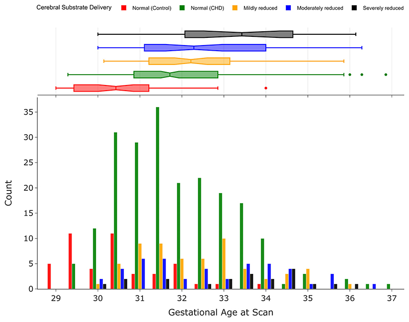
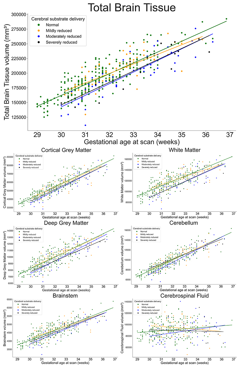
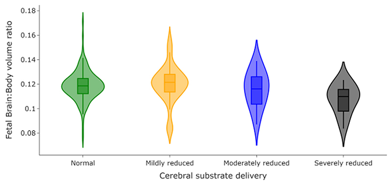
Population: Three hundred eighty fetuses (188 male), comprising 45 healthy controls and 335 with isolated CHD, scanned between 29 and 37 weeks gestation. Fetuses with CHD were assigned into one of four groups based on expected cerebral substrate delivery.
Field strength/sequence: T2-weighted single-shot fast-spin-echo sequences and a balanced steady-state free precession gradient echo sequence were obtained on a 1.5 T scanner.
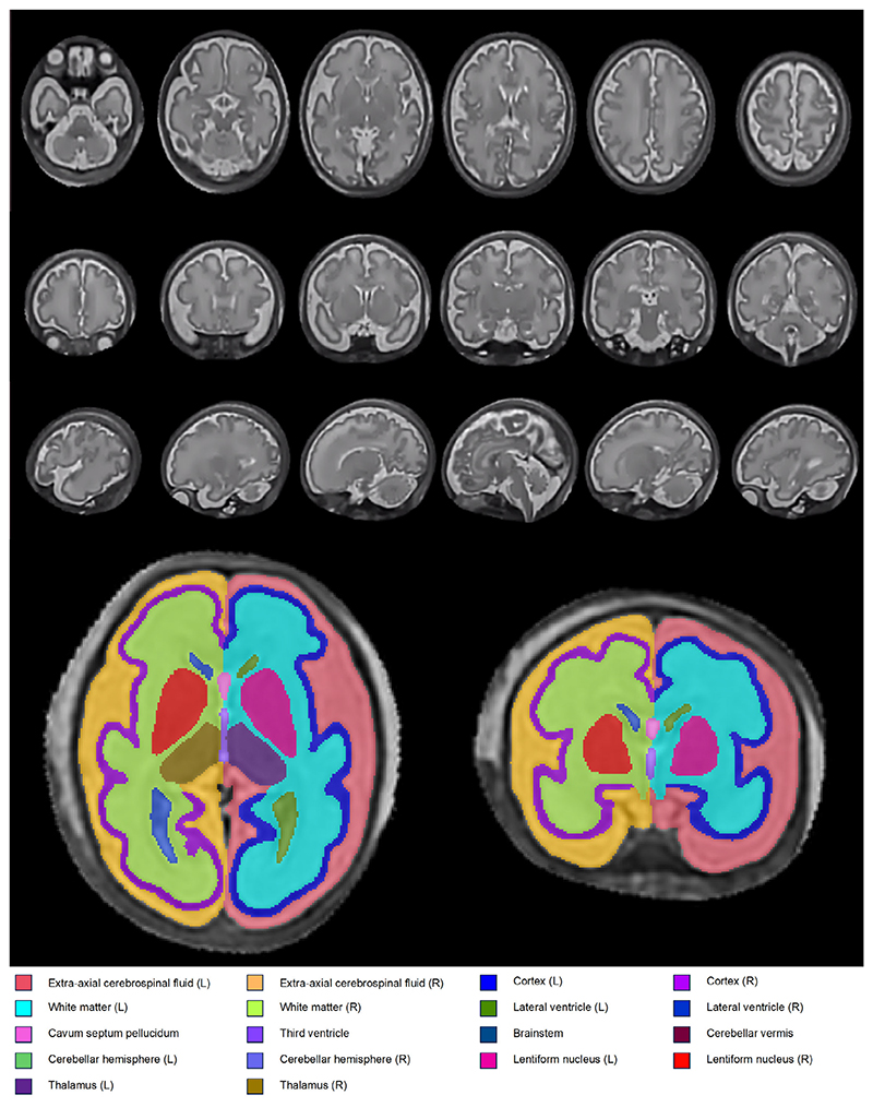
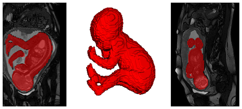
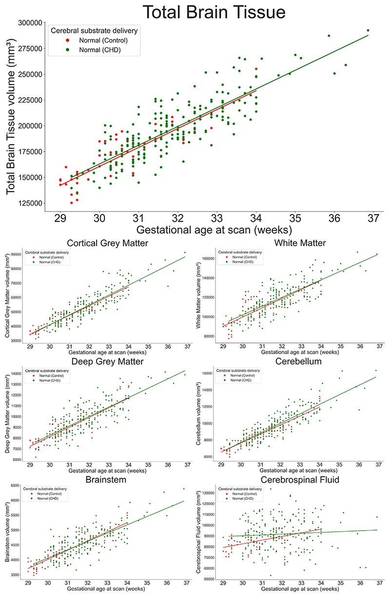
Assessment: Images were motion-corrected and reconstructed using an automated slice-to-volume registration reconstruction technique, before undergoing segmentation using an automated pipeline and convolutional neural network that had undergone semi-supervised training. Differences in total, regional brain (cortical gray matter, white matter, deep gray matter, cerebellum, and brainstem) and brain:body volumes were compared between groups.
Statistical tests: ANOVA was used to test for differences in brain volumes between groups, after accounting for sex and gestational age at scan. PFDR-values <0.05 were considered statistically significant.
Results: Total and regional brain volumes were smaller in fetuses where cerebral substrate delivery is reduced. No significant differences were observed in total or regional brain volumes between control fetuses and fetuses with CHD but normal cerebral substrate delivery (all PFDR > 0.12). Severely reduced cerebral substrate delivery is associated with lower brain:body volume ratios.
Data conclusion: Total and regional brain volumes are smaller in fetuses with CHD where there is a reduction in cerebral substrate delivery, but not in those where cerebral substrate delivery is expected to be normal.
Evidence level: 3 TECHNICAL EFFICACY: Stage 3.
Keywords: MRI; congenital heart disease; fetal brain development.
© 2023 The Authors. Journal of Magnetic Resonance Imaging published by Wiley Periodicals LLC on behalf of International Society for Magnetic Resonance in Medicine.
Figures

References
-
- EUROCAT. European platform on rare disease registration. 2020
-
- Latal B. Neurodevelopmental outcomes of the child with congenital heart disease. Clin Perinatol. 2016;43:173–185. - PubMed
-
- von Rhein M, Buchmann A, Hagmann C, et al. Brain volumes predict neurodevelopment in adolescents after surgery for congenital heart disease. Brain J Neurol. 2014;137(Pt 1):268–276. - PubMed
-
- von Rhein M, Buchmann A, Hagmann C, et al. Severe congenital heart defects are associated with global reduction of neonatal brain volumes. J Pediatr. 2015;167:1259–1263.:e1. - PubMed
Publication types
MeSH terms
Grants and funding
- 102431/WT_/Wellcome Trust/United Kingdom
- 203148/WT_/Wellcome Trust/United Kingdom
- National Institute for Health Research (NIHR) Biomedical Research Centre based at Guy's and St Thomas' NHS Foundation Trust and King's College London
- MR/L011530/1/MRC_/Medical Research Council/United Kingdom
- FS/ICRF/22/26028/BHF_/British Heart Foundation/United Kingdom
LinkOut - more resources
Full Text Sources
Medical
