Review
. 2023 Nov 1;145(43):23397-23415.
doi: 10.1021/jacs.3c01135. Epub 2023 Oct 17.

Ruthenium-Based Photoactivated Chemotherapy

Affiliations
Review

Ruthenium-Based Photoactivated Chemotherapy

Sylvestre Bonnet. J Am Chem Soc. .

Abstract

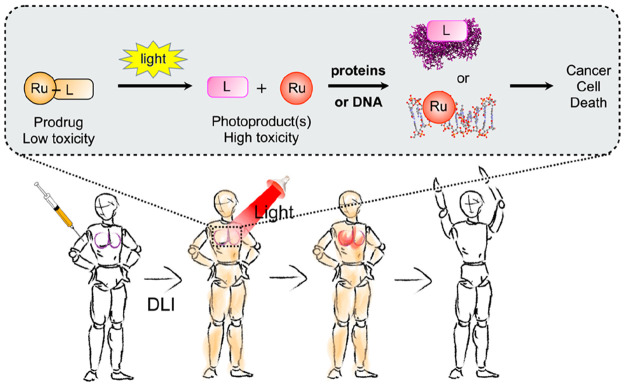
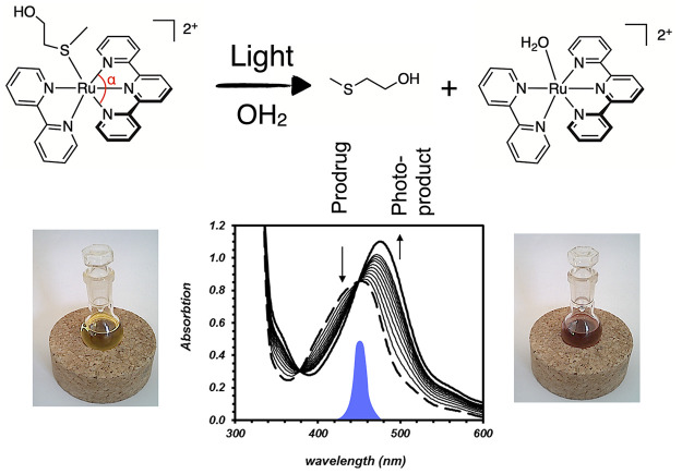
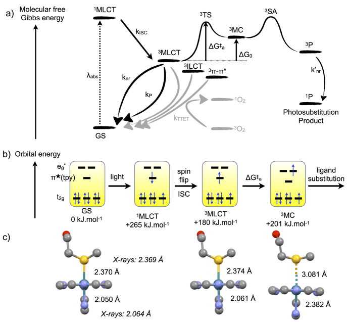
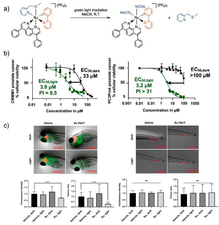
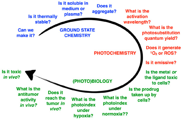
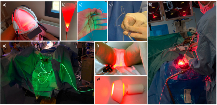
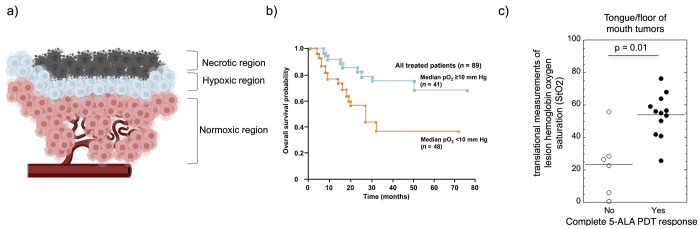
Ruthenium(II) polypyridyl complexes form a vast family of molecules characterized by their finely tuned photochemical and photophysical properties. Their ability to undergo excited-state deactivation via photosubstitution reactions makes them quite unique in inorganic photochemistry. As a consequence, they have been used, in general, for building dynamic molecular systems responsive to light but, more particularly, in the field of oncology, as prodrugs for a new cancer treatment modality called photoactivated chemotherapy (PACT). Indeed, the ability of a coordination bond to be selectively broken under visible light irradiation offers fascinating perspectives in oncology: it is possible to make poorly toxic agents in the dark that become activated toward cancer cell killing by simple visible light irradiation of the compound inside a tumor. In this Perspective, we review the most important concepts behind the PACT idea, the relationship between ruthenium compounds used for PACT and those used for a related phototherapeutic approach called photodynamic therapy (PDT), and we discuss important questions about real-life applications of PACT in the clinic. We conclude this Perspective with important challenges in the field and an outlook.


Conflict of interest statement

The author declares no competing financial interest.

Figures

Figure 1
Figure 1
Principle of ruthenium-based photoactivated chemotherapy (PACT). Top: light-induced bond cleavage reaction in the prodrug. Either the photoreleased ligand (L) or the metal fragment (Ru), or both, interact(s) with biomolecules, leading to cell death. Bottom: PACT treatment of a patient with a lung tumor (in purple). The prodrug (orange) is injected intravenously, distributes in the body, and reaches the tumor in its non-toxic form. After the drug-to-light interval (DLI), light is shone onto the tumor, activating the prodrug and destroying the tumor. Finally, the body excretes the excess drug. Image courtesy Bianka Siewert.
Figure 2
Figure 2
Example of a photosubstitution reaction used in PACT with ruthenium photocage [Ru(tpy)(bpy)(Hmte)]2+. The blue peak in the UV–vis spectrum shows the emission of the light source used to trigger photosubstitution, centered at 450 nm. The bottom graph shows the time evolution of the absorption spectrum of the solution during light irradiation. The low α angle (∼160°) in the terpyridine ligand distorts the first coordination sphere of the metal center compared to a perfect octahedron (180°), which facilitates photosubstitution. Data adapted from ref (17).
Figure 3
Figure 3
Classical model for photosubstitution reactions in ruthenium(II) polypyridyl complexes. (a) Molecular energy of the different states involved in the photochemistry of ruthenium(II) polypyridyl complexes. Gray pathways generate 1O2 in the presence of dioxygen; black pathways remain in the absence of O2. kISC, knr, knr, kP, and kTTET are rate constants for intersystem crossing, non-radiative decay, phosphorescence, and triplet–triplet energy transfer, respectively. ΔGa is the activation barrier for the conversion of the 3MLCT to the 3MC state, and ΔG0 = G(3MC) – G(3MCLT). 3SA represents a Solvent Adduct of the complex in the triplet state. (b) Orbital energy scheme of the excited states involved in photosubstitution. Numerical values for bond lengths are indicated for [Ru(tpy)(bpy)(Hmte)]2+ (Figure 2), as reported in ref (50).
Figure 4
Figure 4
Relationship between the photosubstitution quantum yield in water (φPS) and the activation energy (Ea) to promote the 3MCLT state to the 3MC state, in [Ru(tpy)(L)(MeCN)]n+ (n = 1 or 2), where L is a bidentate ligand. Each dot represents a metal complex. Adapted from ref (54). Copyright 2022 American Chemical Society.
Figure 5
Figure 5
Selection of ruthenium-based PACT compounds. The first photosubstituted ligand is highlighted in blue.
Figure 6
Figure 6
For ruthenium-based PACT compounds, the activation wavelength λexc does not have to coincide with the absorption maximum λmax. Top: 132+ (see Figure 5) has a maximum in the blue (λmax = 452 nm) but was activated with green light (λexc = 520 nm, εexc = 1510 M–1·cm–1) in vitro and in vivo. Red light (630 nm) hardly activated the compound. Image developed using data from ref (76). Copyright 2019 American Chemical Society. Bottom: 122+ (see Figure 5) has λmax shifted to the green (531 nm), which allowed red light activation (λexc = 625 nm, εexc = 379 M–1·cm–1). Image developed using data from ref (78). Copyright 2017 Wiley-VCH.
Figure 7
Figure 7
Examples of cyclometalated complexes investigated in Ru-based PACT. The ligand that is photosubstituted first is colored in blue and the carbon atom bound to ruthenium in red.
Figure 8
Figure 8
(a) Formula of the CYP3A4-inhibiting ritonavir analogue 24 photocaged by Turro et al. with the [Ru(tpy)(dmbpy)]2+ moiety. (b) X-ray structure of the uncaged CYP3A4 inhibitor shown in (a) bound via pyridine coordination to the heme iron center (PDB: 4D78). (c) Protein activity dose–response curves for the uncaged inhibitor 24, the ruthenium-caged inhibitor 112+ (Figure 5) in the dark and after light activation, and the control ruthenium caging group [Ru(tpy)(dmbpy)Cl]Cl. (d) X-ray structure of the caged CYP3A4 inhibitor 112+ interacting with CYP3A4 without pyridine coordination to the heme iron center (PDB: 7KS8). Color code: pyridine nitrogen atoms are in green, heme is in red, the inhibitor 24 is in blue, and the ruthenium caging group of 112+ is in orange. Adapted from ref (80). Copyright 2021 American Chemical Society.
Figure 9
Figure 9
Non-trivial relationship between in vitro and in vivo performances of the PACT compound [5](PF6)2. (a) Photosubstitution reaction for 52+ irradiated with green light in acetonitrile. (b) In vitro dose–response curves for 52+ in CRMM1 eye cancer cell (PI ≈ 8.5) and PC3Pro4 prostate cancer cells (PI > 31). (c) In vivo performance of 52+ under green light activation (520 nm, 114 J/cm2) in an orthotopic CRMM1 eye tumor model in zebrafish embryo (left) and in a PC3Pro4 ectopic prostate zebrafish tumor model (right). Green fluorescence shows blood vessels, and red fluorescence shows the tumor cells. ****p < 0.0001. Reproduced with permission from ref (64). Copyright 2022 Royal Society of Chemistry.
Figure 10
Figure 10
General design aspects for the ruthenium-based PACT compound.
Figure 11
Figure 11
Lasers and devices used to shine light in patients for anticancer phototherapy. (a) Fabric-based biophotonic device used for Phosistos photodynamic therapy of actinic keratosis. (b) Frontal light delivery using an optical fiber, for example, for irradiation of skin tumors. (c) Radial light distribution for interstitial photodynamic therapy using an optical fiber terminated by a light diffuser. (d) The TLC-3200 system for simultaneous green light irradiation of bladder tumors (532 nm) and light dosimetry during PDT treatment with TLD-1433. (e) Clinical setup of PDT bladder cancer treatment using TLD-1433 and green light. Image courtesy Lothar Lilge. (f) PAGETEX device for controlled vulvar illumination with 635 nm light in the PDT treatment of primary extramammary Paget’s disease of the vulva. Reprinted with permission under a Creative Commons CC BY 4.0 from ref (109). Copyright 2020 The Authors, published by Wiley-VCH. (g) Cevira device for cervix illumination with light. Image courtesy Serge Mordon. (h) Homogeneous light diffusion with a light-scattering balloon inflated in the excised primary tumor cavity for intraoperative PDT treatment of glioblastoma (INDYGO trial). Reprinted with permission under a Creative Commons CC BY 4.0 from ref (115). Copyright 2020 The Authors, published by Springer.
Figure 12
Figure 12
Hypoxia in oncology. (a) Three regions around a blood vessel in a tumor. Image courtesy Iris Kort. (b) Association between tumor hypoxia and overall survival in advanced cancer of the uterine cervix. Reprinted with permission from Vaupel et al., Association between Tumor Hypoxia and Malignant Progression in Advanced Cancer of the Uterine Cervix. Cancer Res., 1996, 56, 4509–4515. Copyright 1996 American Association for Cancer Research. (c) Clinical response to 5-ALA PDT treatment in tongue/floor of mouth tumor patients. Reproduced with permission from Busch et al., Lesion oxygenation associates with clinical outcomes in premalignant and early stage head and neck tumors treated on a phase 1 trial of photodynamic therapy. Photodiagnosis Photodynamic Therapy2018, 21, 28–35. Copyright 2018 Elsevier.
Figure 13
Figure 13
Heterogeneity of hypoxia in hind limb SQ20b human squamous cell carcinoma subcutaneous xenograft in mice shown by three-color hypoxia imaging. Blue is Hoechst 33342 (nuclei), green is pimonidazole (hypoxia marker 1), and red is carbonic anhydrase (hypoxia marker 2). Left bar is 2 mm, right bar is 500 μm. The right image shows high magnification of the region of interest shown on the left by a white rectangle. The circular hole was caused by angiocatheter placement before tumor sectioning. This research was originally published in ref (135). Copyright 2014 Society of Nuclear Medicine and Molecular Imaging, Inc.

References

    1. Finsen N. R. Remarks on the Red-Light Treatment of Small-Pox: Is the Treatment of Small-Pox Patients in Broad Daylight Warrantable?. BMJ 1903, 1 (2214), 1297–1298. 10.1136/bmj.1.2214.1297. - DOI - PMC - PubMed
    1. Ip S.; Chung M.; Kulig J.; O’Brien R.; Sege R.; Glicken S.; Maisels M. J.; Lau J. Subcommittee on Hyperbilirubinemia. An Evidence-Based Review of Important Issues Concerning Neonatal Hyperbilirubinemia. Pediatrics 2004, 114 (1), e130–e153. 10.1542/peds.114.1.e130. - DOI - PubMed
    1. Abdel-kader M. H.The Journey of PDT Throughout History: PDT from Pharos to Present. In Comprehensive Series in Photochemical & Photobiological Sciences; Kostron H., Hasan T., Eds.; Royal Society of Chemistry: Cambridge, 2016; Chap. 1, pp 1–21. 10.1039/9781782626824-00001. - DOI
    1. Hammerle F.; Bingger I.; Pannwitz A.; Magnutzki A.; Gstir R.; Rutz A.; Wolfender J.-L.; Peintner U.; Siewert B. Targeted Isolation of Photoactive Pigments from Mushrooms Yielded a Highly Potent New Photosensitizer: 7,7′-Biphyscion. Sci. Rep. 2022, 12 (1), 1108.10.1038/s41598-022-04975-9. - DOI - PMC - PubMed
    1. Youf R.; Müller M.; Balasini A.; Thétiot F.; Müller M.; Hascoët A.; Jonas U.; Schönherr H.; Lemercier G.; Montier T.; Le Gall T. Antimicrobial Photodynamic Therapy: Latest Developments with a Focus on Combinatory Strategies. Pharmaceutics 2021, 13 (12), 1995.10.3390/pharmaceutics13121995. - DOI - PMC - PubMed

Publication types

MeSH terms