. 2023 Dec;12(32):e2301571.
doi: 10.1002/adhm.202301571. Epub 2023 Nov 1.

Selective Immunosuppression Targeting the NLRP3 Inflammasome Mitigates the Foreign Body Response to Implanted Biomaterials While Preserving Angiogenesis

Affiliations

Selective Immunosuppression Targeting the NLRP3 Inflammasome Mitigates the Foreign Body Response to Implanted Biomaterials While Preserving Angiogenesis

Alex H P Chan et al. Adv Healthc Mater. 2023 Dec.

Abstract

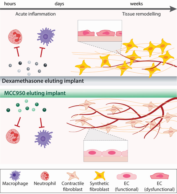
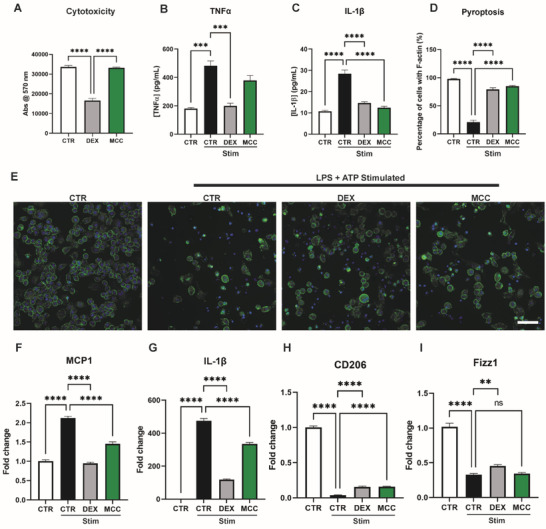
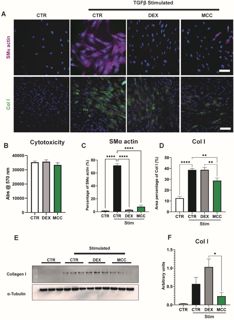
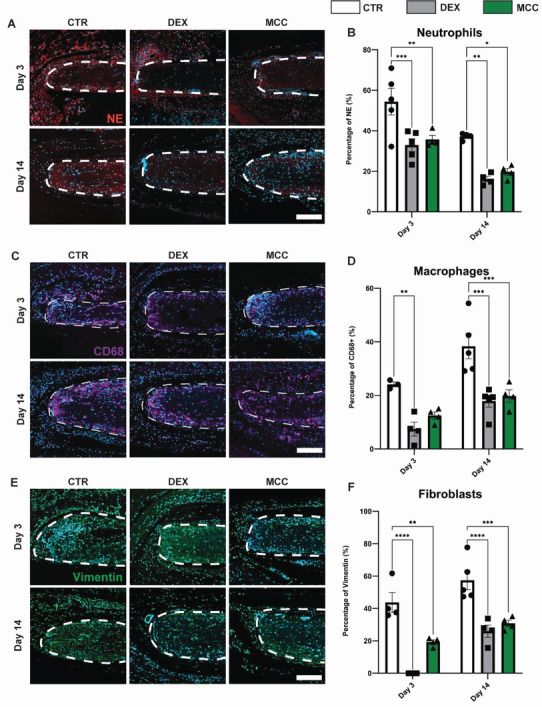
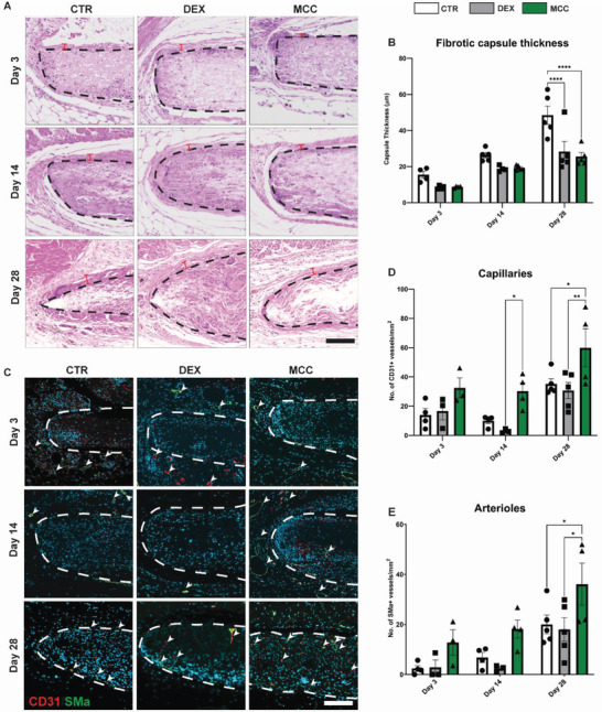
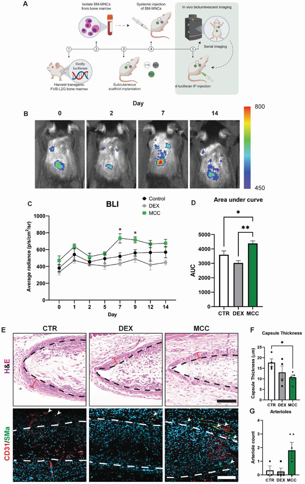
Medical devices are a mainstay of the healthcare industry, providing clinicians with innovative tools to diagnose, monitor, and treat a range of medical conditions. For implantable devices, it is widely regarded that chronic inflammation during the foreign body response (FBR) is detrimental to device performance, but also required for tissue regeneration and host integration. Current strategies to mitigate the FBR rely on broad acting anti-inflammatory drugs, most commonly, dexamethasone (DEX), which can inhibit angiogenesis and compromise long-term device function. This study challenges prevailing assumptions by suggesting that FBR inflammation is multifaceted, and selectively targeting its individual pathways can stop implant fibrosis while preserving beneficial repair pathways linked to improved device performance. MCC950, an anti-inflammatory drug that selectively inhibits the NLRP3 inflammasome, targets pathological inflammation without compromising global immune function. The effects of MCC950 and DEX on the FBR are compared using implanted polycaprolactone (PCL) scaffolds. The results demonstrate that both DEX and MCC950 halt immune cell recruitment and cytokine release, leading to reduced FBR. However, MCC950 achieves this while supporting capillary growth and enhancing tissue angiogenesis. These findings support selective immunosuppression approaches as a potential future direction for treating the FBR and enhancing the longevity and safety of implantable devices.

Keywords: NLRP3 inflammasome; biomaterials; dexamethasone; foreign body responses; implantable devices.


Conflict of interest statement

The authors declare no conflict of interest.

Figures

Figure 1
Figure 1
Schematic comparison of dexamethasone (DEX) and MCC950 as pharmacological approaches to mitigating the foreign body response (FBR). DEX and MCC950 exhibit similar immunosuppressive effects in the acute stages of the FBR, reducing the recruitment of neutrophils and macrophages. End‐stage remodeling outcomes show both drugs can effectively reduce fibrotic capsule formation, however MCC950 shows evidence of increased suppression of collagen‐producing (synthetic) fibroblast differentiation. Importantly these effects of MCC950 occur without compromising endothelial integrity/function and preserves angiogenesis. Abbreviations: EC, endothelial cell.
Figure 2
Figure 2
Immunosuppresive effects of DEX and MCC950 on cultured J774a.1 murine macrophages. A) alamarBlue cytotoxicity assay following 3 day culture with DEX and MCC950. B,C) Levels of TNF‐α and IL‐1β cytokines following NLRP3 inflammasome stimulation (STIM; LPS+ATP) measured by ELISA. D,E) Quantification and representative images of pyroptosis. Cells stained with DAPI (blue) and rhodamine phalloidin (green) to visualise cell nuclei and F‐actin, respectively. Data represented as percentage of cells with concentric F‐actin membrane. F–I) qPCR of M1 (MCP1, IL‐1β) and M2 (CD206, Fizz1) gene sets 24 h post‐stimulation (STIM; LPS+ATP). Data represented as mean ± SEM (n = 3–4). Statistical significance was determined using Dunnett's multiple comparison one‐way ANOVA test relative to stimulated group (**p < 0.01, ***p < 0.001, ****p < 0.0001). Scale bar represents 50 µm.
Figure 3
Figure 3
MCC950 possessed anti‐fibrotic effects on human dermal fibroblasts. A) Representative images of human dermal fibroblasts stimulated with TGF‐β to induce fibroblast‐to‐myofibroblast differentiation, indicated by increased SMα actin (purple) and Collagen I (Col I, green) intracellular staining. Cells counterstained using DAPI (blue). B) alamarBlue cytotoxicity assay following 3 day culture with DEX and MCC950. C,D) Quantification of intracellular staining of SMα actin and Col I in fibroblasts after 3 days of TGF‐β stimulation in the presence of DEX and MCC950. E,F) Western Blots and quantification of Col I. Data represented as mean ± SEM (n = 3–4). Statistical significance was determined using Dunnett's multiple comparison one‐way ANOVA test relative to stimulated group (*p < 0.05, **p < 0.01, ****p < 0.0001). Scale bar represents 100 µm.
Figure 4
Figure 4
MCC950 promoted integrity and function of human endothelial cells. A) Representative images of endothelial cells stained for vascular endothelial‐cadherin (VE‐cadherin, orange) and endothelial nitric oxide synthase (eNOS, green) after 3 days in culture with DEX and MCC950. Cells counterstained using DAPI (blue). B) alamarBlue cytotoxicity assay following 3 day culture with DEX and MCC950. C,D) Quantification of VE‐cadherin and eNOS staining following 3 day treatment with DEX and MCC950. E–H) qPCR of pro‐angiogenic gene set (CDH5, PECAM, eNOS, and KDR) 24 and 48 h post‐stimulation with DEX and MCC950. Data represented as mean ± SEM (n = 3–4). Statistical significance was determined using Dunnett's multiple comparison one‐way ANOVA test relative to stimulated group (**p < 0.01, ***p < 0.001, ****p < 0.0001). Scale bar represents 50 µm.
Figure 5
Figure 5
MCC950 and DEX possessed similar levels of immunosuppressive and anti‐fibrotic effects on subcutaneously implanted PCL scaffolds at 3 and 14 days post‐implantation. A) Representative images of neutrophils stained with neutrophil elastase (red), counterstained for nuclei with DAPI (blue). B) Quantification of area stained with neutrophil elastase. C) Representative images of macrophages stained with CD68 (purple), counterstained for nuclei with DAPI (blue). D) Quantification of area stained with CD68. E) Representative images of fibroblasts stained with vimentin (green), counterstained for nuclei with DAPI (blue). F) Quantification of area stained with vimentin. Data represented as mean ± SEM (n = 4–5). Statistical significance was determined using Tukey's multiple comparison two‐way ANOVA test relative to control group (*p < 0.05, **p < 0.01, ***p < 0.001, ****p < 0.0001). Scale bar represents 100 µm.
Figure 6
Figure 6
MCC950 and DEX equally suppressed fibrotic capsule formation, however only MCC950 enhances angiogenesis around subcutaneously implanted PCL scaffolds at 3, 14, and 28 days post‐implantation. A) Representative images of implant cross‐sections (black dotted line) stained H&E to visualize fibrotic capsule (red bars). B) Quantification of fibrotic capsule thickness. C) Representative images of scaffold cross‐sections co‐stained with CD31 (red) and SMα actin (green). Cell nuclei counterstained with DAPI (blue). D) Quantification of capillaries mm−2 indicated by CD31 staining alone. E) Quantification of arterioles mm−2 indicated by co‐staining of CD31 and SMα actin. Data represented as mean ± SEM (n = 4–5). Statistical significance was determined using Tukey's multiple comparison two‐way ANOVA test relative to control group (*p < 0.05, **p < 0.01, ***p < 0.001). Scale bar represents 100 µm.
Figure 7
Figure 7
MCC950 enhanced the engraftment of injected immune cell fractions yet had the thinnest fibrotic capsules and highest levels of native arteriole formation around PCL scaffolds subcutaneously implanted for 14 days. A) Schematic representation of systemic (tail‐vein) injected bioluminescent (BLI) bone marrow mononuclear cells (BM‐MNCs). B) Representative BLI images of mouse backs with subcutaneously implanted scaffolds at days 0, 2, 7, and 14. Heat bar represents BLI signal in units of radiance (p s−1 cm−2 sr−1). C) Temporal measurements of BLI signal over 14 days across control, DEX and MCC950 implants. Statistical significance was determined using two‐way ANOVA test relative to control group (*p < 0.05). D) Representation of temporal measurements as area under the curve (AUC) over 14 days. E) Representative images of H&E, and CD31 (red)/SMα (green) stained scaffold cross sections at 14 days post‐implantation and BM‐MNC injection. Dotted lines represent scaffold borders. Red bars indicated fibrotic capsule thickness. Quantification of F) fibrotic capsule thickness and G) native arteriole count mm−2. Data represented as mean ± SEM (n = 4–5). Statistical significance was determined using Dunnett's multiple comparison one‐way ANOVA test relative to control group (*p < 0.05, **p < 0.01). Scale bar represents 100 µm.

Similar articles

References

    1. Zhang D. H., Chen Q., Shi C., Chen M. Z., Ma K. Q., Wan J. L., Liu R. H., Adv. Funct. Mater. 2021, 31, 2007226.
    1. a) Shah J., Smith R. H., Al‐Gharaibeh A., Front. Surg. 2020, 6, 201900073;
    2. b) Fernández E., Greger B., House P. A., Aranda I., Botella C., Albisua J., Soto‐Sánchez C., Alfaro A., R. A., Normann, Front. Neuroeng. 2014, 7, 201400024. - PMC - PubMed
    1. Chandorkar Y., Ravikumar K., Basu B., ACS Biomater. Sci. Eng. 2019, 5, 19. - PubMed
    1. Veiseh O., Doloff J. C., Ma M., Vegas A. J., Tam H. H., Bader A. R., Li J., Langan E., Wyckoff J., Loo W. S., Jhunjhunwala S., Chiu A., Siebert S., Tang K., Hollister‐Lock J., Aresta‐Dasilva S., Bochenek M., Mendoza‐Elias J., Wang Y., Qi M., Lavin D. M., Chen M., Dholakia N., Thakrar R., Lacík I., Weir G. C., Oberholzer J., Greiner D. L., Langer R., Anderson D. G., Nat. Mater. 2015, 14, 643. - PMC - PubMed
    1. Wang Y., Vaddiraju S., Gu B., Papadimitrakopoulos F., Burgess D. J., J. Diabetes Sci. Technol. 2015, 9, 966. - PMC - PubMed

Publication types