. 2024 Feb 1;22(2):209-220.
doi: 10.1158/1541-7786.MCR-23-0290.

SOX10 Loss Sensitizes Melanoma Cells to Cytokine-Mediated Inflammatory Cell Death

Affiliations

SOX10 Loss Sensitizes Melanoma Cells to Cytokine-Mediated Inflammatory Cell Death

Sheera R Rosenbaum et al. Mol Cancer Res. .

Abstract

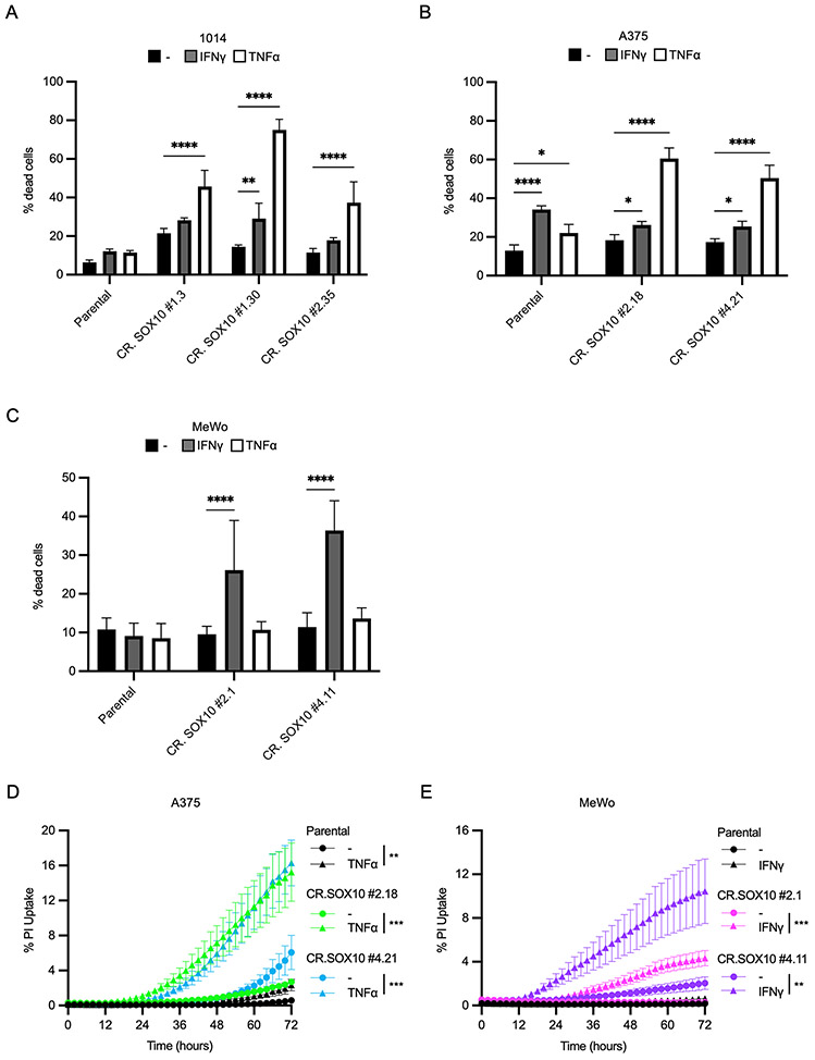
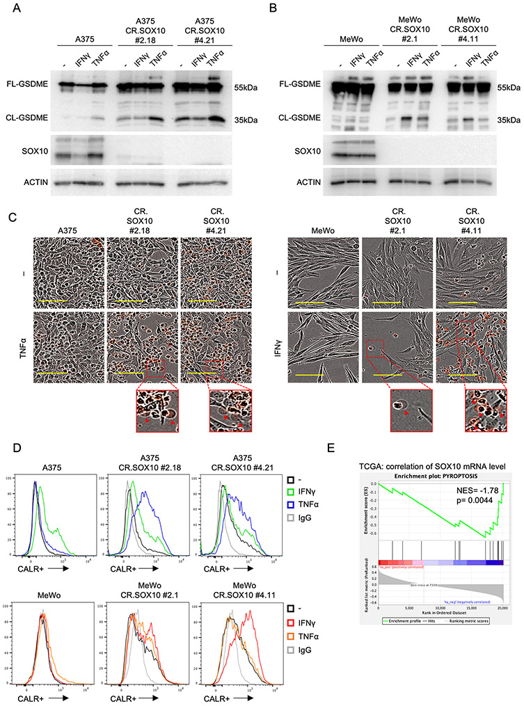
The transcription factor, SOX10, plays an important role in the differentiation of neural crest precursors to the melanocytic lineage. Malignant transformation of melanocytes leads to the development of melanoma, and SOX10 promotes melanoma cell proliferation and tumor formation. SOX10 expression in melanomas is heterogeneous, and loss of SOX10 causes a phenotypic switch toward an invasive, mesenchymal-like cell state and therapy resistance; hence, strategies to target SOX10-deficient cells are an active area of investigation. The impact of cell state and SOX10 expression on antitumor immunity is not well understood but will likely have important implications for immunotherapeutic interventions. To this end, we tested whether SOX10 status affects the response to CD8+ T cell-mediated killing and T cell-secreted cytokines, TNFα and IFNγ, which are critical effectors in the cytotoxic killing of cancer cells. We observed that genetic ablation of SOX10 rendered melanoma cells more sensitive to CD8+ T cell-mediated killing and cell death induction by either TNFα or IFNγ. Cytokine-mediated cell death in SOX10-deficient cells was associated with features of caspase-dependent pyroptosis, an inflammatory form of cell death that has the potential to increase immune responses.

Implications: These data support a role for SOX10 expression altering the response to T cell-mediated cell death and contribute to a broader understanding of the interaction between immune cells and melanoma cells.


Conflict of interest statement

Conflict of Interest: A.E. Aplin has ownership interest in patent number 9880150 and has a pending patent, PCT/US22/76492. No potential conflicts of interest were declared by the other authors.

Figures

Figure 1:
Figure 1:. Cytotoxic T cell-secreted cytokines reduce the growth of SOX10/Sox10-knockout cells
A) CRISPR/Cas9 knockout of Sox10/SOX10 in 1014, A375, and MeWo cells was verified by Western blot. B) 1014 parental or Sox10-knockout cells were peptide-pulsed for 3 hours and incubated with Pmel-1 T cells for 48 hours. Cancer cells were washed and stained by crystal violet. Wells were imaged, well coverage was quantified on ImageJ, and % surviving cells was graphed. C) Images representative of three independent experiments from (B). Scale bars represent 250 μm D) 1014 parental or Sox10-knockout cells were treated with 100 ng/mL IFNγ or 50 ng/mL TNFα for 72 hours. Cancer cells were washed and stained with crystal violet. Wells were imaged, and percent well coverage was quantified on ImageJ. E) As in D, for A375 parental or SOX10-knockout cells. F) As in D, for MeWo parental or SOX10-knockout cells. G) Images representative of three independent experiments from (D). Scale bars represent 500 μm. H) As in G, for data shown in (E). I) As in G, for data shown in (F). All data are representative of three independent experiments. Bar graphs show mean + SD. *p<0.05, **p<0.01, ***p<0.001, ****p<0.0001.
Figure 2:
Figure 2:. SOX10/Sox10-knockout sensitizes melanoma cells to cytokine-mediated cell death
A) 1014 parental or Sox10-knockout cells were treated with 100 ng/mL IFNγ or 50 ng/mL TNFα for 72 hours. Cell death was evaluated by Annexin V, propidium iodide (PI), and dual Annexin V/PI staining by flow cytometry. B) As in A, for A375 parental or SOX10-knockout cells. C) As in A, for MeWo parental or SOX10-knockout cells. D) A375 parental or SOX10-knockout cells were treated with 50 ng/mL TNFα for 72 hours. PI was added to a final concentration of 0.5 μM at the time of treatment, and phase contrast and RFP images were taken every 2 hours at 10x using live-cell imaging. Percent PI uptake at each time point was calculated using the formula (percent red confluence/percent phase confluence) x 100. E) As in D, for MeWo parental or SOX10-knockout cells treated with 100 ng/mL IFNγ. All data are representative of three independent experiments, except in (C) and (E) where six and four independent experiments were conducted, respectively. Bar graphs show mean + SD. Line graphs show mean ± SEM. *p<0.05, **p<0.01, ***p<0.001, ****p<0.0001.
Figure 3:
Figure 3:. Analysis of canonical cytokine signaling pathways in SOX10-deficient cells
A) A375 parental or SOX10-knockout cells were treated with 100 ng/mL IFNγ or 50 ng/mL TNFα for 72 hours. Cells were lysed and lysates were probed for canonical signaling downstream of TNFα receptor activation by Western blot. B) As in A, for MeWo parental or SOX10-knockout cells. C) A375 parental or SOX10-knockout cells were treated with 100 ng/mL IFNγ or 50 ng/mL TNFα for 72 hours. Cells were lysed and lysates were probed for canonical signaling downstream of IFNγ receptor activation by Western blot. D) As in C, for MeWo parental or SOX10-knockout cells. All data are representative of three independent experiments.
Figure 4:
Figure 4:. Cytokine-induced cell death in SOX10-knockout cells is caspase-dependent
A) A375 parental or SOX10-knockout cells were treated with 50 ng/mL TNFα for 72 hours and lysates were collected for RPPA analysis. A heat map displays median-centered, log2-transformed group average expression data for cell death pathway proteins with a p-value < 0.05 for any comparison. B) As in A, except MeWo parental or SOX10-knockout cells were treated with 100 ng/mL IFNγ for 72 hours. C) A375 parental or SOX10-knockout cells were treated with 100 ng/mL IFNγ or 50 ng/mL TNFα for 72 hours. Cells were lysed and lysates were probed for cleavage of caspases by Western blot. D) As in C, for MeWo parental or SOX10-knockout cells. E) A375 parental or SOX10-knockout cells were pre-treated with 50 μM of the pan-caspase inhibitor Z-VAD-FMK for 30 minutes prior to treatment with 50 ng/mL TNFα for 72 hours. Cell death was then evaluated by Annexin V, propidium iodide (PI), and dual Annexin V/PI staining by flow cytometry. F) As in E, except MeWo parental or SOX10-knockout cells were treated with 100 ng/mL IFNγ after 50 μM Z-VAD-FMK pre-treatment. All data are representative of three independent experiments. Bar graphs show mean + SD. *p<0.05, **p<0.01, ***p<0.001, ****p<0.0001.
Figure 5:
Figure 5:. Cytokine-induced cell death in SOX10-knockout cells is associated with markers of pyroptosis
A) A375 parental or SOX10-knockout cells were treated with 100 ng/mL IFNγ or 50 ng/mL TNFα for 72 hours. Cells were lysed and lysates were probed for cleavage of GSDME by Western blot. Data are representative of three independent experiments. B) As in A, for MeWo parental or SOX10-knockout cells. C) A375 or MeWo, parental or SOX10-knockout cells were treated with 50 ng/mL TNFα or 100 ng/mL IFNγ, respectively. 0.5 μM PI was added at the time of treatment, and phase contrast and RFP images were taken after 72 hours at 10x using live-cell imaging. Examples of plasma membrane swelling are indicated with red arrows in inset images. Scale bars represent 100 μm. Images are representative of three independent experiments. D) A375 or MeWo, parental or SOX10-knockout cells, were treated with 100 ng/mL IFNγ or 50 ng/mL TNFα for 72 hours, and calreticulin expression was evaluated by flow cytometry staining. Data are representative of three independent experiments. E) A TCGA melanoma dataset was evaluated for the correlation between SOX10 mRNA levels and a pyroptosis gene signature.

References

    1. Simões-Costa M, and Bronner ME. Establishing neural crest identity: a gene regulatory recipe. Development 2015;142:242–257 - PMC - PubMed
    1. Shakhova O, Zingg D, Schaefer SM, Hari L, Civenni G, Blunschi J, et al. Sox10 promotes the formation and maintenance of giant congenital naevi and melanoma. Nat Cell Biol 2012;14:882–890 - PubMed
    1. Cronin JC, Watkins-Chow DE, Incao A, Hasskamp JH, Schönewolf N, Aoude LG, et al. SOX10 ablation arrests cell cycle, induces senescence, and suppresses melanomagenesis. Cancer Res 2013;73:5709–5718 - PMC - PubMed
    1. Dunn GP, Bruce AT, Ikeda H, Old LJ, and Schreiber RD. Cancer immunoediting: from immunosurveillance to tumor escape. Nat Immunol 2002;3:991–998 - PubMed
    1. Wouters J, Kalender-Atak Z, Minnoye L, Spanier KI, De Waegeneer M, Bravo González-Blas C, et al. Robust gene expression programs underlie recurrent cell states and phenotype switching in melanoma. Nat Cell Biol 2020;22:986–998 - PubMed

Publication types