

Challenging Tumor Heterogeneity with HER2, p16 and Somatostatin Receptor 2 Expression in a Case of EBV-Associated Lymphoepithelial Carcinoma of the Salivary Gland
- PMID: 37847488
- PMCID: PMC10739679
- DOI: 10.1007/s12105-023-01592-4
Challenging Tumor Heterogeneity with HER2, p16 and Somatostatin Receptor 2 Expression in a Case of EBV-Associated Lymphoepithelial Carcinoma of the Salivary Gland
Abstract
Background: Lymphoepithelial carcinoma of the salivary glands (LECSG) is a rare disease in the Western hemisphere that is typically associated with an EBV infection. The molecular mechanisms of LECSG tumorigenesis are poorly understood.
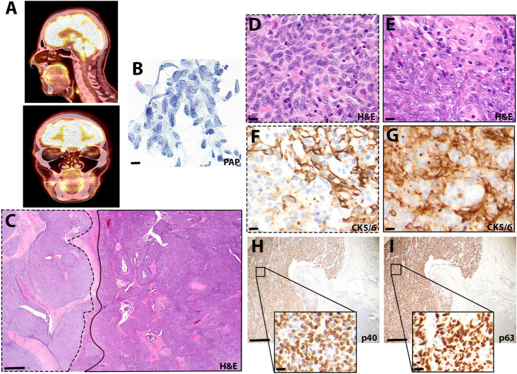
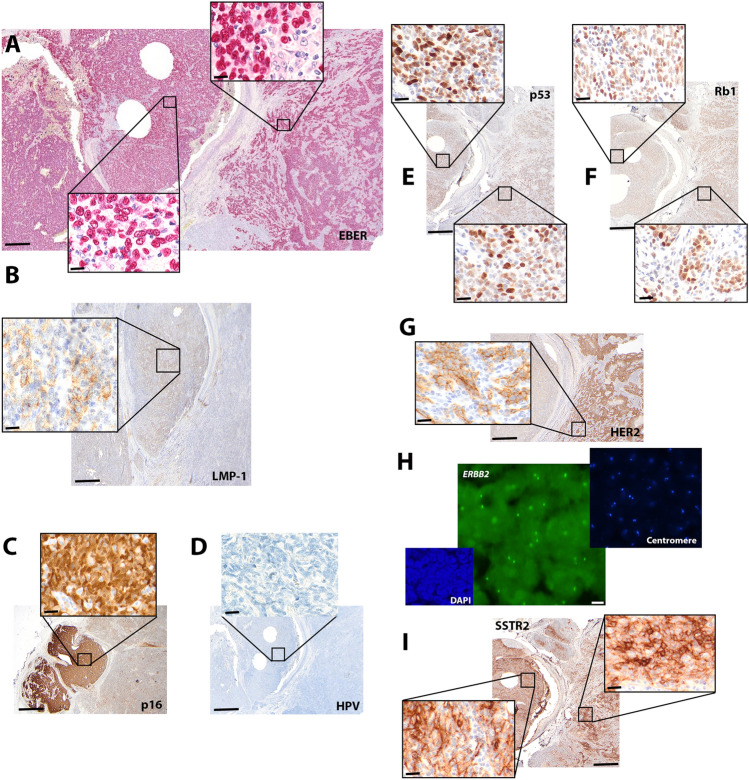
Results: Here we report a case of EBV-associated LECSG with an unusual immunophenotype. The tumor exhibited bi-morphic histological features with a mutually exclusive expression of HER2 and p16. The p16-positive domain of the tumor immunohistochemically co-expressed late membrane protein 1 (LMP-1), while the HER2 positive domain did not. Both tumor regions expressed SSTR2.
Methods: In situ hybridization confirmed the EBV origin of the tumor while extensive immunohistochemical characterization and the recently established RNA-based next generation sequencing panel ("SalvGlandDx" panel) did not reveal evidence for another salivary gland neoplasm. No HPV co-infection was detected by in situ hybridization or PCR-based screenings and no ERBB2 gene amplification was detected by fluorescence in situ hybridization.
Conclusion: These findings suggest tumor heterogeneity and lack of genomic aberrations in EBV-associated LECSGs. The heterogenous and unusual immunohistochemical features explain the diagnostic difficulties and simultaneously extend the immunophenotype spectrum of this tumor entity.
Keywords: EBV; HER2; Lymphoepithelial carcinoma; SSTR2; Salivary gland; p16.
© 2023. The Author(s).
Conflict of interest statement
SNF is an employee of Oncobit AG. All the authors declare no conflict of interest.
Figures
Similar articles
-
Lymphoepithelial carcinoma of the salivary gland: in situ detection of Epstein-Barr virus.J Clin Pathol. 1995 Nov;48(11):1022-7. doi: 10.1136/jcp.48.11.1022. J Clin Pathol. 1995. PMID: 8543624 Free PMC article.
-
HPV Infection, but Not EBV or HHV-8 Infection, Is Associated with Salivary Gland Tumours.Biomed Res Int. 2015;2015:829349. doi: 10.1155/2015/829349. Epub 2015 Nov 5. Biomed Res Int. 2015. PMID: 26618178 Free PMC article.
-
Lymphoepithelial Carcinoma of Salivary Gland EBV-association in Endemic versus Non-Endemic Patients: A Report of 16 Cases.Head Neck Pathol. 2020 Dec;14(4):1001-1012. doi: 10.1007/s12105-020-01172-w. Epub 2020 May 27. Head Neck Pathol. 2020. PMID: 32462279 Free PMC article.
-
Lymphoepithelial Carcinoma of Salivary Glands.Surg Pathol Clin. 2021 Mar;14(1):75-96. doi: 10.1016/j.path.2020.09.009. Surg Pathol Clin. 2021. PMID: 33526225 Review.
-
High-grade salivary gland carcinoma with the ETV6-NTRK3 gene fusion: A case report and literature review of secretory carcinoma with high-grade transformation.Pathol Int. 2021 Jun;71(6):427-434. doi: 10.1111/pin.13100. Epub 2021 Apr 13. Pathol Int. 2021. PMID: 33848386 Review.
Cited by
-
Top IHC/ISH Hacks for and Molecular Surrogates of Poorly Differentiated Sinonasal Small Round Cell Tumors.Head Neck Pathol. 2024 Feb 5;18(1):2. doi: 10.1007/s12105-023-01608-z. Head Neck Pathol. 2024. PMID: 38315310 Free PMC article. Review.
References
Publication types
MeSH terms
Substances
LinkOut - more resources
Full Text Sources
Medical
Research Materials
Miscellaneous
