. 2023 Oct 23;3(10):100620.
doi: 10.1016/j.crmeth.2023.100620. Epub 2023 Oct 16.

Rapid and sensitive one-tube detection of mpox virus using RPA-coupled CRISPR-Cas12 assay

Affiliations

Rapid and sensitive one-tube detection of mpox virus using RPA-coupled CRISPR-Cas12 assay

Fei Zhao et al. Cell Rep Methods. .

Abstract

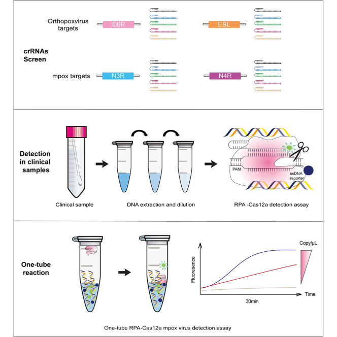
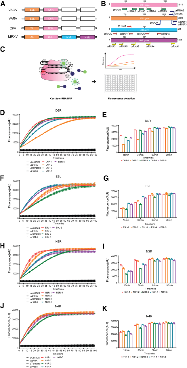
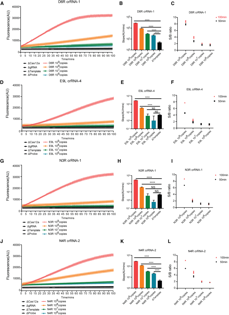
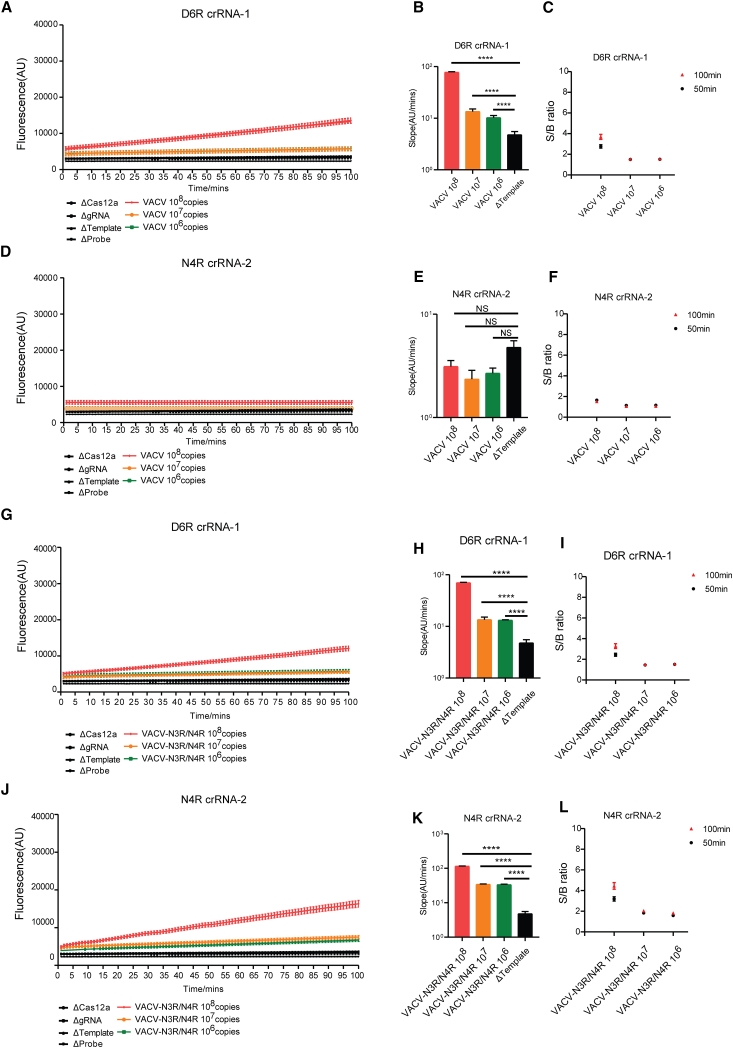
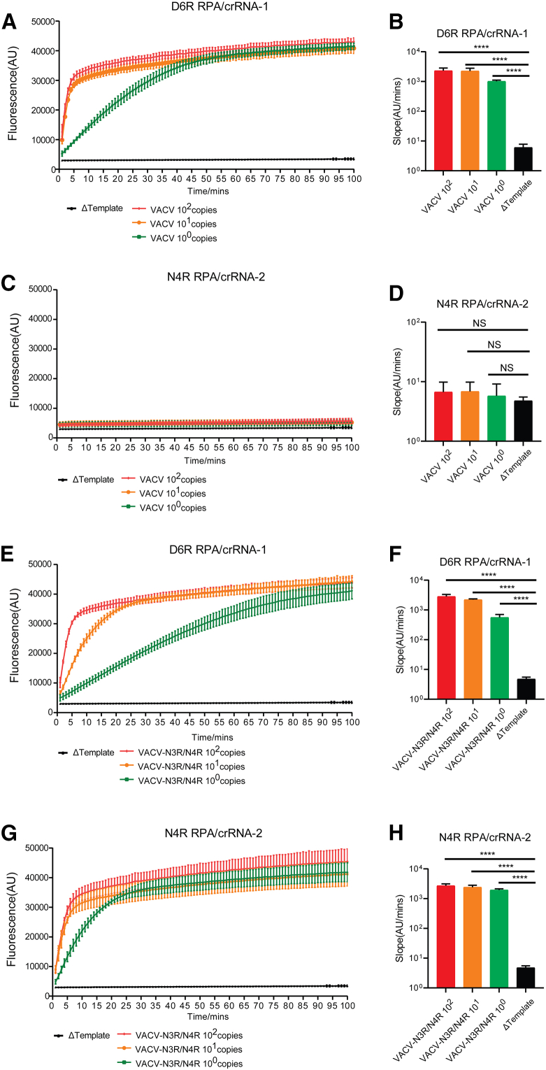
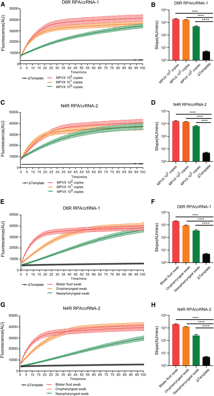
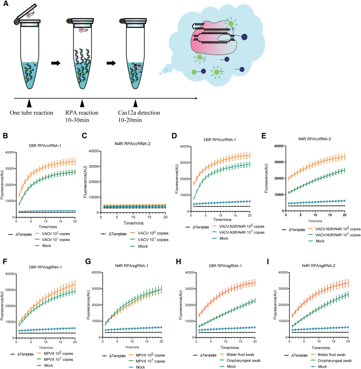
Mpox is caused by a zoonotic virus belonging to the Orthopoxvirus genus and the Poxviridae family. In this study, we develop a recombinase polymerase amplification (RPA)-coupled CRISPR-Cas12a detection assay for the mpox virus. We design and test a series of CRISPR-derived RNAs(crRNAs) targeting the conserved D6R and E9L genes for orthopoxvirus and the unique N3R and N4R genes for mpox viruses. D6R crRNA-1 exhibits the most robust activity in detecting orthopoxviruses, and N4R crRNA-2 is able to distinguish the mpox virus from other orthopoxviruses. The Cas12a/crRNA assay alone presents a detection limit of 108 copies of viral DNA, whereas coupling RPA increases the detection limit to 1-10 copies. The one-tube RPA-Cas12a assay can, therefore, detect viral DNA as low as 1 copy within 30 min and holds the promise of providing point-of-care detection for mpox viral infection.

Keywords: AsCas12a; CP: Biotechnology; CP: Microbiology; CRISPR; RPA; mpox detection; one-tube.


Conflict of interest statement

Declaration of interests F.G., F.Z., Y. Hu, F.X., and S.M. are inventors of pending and issued patents on CRISPR-Cas12a detection of mpox.

Figures

None
Graphical abstract
Figure 1
Figure 1
Screening crRNAs for detection of orthopoxviruses and mpox viruses together with Cas12a (A) Illustration of D6R and E9L genes that are conserved among four orthopoxviruses and genes N3R and N4R that are unique for mpox viruses. VACV, vaccinia virus; VARV, variola virus; CPV, cowpox virus; MPXV, mpox virus. (B) Depiction of the target sites of the designed crRNAs. All target genes are shown in the 5′ to 3′ direction. (C) Illustration of Cas12a-crRNA ribonucleoprotein (RNP) complex binding target DNA, resulting a cleavage of target DNA with its RuvC domain. Upon Cas12a activation, quenched-fluorophore DNA reporters were cleaved, and fluorescence signals were detected. (D, F, H, and J) Each crRNA was tested individually together with Cas12a against 1010 copies/reaction (final concentration: 0.05 nM) of each target plasmid DNA. Fluorescence signals were recorded every minute for 100 min. Background fluorescence in the absence of Cas12a, gRNA, template, or probe DNA is shown as ΔCas12a, ΔgRNA, Δtemplate, and Δprobe, respectively. Data are presented as mean ± standard deviation (SD) of three technical replicates. (E, G, I, and K) Shown are fluorescence signals at 15, 30, 60, and 90 min corresponding to (D), (F), (H), and (J). See also Figures S1 and S2 and Table S1.
Figure 2
Figure 2
The detection limit of Cas12a/crRNA for target plasmid DNA (A, D, G, and J) detection limits of crRNAs D6R-1, E9L-4, N3R-1, and N4R-2 were determined by testing 10-fold dilution of each target plasmid DNA ranging from 109 to 106 copies. Fluorescence signals over 100 min were recorded. Data are presented as mean ± SD of three technical replicates. (B, E, H, and K) Slopes of the curves over 100 min shown in (A), (D), (G), and (J) were calculated by performing simple linear regression to data merged from replicates and are shown as slope ±95% confidence interval. Slopes were compared to the Δtemplate alone control through an analysis of covariance (ANCOVA). NS, not significant; ∗∗∗∗p < 0.0001. (C, F, I, and L) Signal-to-background ratios (S/B ratios) at 50 (black) and 100 min (red) reaction times were calculated and are shown. Background fluorescence is the Δtemplate group. Data are presented as mean ± SD of three technical replicates. See also Figure S3.
Figure 3
Figure 3
Detection limit of Cas12a/crRNA for viral genomic DNA (A, D, G, and J) Detection limits of crRNAs D6R-1 and N4R-2 were determined by testing 10-fold dilution of each target viral genome ranging from 108 to 106 copies. Fluorescence signals over 100 min were recorded. Results are presented as mean ± SD of three technical replicates. VACV, vaccinia virus; VACV-N3R/N4R, recombinant VACV containing mpox virus N3R and N4R genes. (B, E, H, and K) Slopes of the curves over 100 min shown in (A), (D), (G), and (J) were calculated by performing simple linear regression to data merged from replicates and are shown as slope ±95% confidence interval. Slopes were compared to the Δtemplate alone control through an ANCOVA. NS, not significant; ∗∗∗∗p < 0.0001. (C, F, I, and L) S/B ratios at 50 (black) and 100 min (red) reaction times were calculated and are shown. Background fluorescence is the Δtemplate group. Data are presented as mean ± SD of three technical replicates. See also Figure S4.
Figure 4
Figure 4
Detection of viral genomic DNA with RPA-coupled Cas12a/crRNA (A, C, E, and G) VACV and VACV-N3R/N4R viral genomes were diluted to 102 copies, 101 copies, and 1 copy. Viral DNA samples were first amplified with RPA using both D6R primers and N4R primers and then added into the Cas12a/crRNA detection system. Fluorescence signals over 100 min were recorded. Background fluorescence is shown as Δtemplate. Data are presented as mean ± SD of three technical replicates. (B, D, F, and H) Slopes of the curves over 20 min shown in (A), (C), (E), and (G) were calculated by performing simple linear regression to data merged from replicates and are shown as slope ±95% confidence interval. Slopes were compared to the Δtemplate alone control through an ANCOVA. NS, not significant; ∗∗∗∗p < 0.0001. See also Figure S5.
Figure 5
Figure 5
Detection of authentic MPXV and clinical samples with RPA-Cas12a (A, C, E, and G) Viral DNA of MPXV and clinical samples was detected with the RPA-Cas12a system as shown in Figure 4. Fluorescence signals over 100 min are shown. Background fluorescence is shown as Δtemplate. Data are represented as mean ± SD of three technical replicates. (B, D, F, and H) Slopes of the curves over 20 min shown in (A), (C), (E), and (G) were calculated by performing simple linear regression to data merged from replicates and are shown as slope ±95% confidence interval. Slopes were compared to the Δtemplate alone control through an ANCOVA. NS, not significant; ∗∗∗∗p < 0.0001. See also Figures S6 and S7.
Figure 6
Figure 6
Detection of viral DNA with the one-tube RPA-Cas12a system (A) Illustration of the one-tube assay. Cas12a/crRNA RNP was added to the cap of the tube, and the RPA reagent was added to the tube bottom. After 15 min incubation at 37°C for the RPA reaction to complete, Cas12a/crRNA RNP was centrifuged to the bottom and mixed with the RPA products. Fluorescence signals were recorded for 20 min. (B–E) VACV and VACV-N3R/N4R viral DNA genomes were diluted to 102 and 101 copies/μL. RPA-Cas12a/crRNA reactions were performed as shown in (A). Viral DNA was amplified with RPA using both the D6R primers and the N4R primers. Data are presented as mean ± SD of three technical replicates. (F–I) MPXV viral DNA genomes and clinical samples in Figure 5. RPA-Cas12a/crRNA reactions were performed as shown in (A). Viral DNA was amplified with RPA using both the D6R primers and the N4R primers. Data are presented as mean ± SD of three technical replicates.

Similar articles

Cited by

References

    1. Aljabali A.A., Obeid M.A., Nusair M.B., Hmedat A., Tambuwala M.M. Monkeypox virus: An emerging epidemic. Microb. Pathog. 2022;173 doi: 10.1016/j.micpath.2022.105794. - DOI - PMC - PubMed
    1. Alakunle E., Moens U., Nchinda G., Okeke M.I. Monkeypox Virus in Nigeria: Infection Biology, Epidemiology, and Evolution. Viruses. 2020;12 doi: 10.3390/v12111257. - DOI - PMC - PubMed
    1. Dou D., Revol R., Östbye H., Wang H., Daniels R. Influenza A Virus Cell Entry, Replication, Virion Assembly and Movement. Front. Immunol. 2018;9:1581. doi: 10.3389/fimmu.2018.01581. - DOI - PMC - PubMed
    1. Lansiaux E., Jain N., Laivacuma S., Reinis A. The virology of human monkeypox virus (hMPXV): A brief overview. Virus Res. 2022;322 doi: 10.1016/j.virusres.2022.198932. - DOI - PMC - PubMed
    1. Breman J.G., Kalisa R., Steniowski M.V., Zanotto E., Gromyko A.I., Arita I. Human monkeypox, 1970-79. Bull. World Health Organ. 1980;58:165–182. - PMC - PubMed

Publication types