

Shaping transverse-tubules: central mechanisms that play a role in the cytosol zoning for muscle contraction
- PMID: 37848047
- PMCID: PMC10873525
- DOI: 10.1093/jb/mvad083
Shaping transverse-tubules: central mechanisms that play a role in the cytosol zoning for muscle contraction
Abstract
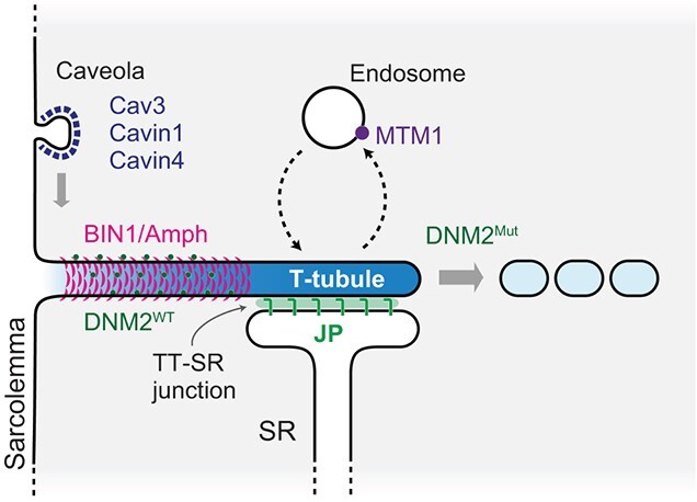
A transverse-tubule (T-tubule) is an invagination of the plasma membrane penetrating deep into muscle cells. An extensive membrane network of T-tubules is crucial for rapid and synchronized signal transmission from the cell surface to the entire sarcoplasmic reticulum for Ca2+ release, leading to muscle contraction. T-tubules are also indispensable for the formation and positioning of other muscle organelles. Their structure and physiological roles are relatively well established; however, the mechanisms shaping T-tubules require further elucidation. Centronuclear myopathy (CNM), an inherited muscular disorder, accompanies structural defects in T-tubules. Membrane traffic-related genes, including MTM1 (Myotubularin 1), DNM2 (Dynamin 2), and BIN1 (Bridging Integrator-1), were identified as causative genes of CNM. In addition, causative genes for other muscle diseases are also reported to be involved in the formation and maintenance of T-tubules. This review summarizes current knowledge on the mechanisms of how T-tubule formation and maintenance is regulated.
Keywords: BIN1/Amph; MTM; T-tubule; dynamin; tubulation.
© The Author(s) 2023. Published by Oxford University Press on behalf of the Japanese Biochemical Society.
Figures


References
-
- Simunovic, M., Evergren, E., Callan-Jones, A., and Bassereau, P. (2019) Curving cells inside and out: roles of BAR domain proteins in membrane shaping and its cellular implications. Annu. Rev. Cell Dev. Biol. 35, 111–129 - PubMed
Publication types
MeSH terms
Grants and funding
LinkOut - more resources
Full Text Sources
Miscellaneous
