. 2023 Oct 2:10:1189293.
doi: 10.3389/fcvm.2023.1189293. eCollection 2023.

Segmenting computed tomograms for cardiac ablation using machine learning leveraged by domain knowledge encoding

Affiliations

Segmenting computed tomograms for cardiac ablation using machine learning leveraged by domain knowledge encoding

Ruibin Feng et al. Front Cardiovasc Med. .

Abstract

Background: Segmentation of computed tomography (CT) is important for many clinical procedures including personalized cardiac ablation for the management of cardiac arrhythmias. While segmentation can be automated by machine learning (ML), it is limited by the need for large, labeled training data that may be difficult to obtain. We set out to combine ML of cardiac CT with domain knowledge, which reduces the need for large training datasets by encoding cardiac geometry, which we then tested in independent datasets and in a prospective study of atrial fibrillation (AF) ablation.

Methods: We mathematically represented atrial anatomy with simple geometric shapes and derived a model to parse cardiac structures in a small set of N = 6 digital hearts. The model, termed "virtual dissection," was used to train ML to segment cardiac CT in N = 20 patients, then tested in independent datasets and in a prospective study.

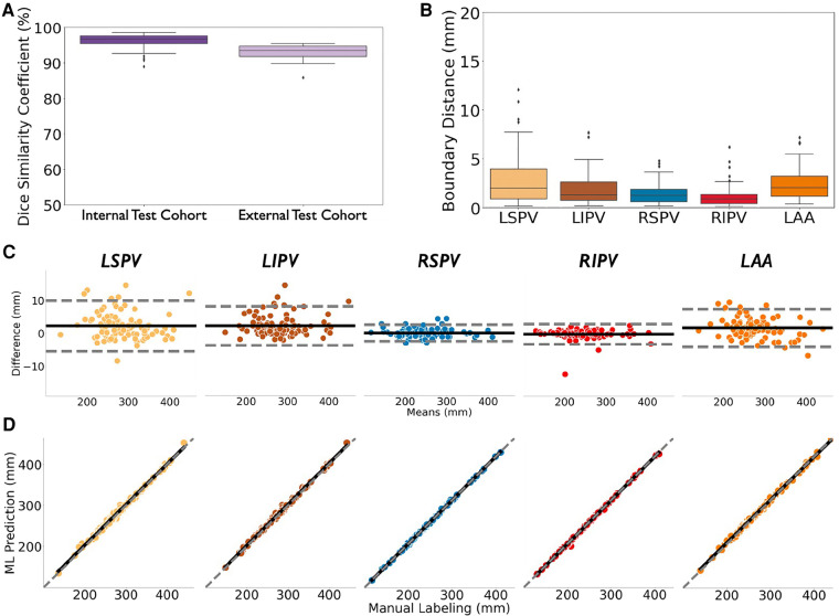
Results: In independent test cohorts (N = 160) from 2 Institutions with different CT scanners, atrial structures were accurately segmented with Dice scores of 96.7% in internal (IQR: 95.3%-97.7%) and 93.5% in external (IQR: 91.9%-94.7%) test data, with good agreement with experts (r = 0.99; p < 0.0001). In a prospective study of 42 patients at ablation, this approach reduced segmentation time by 85% (2.3 ± 0.8 vs. 15.0 ± 6.9 min, p < 0.0001), yet provided similar Dice scores to experts (93.9% (IQR: 93.0%-94.6%) vs. 94.4% (IQR: 92.8%-95.7%), p = NS).

Conclusions: Encoding cardiac geometry using mathematical models greatly accelerated training of ML to segment CT, reducing the need for large training sets while retaining accuracy in independent test data. Combining ML with domain knowledge may have broad applications.

Keywords: ablation; atrial fibrillation; cardiac CT segmentation; domain knowledge; machine learning; mathematical modeling.


Conflict of interest statement

SN reports grant support from the National Institutes of Health (R01 HL149134 and R01 HL83359), consulting from Uptodate Inc., and TDK Inc., intellectual property owned by University of California Regents and Stanford University. FT: Consulting honoraria to institution from Abbott, Boston Scientific, Daiichi Sankyo; no personal gain. AR: grants from NIH (F32HL144101), NIH LRP, and Stanford SSPS. PC: consulting at American College of Cardiology. MR: equity interests in Corify Health Care. The remaining authors declare that the research was conducted in the absence of any commercial or financial relationships that could be construed as a potential conflict of interest.

Figures

Figure 1
Figure 1
Concept and overview. (A) Conventional machine learning (top) can learn patterns in complex data, but requires laborious manual labeling, in large datasets which may be difficult to obtain. Conversely, our proposed approach (bottom) used natural intelligence to replace manual labeling with anatomical concepts encoded mathematically of domain knowledge, to learn rapidly from small datasets. (B) We applied mathematical encoding to segment heart CT scans via ML of small datasets. We represented heart structures as geometric primitives (“virtual dissection”). This was used to train ML on a small dataset (N = 20) and was able to accurately segment hearts in 2 larger cohorts from different institutions (N = 100, 60). In a prospective study (N = 42), the model segmented cardiac CT scans faster, but as accurately as experts. Acronyms: LA, left atrium; LSPV, left superior pulmonary vein; LIPV, left inferior pulmonary vein; RSPV, right superior pulmonary vein; RIPV, right inferior pulmonary vein; LAA, left atrial appendage.
Figure 2
Figure 2
Virtual Dissection algorithm. (A) The detailed pipeline. (B) The progress of the iterative erosion. The automatically selected iteration for erosion is highlighted in red. (C) The progress of the iterative dilation. The automatically selected iteration for dilation is highlighted in red. Acronyms: LA, left atrium; LSPV, left superior pulmonary vein; LIPV, left inferior pulmonary vein; RSPV, right superior pulmonary vein; RIPV, right inferior pulmonary vein; LAA, left atrial appendage.
Figure 3
Figure 3
Virtual dissection performance. (A) Representative samples of digital atria geometrically parsed by un-optimized algorithm. (B) Bland-Altman plots of the centroid-to-boundary of un-optimized algorithm vs. experts in 6 digital atria. After optimizing Virtual Dissection with N = 5 patient cases from the development cohort, (C) Representative patient atria from optimized algorithm in independent Test cohort (N = 100). (D) Bland–Altman plots of the centroid-to-boundary distance of optimized algorithm vs. experts in the Test cohort. (E) Success rate of virtual dissection algorithm using N={0, 5, 10, 20, 30} cases. Acronyms: LSPV, left superior pulmonary vein; LIPV, left inferior pulmonary vein; RSPV, right superior pulmonary vein; RIPV, right inferior pulmonary vein; LAA, left atrial appendage.
Figure 4
Figure 4
Comparison between the ML model predicted CT segmentation (left) and ground truth manual outlining (right) overlaid on the input CT scans in representative samples selected using 25th, 50th and 75th percentiles of segmentation accuracy in an independent test cohort (N = 100). Our ML model effectively captured the LA geometry, highlighting key features of PVs, LAA, and their ostia. The mitral valve plane represented in the 50th- and 25th- percentile samples showed slight variation between ML prediction and manual labeling, likely from limited image quality. Slight differences in PV measurements were found in the 25th-percentile sample, which may not be clinically relevant. Acronyms: LA, left atrium; LSPV, left superior pulmonary vein; LIPV, left inferior pulmonary vein; RSPV, right superior pulmonary vein; RIPV, right inferior pulmonary vein; LAA, left atrial appendage.
Figure 5
Figure 5
Accuracy CT segmentation using ML of optimized virtual dissection in two test cohorts (A) dice score of ML-based CT segmentation in the internal test cohort (N = 100; left) and an external test cohort from a different institution with different CT scanners (N = 60; right). (B) Boundary surface distances between ML-prediction and expert labelling in the Test Dataset (N = 100). (C) and (D) are Bland–Altman plots and linear regression plots of the centroid-to-boundary distance in the Test Dataset (N = 100). Acronyms: LSPV, left superior pulmonary vein; LIPV, left inferior pulmonary vein; RSPV, right superior pulmonary vein; RIPV, right inferior pulmonary vein; LAA, left atrial appendage.
Figure 6
Figure 6
Prospective segmentation of cardiac CT scans in 42 consecutive patients undergoing AF ablation by virtual-dissection trained ML vs. experts. (A,B) Virtual dissection trained ML significantly shortens segmentation time compared to experts. (C) Box plots of Dice similarity coefficient between ML and experts were similar. (D) and (E) LA volume and LA sphericity index marked by Virtual Dissection (red cross) accurately tracks the mean between experts (black cross).

Similar articles

Cited by

References

    1. Kirchhof P, Camm AJ, Goette A, Brandes A, Eckardt L, Elvan A, et al. Early rhythm-control therapy in patients with atrial fibrillation. N Engl J Med. (2020) 383(14):1305–16. 10.1056/NEJMoa2019422 - DOI - PubMed
    1. Narayan SM, Baykaner T, Clopton P, Schricker A, Lalani GG, Krummen DE, et al. Ablation of rotor and focal sources reduces late recurrence of atrial fibrillation compared with trigger ablation alone: extended follow-up of the confirm trial (conventional ablation for atrial fibrillation with or without focal impulse and rotor modulation). J Am Coll Cardiol. (2014) 63(17):1761–8. 10.1016/j.jacc.2014.02.543 - DOI - PMC - PubMed
    1. Biosense Webster I. Carto® 3 System Instructions for Use (V3.2.3) (2015). Available from: https://www.e-ifu.com/search-document-metadata/CARTO%C2%AE%203%20SYSTEM%...
    1. Zhu J, Liu Y, Zhang J, Wang Y, Chen L. Preliminary clinical study of the differences between interobserver evaluation and deep convolutional neural network-based segmentation of multiple organs at risk in Ct images of lung cancer. Front Oncol. (2019) 9:627. 10.3389/fonc.2019.00627. - DOI - PMC - PubMed
    1. LeCun Y, Bengio Y, Hinton G. Deep learning. Nature. (2015) 521(7553):436–44. 10.1038/nature14539 - DOI - PubMed