Diagnostic accuracy of noninvasive fractional flow reserve derived from computed tomography angiography in ischemia-specific coronary artery stenosis and indeterminate lesions: results from a multicenter study in China
- PMID: 37849942
- PMCID: PMC10577408
- DOI: 10.3389/fcvm.2023.1236405
Diagnostic accuracy of noninvasive fractional flow reserve derived from computed tomography angiography in ischemia-specific coronary artery stenosis and indeterminate lesions: results from a multicenter study in China
Abstract
Background: To determine the diagnostic performance of a novel computational fluid dynamics (CFD)-based algorithm for in situ CT-FFR in patients with ischemia-induced coronary artery stenosis. Additionally, we investigated whether the diagnostic accuracy of CT-FFR differs significantly across the spectrum of disease and analyzed the influencing factors that contribute to misdiagnosis.
Methods: Coronary computed tomography angiography (CCTA), invasive coronary angiography (ICA), and FFR were performed on 324 vessels from 301 patients from six clinical medical centers. Local investigators used CCTA and ICA to conduct assessments of stenosis, and CT-FFR calculations were performed in the core laboratory. For CCTA and ICA, CT-FFR ≤ 0.8 and a stenosis diameter ≥ 50% were identified as lesion-specific ischemia. Univariate logistic regression models were used to assess the effect of features on discordant lesions (false negative and false positive) in different CT-FFR categories. The diagnostic performance of CT-FFR was analyzed using an invasive FFR ≤ 0.8 as the gold standard.
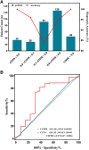
Results: The Youden index indicated an optimal threshold of 0.80 for CT-FFR to identify functionally ischemic lesions. On a per-patient basis, the diagnostic sensitivity, specificity, accuracy, positive predictive value (PPV), and negative predictive value (NPV) for CT-FFR were 96% (91%-98%), 92% (87%-96%), 94% (90%-96%), 91% (85%-95%), and 96% (92%-99%), respectively. The diagnostic efficacy of CT-FFR was higher than that of CCTA without the influence of calcification. Closer to the cut point, there was less certainty, with the agreement between the invasive FFR and the CT-FFR being at its lowest in the CT-FFR range of 0.7-0.8. In all lesions, luminal stenosis ≥ 50% significantly affected the risk of reduced false negatives (FN) and false positives (FP) results by CT-FFR, irrespective of the association with calcified plaque.
Conclusions: In summary, CT-FFR based on the new parameter-optimized CFD model has a better diagnostic performance than CTA for lesion-specific ischemia. The presence of calcified plaque has no significant effect on the diagnostic performance of CT-FFR and is independent of the degree of calcification. Given the range of applicability of our software, its use at a CT-FFR of 0.7-0.8 requires caution and must be considered in the context of multiple factors.
Keywords: computational fluid dynamics (CFD); computed tomography (CTA); coronary artery disease (CAD); coronary computed tomography-derived fractional flow reserve (CT-FFR); indeterminate lesions.
© 2023 Ding, Li, Zhang, Tang, Zhang, Yang, Shou, Ye, Zhao, Ye, Zhang, Liu and Zeng.
Conflict of interest statement
CZ and YL were employed by Shenzhen Escope Technology Ltd. The remaining authors declare that the research was conducted in the absence of any commercial or financial relationships that could be construed as a potential conflict of interest.
Figures


Similar articles
-
Diagnostic performance of coronary computed tomography (CT) angiography derived fractional flow reserve (CTFFR) in patients with coronary artery calcification: insights from multi-center experiments in China.Ann Transl Med. 2022 Jul;10(14):788. doi: 10.21037/atm-22-3180. Ann Transl Med. 2022. PMID: 35965817 Free PMC article.
-
Noninvasive diagnosis of ischemia-causing coronary stenosis using CT angiography: diagnostic value of transluminal attenuation gradient and fractional flow reserve computed from coronary CT angiography compared to invasively measured fractional flow reserve.JACC Cardiovasc Imaging. 2012 Nov;5(11):1088-96. doi: 10.1016/j.jcmg.2012.09.002. JACC Cardiovasc Imaging. 2012. PMID: 23153908
-
Diagnosis of ischemia-causing coronary stenoses by noninvasive fractional flow reserve computed from coronary computed tomographic angiograms. Results from the prospective multicenter DISCOVER-FLOW (Diagnosis of Ischemia-Causing Stenoses Obtained Via Noninvasive Fractional Flow Reserve) study.J Am Coll Cardiol. 2011 Nov 1;58(19):1989-97. doi: 10.1016/j.jacc.2011.06.066. J Am Coll Cardiol. 2011. PMID: 22032711 Clinical Trial.
-
Meta-Analysis of Diagnostic Performance of Coronary Computed Tomography Angiography, Computed Tomography Perfusion, and Computed Tomography-Fractional Flow Reserve in Functional Myocardial Ischemia Assessment Versus Invasive Fractional Flow Reserve.Am J Cardiol. 2015 Nov 1;116(9):1469-78. doi: 10.1016/j.amjcard.2015.07.078. Epub 2015 Aug 14. Am J Cardiol. 2015. PMID: 26347004 Free PMC article. Review.
-
Computed tomography angiography-derived fractional flow reserve (CT-FFR) for the detection of myocardial ischemia with invasive fractional flow reserve as reference: systematic review and meta-analysis.Eur Radiol. 2020 Feb;30(2):712-725. doi: 10.1007/s00330-019-06470-8. Epub 2019 Nov 6. Eur Radiol. 2020. PMID: 31696294
Cited by
-
Computed Tomography-Derived Fractional Flow Reserve: Developing A Gold Standard for Coronary Artery Disease Diagnostics.Rev Cardiovasc Med. 2024 Oct 22;25(10):372. doi: 10.31083/j.rcm2510372. eCollection 2024 Oct. Rev Cardiovasc Med. 2024. PMID: 39484113 Free PMC article. Review.
-
Diagnostic performance of the quantitative flow ratio and CT-FFR for coronary lesion-specific ischemia.Sci Rep. 2024 Jul 23;14(1):16969. doi: 10.1038/s41598-024-68212-1. Sci Rep. 2024. PMID: 39043839 Free PMC article.
-
Artificial intelligence-enhanced detection of subclinical coronary artery disease in athletes: diagnostic performance and limitations.Int J Cardiovasc Imaging. 2024 Dec;40(12):2503-2511. doi: 10.1007/s10554-024-03256-y. Epub 2024 Oct 7. Int J Cardiovasc Imaging. 2024. PMID: 39373817 Free PMC article.
References
-
- Leipsic J, Abbara S, Achenbach S, Cury R, Earls JP, Mancini GBJ, et al. SCCT guidelines for the interpretation and reporting of coronary CT angiography: a report of the society of cardiovascular computed tomography guidelines committee. J Cardiovasc Comput Tomogr. (2014) 8(5):342–58. 10.1016/j.jcct.2014.07.003 - DOI - PubMed
-
- Budoff MJ, Dowe D, Jollis JG, Gitter M, Sutherland J, Halamert E, et al. Diagnostic performance of 64-multidetector row coronary computed tomographic angiography for evaluation of coronary artery stenosis in individuals without known coronary artery disease: results from the prospective multicenter ACCURACY (assessment by coronary computed tomographic angiography of individuals undergoing invasive coronary angiography) trial. J Am Coll Cardiol. (2008) 52(21):1724–32. 10.1016/j.jacc.2008.07.031 - DOI - PubMed
-
- Montalescot G, Sechtem U, Achenbach S, Andreotti F, Arden C, Budaj A, et al. 2013 ESC guidelines on the management of stable coronary artery disease: the task force on the management of stable coronary artery disease of the European Society of Cardiology. Eur Heart J (2013) 34(38):2949–3003. 10.1093/eurheartj/eht296 - DOI - PubMed
LinkOut - more resources
Full Text Sources
Miscellaneous

